

Management of periapical lesion with persistent exsudate
- PMID: 35262549
- PMCID: PMC9645141
- DOI: 10.1590/0103-6440202204818
Management of periapical lesion with persistent exsudate
Abstract
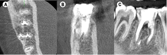
This case describes the treatment and follow-up of a mandibular molar in an 18-year-old female with a periapical cyst. Thus, it becomes important to know which options should we take when faced with a clinical situation that we cannot resolve through conventional methods, and which techniques and approaches we have to achieve treatment success. This case showed the treatment plan and follow-up, by the use of CBCT images, from a previously treated mandibular molar with a large periapical abscess and cystic lesion, in which, the first treatment plan approach was to make the endodontic retreatment. During the chemo-mechanical preparation the presence of permanent intracanal purulent exudate made it impossible to dry the canals, impeding obturation of the root canal system. Due to this clinical situation, a surgical approach was performed with the intention of reduce this permanent exudate and to execute a decompression technique. Clinical findings, periapical radiographs and cone beam computed tomographic, indicated almost complete resolution of the radiolucency, after a one-year follow-up.
Este relato de caso descreve o tratamento e acompanhamento de um molar inferior em uma jovem de 18 anos com cisto periapical. Assim, torna-se importante saber quais opções devemos tomar diante de uma situação clínica que não podemos resolver pelos métodos convencionais, e quais técnicas e abordagens temos para alcançar o sucesso do tratamento. Este caso mostrou o plano de tratamento e acompanhamento, por meio de imagens de tomografia computadorizada de feixe cônico (TCFC), de um molar inferior previamente tratado com grande abscesso periapical e lesão cística, no qual, a primeira abordagem do plano de tratamento foi fazer o retratamento endodôntico. Durante o preparo químico-mecânico a presença de exsudato purulento intracanal permanente impossibilitou a secagem dos canais, impedindo a obturação do sistema de canais radiculares. Devido a essa situação clínica, foi realizada abordagem cirúrgica com a intenção de reduzir esse exsudato permanente e executar uma técnica de descompressão. Após um ano de acompanhamento, os achados clínicos e radiográficos indicaram processo de reparo.
Conflict of interest statement
The authors have stated explicitly that there are no conflicts of interest in connection with this article
Figures


References
-
- Ricucci D, Rôças IN, Hernández S, Siqueira JF., Jr "True" Versus "Bay" Apical Cysts: Clinical, Radiographic, Histopathologic, and Histobacteriologic Features. J Endod. 2020;9:1217–1227. - PubMed
-
- Berretta LM, Melo G, Mello FW, Lizio G, Rivero ERC. Effectiveness of marsupialisation and decompression on the reduction of cystic jaw lesions: a systematic review. Br J Oral Maxillofac Surg. 2021;23 S0266-4356(21)00109-1. - PubMed
-
- El-Naggar AK, Chan JKC, Grandis JR, Takata T, Slootweg PJ. Classification of Head and Neck Tumours. (4th) 2017;9
Publication types
MeSH terms
LinkOut - more resources
Full Text Sources
