. 2022 Feb;118(2):488-502.
doi: 10.36660/abc.20201233.

Ventricular Synchrony in Para-Hisian Cardiac Pacing as an Alternative for Physiological Cardiac Activation (Indirect Recruitment of the His Bundle?)

[Article in English, Portuguese]
Affiliations

Ventricular Synchrony in Para-Hisian Cardiac Pacing as an Alternative for Physiological Cardiac Activation (Indirect Recruitment of the His Bundle?)

[Article in English, Portuguese]
Andres Di Leoni Ferrari et al. Arq Bras Cardiol. 2022 Feb.

Abstract

Background: Artificial cardiac pacing by direct or indirect His bundle capture results in synchronous ventricular contraction (physiological pacing).

Objectives: To compare cardiac synchronization, technical characteristics, and electronic parameters between two techniques of indirect His-bundle pacing: non-selective (NS-HBP) vs para-Hisian pacing (PHP).

Methods: The experimental intervention (between November 2019 and April 2020) consisted of implanting a DDD pacemaker in patients who had left ventricular ejection fraction (LVEF) > 35%. The resulting cardiac synchronization was compared using an electrocardiographic algorithm that analyzed QRS variation and the technical characteristics of non-selective Hisian pacing (DDD-His) and para-Hisian pacing (DDD-Var).

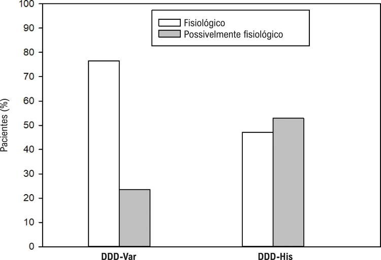
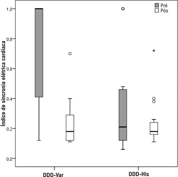
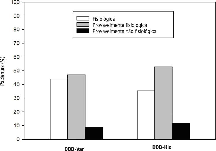
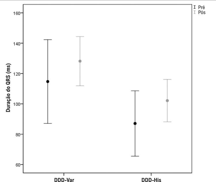
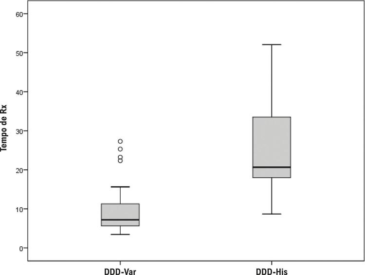
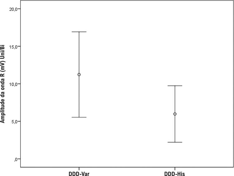
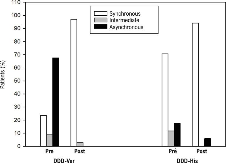
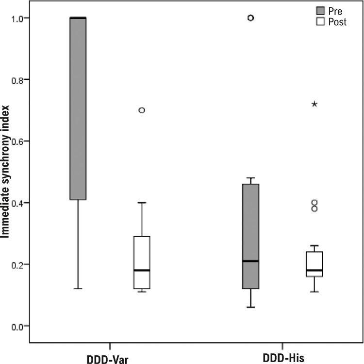
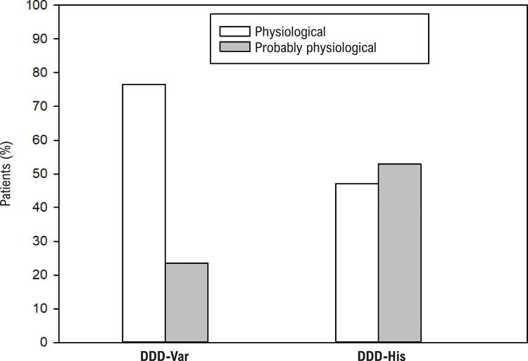
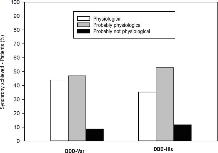
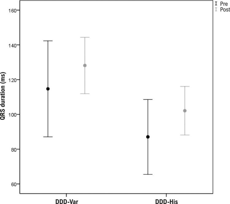
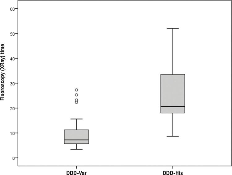
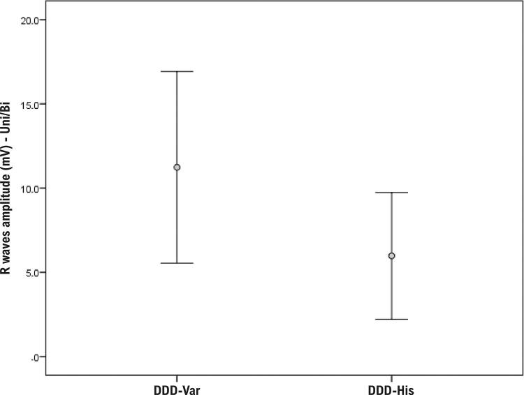
Results: Of 51 total patients (men: 28), 66.7% (34) were allocated to the DDD-Var group and 33.3% (17) to the DDD-His group. The mean ages in each group were 74 and 79 years, respectively. In the DDD-Var group, QRS variation (ventricular synchrony) improved after implantation (p < 0.001). In post-implantation ECG, 91.2% of the DDD-Var group presented a physiological pacing pattern, which was similar to the DDD-His group (88.2%; p = 0.999). The paced QRS axis was also similar (physiological) for both groups. Intraoperative fluoroscopy time (XRay) during implantation was lower for the para-Hisian technique (median 7 min in the DDD-Var group vs 21 min in the DDD-His group, p < 0.001). The mean QRS duration increased in the DDD-Var group (114.7 ms pre-implantation vs 128.2 ms post-implantation, p = 0.044). The mean post-implantation R-wave amplitude was 11.2 mV in the DDD-Var group vs 6.0 mV in the DDD-His group, p = 0.001.

Conclusion: Para-Hisian pacing appears to indirectly recruit the His bundle, which would make this an effective and comparable strategy for physiological pacing, resulting in synchronous ventricular contraction similar to that of non-selective Hisian pacing.

Fundamento: A estimulação cardíaca artificial (ECA) por captura direta ou indireta do feixe de His resulta em contração ventricular sincrônica (ECA fisiológica).

Objetivos: Comparar sincronia cardíaca, características técnicas e resultados de parâmetros eletrônicos entre duas técnicas de ECA indireta do feixe de His: a não seletiva e a parahissiana.

Métodos: Intervenção experimental (novembro de 2019 a abril de 2020) com implante de marca-passo definitivo (MPd) DDD em pacientes com fração de ejeção ventricular esquerda > 35%. Foram comparadas a sincronia cardíaca resultante mediante algoritmo de análise eletrocardiográfica da variância espacial do QRS e as características técnicas associadas a cada método entre ECA hissiana não seletiva (DDD-His) e parahissiana (DDD-Var).

Resultados: De 51 pacientes (28 homens), 34 (66,7%) foram alocados no grupo DDD-Var e 17 (33,3%), no grupo DDD-His, com idade média de 74 e 79 anos, respectivamente. No grupo DDD-Var, a análise da variância espacial do QRS (índice de sincronia ventricular) mostrou melhora após o implante de MPd (p < 0,001). Ao ECG pós-implante, 91,2% dos pacientes do grupo DDD-Var mostraram padrão fisiológico de ECA, comprovando ativação similar à do DDD-His (88,2%; p = 0,999). O eixo do QRS estimulado também foi similar (fisiológico) para ambos os grupos. A mediana do tempo de fluoroscopia do implante foi de 7 minutos no grupo DDD-Var e de 21 minutos no DDD-His (p < 0,001), favorecendo a técnica parahissiana. A duração média do QRS aumentou nos pacientes do DDD-Var (114,7 ms pré-MPd e 128,2 ms pós-implante, p = 0,044). A detecção da onda R foi de 11,2 mV no grupo DDD-Var e de 6,0 mV no DDD-His (p = 0,001).

Conclusão: A ECA parahissiana comprova recrutamento indireto do feixe de His, mostrando-se uma estratégia eficaz e comparável à ECA fisiológica ao resultar em contração ventricular sincrônica similar à obtida por captura hissiana não seletiva.


Conflict of interest statement

Potencial conflito de interesse

Não há conflito com o presente artigo
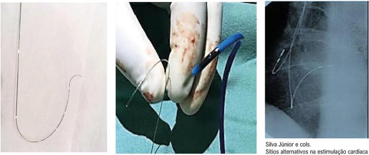
Figures

Figura 1
Figura 1. – Esquerda: estilete guia manualmente moldado para direcionamento e implante de eletrodo do ventrículo direito (VD) no 1/3 proximal do septo interventricular para a estimulação parahissiana. Centro: Operador (ADLF) mostrando comparação do formato obtido pela moldagem da guia com a curvatura de uma das bainhas pré-moldada disponível no Brasil (C315His Medtronic™). Direita: fluoroscopia oblíqua esquerda mostrando a posição final do eletrodo do VD. Nota-se angulação da ponta, perpendicular à coluna. Adaptado de Ortega et al. e Júnior et al.12,19
Figura 2A
Figura 2A. – Correlação da variância espacial do QRS e valores normais para um paciente com condução intraventricular intacta (derivações II e V6). Esquerda: traçados de eletrocardiograma. Centro: segmentos QRS sobrepostos (IIqrs e V6qrs). Direita: análise de correlação cruzada das derivações II e V6. Os picos dos complexos QRS coincidem, e o sinal de correlação cruzada mostra o seu máximo no tempo 0 (CorS = 0). CorS: deslocamento de correlação cruzada (ms); CorW: largura de correlação cruzada (ms); CorA: amplitude de correlação cruzada (mV); AII: área sob a derivação II; AV6: área sob a derivação V6. Adaptado de Bonomini et al.22
Figura 2B
Figura 2B. – Curvas com diferentes morfologias obtidas com o SynchromaxTM de acordo com o índice imediato de sincronia obtido em tempo real a partir do local de estimulação cardíaca artificial. Traços azuis: análise de variância do QRS da derivação II. Traços vermelhos: análise da variância espacial do QRS da derivação V6. BRD: Bloqueio de ramo direito; BRE: Bloqueio de ramo esquerdo; TRC: Terapia de Ressincronização Cardíaca; BDAS: Bloqueio Divisional ânterossuperior esquerdo; VD: ventrículo direito.
Figura 3
Figura 3. – Modelo eletrocardiográfico proposto por Burri et al.26 pela combinação de a) ausência de plateau em D1; b) ausência de entalhe (notch) na derivação V1; c) duração da ativação elétrica até o pico da onda R (RWPT) em V6 < 100 ms.28–30 Os três parâmetros presentes (a, b, c) indicam estimulação cardíaca artificial (ECA) fisiológica semelhante à NS-HBP e descartam ativação ventricular puramente miocárdica. A presença de dois critérios determina ECA provavelmente fisiológica, enquanto a presença de apenas um critério indica ECA provavelmente não fisiológica. A ausência de todos os elementos denota captura puramente do miocárdio inespecífico (ECA miocárdica).28
Figura 4
Figura 4. – Frequência de distribuição das categorias de sincronia cardíaca pré e pós-operatória entre os grupos.
Figura 5
Figura 5. – Comparação do índice imediato de sincronia elétrica cardíaca entre os grupos.
Figura 6
Figura 6. – Gráfico comparativo do eixo do eletrocardiograma entre os grupos.
Figura 7
Figura 7. – Gráfico comparativo da sincronia cardiaca (Iimes) obtida com ambos métodos de estimulação cardíaca artificial
Figura 8
Figura 8. – Diferenças de duração do complexo QRS pré e pós-implante entre os grupos.
Figura 9
Figura 9. – Comparativo das médias de uso de fluoroscopia (Rx) entre os grupos.
Figura 10
Figura 10. – Diferença da amplitude detectada nas ondas R entre os dois grupos.
Figure 1
Figure 1. – Left: a hand-shaped stylet guiding the positioning of the RV lead in the uupermost proximal third of the interventricular septum for para-Hisian pacing. Center: Operator (ADLF) showing a comparison of the shape obtained by molding the guide wire with the curvature of one of the pre-molded sheaths available in Brazil (C315His Medtronic™). Right: fluoroscopy (left oblique projection) showing the final position of the lead in the right ventricle. Note the angulation of the tip, which is perpendicular to the spine. Adapted from12,19
Figure 2A
Figure 2A. – Correlation of QRS variation and normal values for a patient with intact intraventricular conduction (leads II and V6). Left: Conventional ECG traces. Center: overlapping QRS segments (D2 QRS and V6 QRS). Right: Cross-correlation analysis of leads II and V6. The QRS peaks coincide and the maximum cross-correlation signal is at time zero (CorS = 0). CorS: cross-correlation offset (ms). CorW: cross-correlation width (ms), CorA: cross-correlation amplitude (mV), AII: area under lead D2, aV6: area under lead V6. Adapted from Bonomini et al.22
Figure 2B
Figure 2B. – Curves obtained with SynchromaxTM according to the immediate synchrony index performed in real time from the pacing site in relation to cardiac synchrony obtained from the RV pacing site. Blue lines: QRS variation analysis from lead II. Red dashes: QRS variation analysis for lead V6.CRT: cardiac resynchronization therapy; LAH: left anterior hemiblock; RV: right ventricle. LBBB: Left bundle branch block. RBB: Right bundle block
Figure 3
Figure 3. – Electrocardiographic model proposed by Burri et al.26 including a combination of: a) absence of plateau in D1; b) absence of notching in lead V1; c) R-wave peak time in V6 < 100 ms.28–30 The presence of a, b, and c indicates “physiological” pacing in NS-HBP and rule out purely myocardial activation. The presence of 2 of criteria indicates “probably physiological” pacing, while the presence of only one criterion indicates “probably not physiological” pacing. The absence of all criteria indicates purely nonspecific myocardial capture (myocardial pacing).28
Figure 4
Figure 4. – Distribution frequency of pre- and postoperative cardiac synchronization categories between the para-Hisian pacing (DDD-Var) and non-selective His pacing (DDD-His) groups.
Figure 5
Figure 5. – Comparison of the immediate synchrony index between the para-Hisian pacing (DDD-Var) and non-selective His pacing (DDD-His) groups.
Figure 6
Figure 6. – Comparison of the ECG axis between the para-Hisian pacing (DDD-Var) and non-selective His pacing (DDD-His) groups.
Figure 7
Figure 7. – Comparative chart of cardiac synchrony (Iimes) obtained with both methods of artificial cardiac stimulation.
Figure 8
Figure 8. – Pre- and post-implantation differences in QRS complex duration between the para-Hisian pacing (DDD-Var) and non-selective His pacing (DDD-His) groups.
Figure 9
Figure 9. – Comparison of mean intraoperative XRay time between the para-Hisian pacing (DDD-Var) and non-selective His pacing (DDD-His) groups.
Figure 10
Figure 10. – Difference in R wave amplitude between the para-Hisian pacing (DDD-Var) and non-selective His pacing (DDD-His) groups.

Comment in

References

    1. Cho SW, Gwag HB, Hwang JK, Chun KJ, Park KM, On YK, et al. Clinical Features, Predictors, and Long-Term Prognosis of Pacing-Induced Cardiomyopathy. Eur J Heart Fail. 2019;21(5):643-51. doi: 10.1002/ejhf.1427. - PubMed
    1. Safak E, Ince H, Gkouvatsou L, Schultheiss HP, Ortak J, Caglayan E, et al. Pacing-Induced Cardiomyopathy in Chronic right Ventricular Apical Pacing: A Midterm Follow-Up Study. Eur J Med Res. 2019 Jul;24(1):23. doi: 10.1186/s40001-019-0386-5. - PMC - PubMed
    1. Matusik PT. Adverse Clinical Outcomes Related to Right Ventricular Pacing. Eur Heart J. 2019;40(20):1586-88. doi: 10.1093/eurheartj/ehz279. - PubMed
    1. Ferrari AD, Borges AP, Albuquerque LC, Sussenbach CP, Rosa PR, Piantá RM, et al. Cardiomyopathy Induced by Artificial Cardiac Pacing: Myth or Reality Sustained by Evidence? Rev Bras Cir Cardiovasc. 2014;29(3):402-13. doi: 10.5935/1678-9741.20140104. - PMC - PubMed
    1. Ferrari ADL. Avaliação de Dessincronia Atrioventricular em Portadores de Marca-Passo Bicameral Devido a Doença do Nó Sinusal e Bloqueio Atrioventricular de Primeiro Grau [dissertation]. São Paulo: Universidade de São Paulo; 2017. doi: 10.11606/T.98.2017.tde-05102017-074636.