Review
. 2022 Mar 2;63(3):5.
doi: 10.1167/iovs.63.3.5.

The Role of GJD2(Cx36) in Refractive Error Development

Affiliations
Review

The Role of GJD2(Cx36) in Refractive Error Development

Emilie van der Sande et al. Invest Ophthalmol Vis Sci. .

Abstract

Refractive errors are common eye disorders characterized by a mismatch between the focal power of the eye and its axial length. An increased axial length is a common cause of the refractive error myopia (nearsightedness). The substantial increase in myopia prevalence over the last decades has raised public health concerns because myopia can lead to severe ocular complications later in life. Genomewide association studies (GWAS) have made considerable contributions to the understanding of the genetic architecture of refractive errors. Among the hundreds of genetic variants identified, common variants near the gap junction delta-2 (GJD2) gene have consistently been reported as one of the top hits. GJD2 encodes the connexin 36 (Cx36) protein, which forms gap junction channels and is highly expressed in the neural retina. In this review, we provide current evidence that links GJD2(Cx36) to the development of myopia. We summarize the gap junctional communication in the eye and the specific role of GJD2(Cx36) in retinal processing of visual signals. Finally, we discuss the pathways involving dopamine and gap junction phosphorylation and coupling as potential mechanisms that may explain the role of GJD2(Cx36) in refractive error development.


Conflict of interest statement

Disclosure: E. van der Sande, None; A.E.G. Haarman, None; W.H. Quint, None; K.C.D. Tadema, None; M.A. Meester-Smoor, None; M. Kamermans, None; C.I. De Zeeuw, None; C.C.W. Klaver, None; B.H.J. Winkelman, None; A.I. Iglesias, None

Figures

Figure 1.
Figure 1.
Structure and conservation of GJD2(Cx36). Panel (A) shows the GJD2(Cx36) protein consisting of four transmembrane domains (TMD1-4), alternated by two extracellular loops (EL1-2), with the phosphorylation sites on the intracellular loop (IL; Ser110) and on the C-terminus (COOH; Ser293, Ser276 for zebrafish). Panel (B) shows the conservation of GJD2(Cx36) throughout commonly used species for myopia research. The phylogenetic tree in vertebrate lineages of reptiles and birds shows two subfamilies, GJD2(Cx35/Cx36) and GJD1(Cx34). The latter paralog is not present in mammals, whereas in teleost fish four functional orthologs have been identified (gjd1a(Cx34.1), gjd1b(Cx34.7), gjd2b(Cx35.1), and gjd2a(Cx35.5)). Panel (C) shows that for mammalian species 98% to 100% and for zebrafish 70% to 83% of the GJD2(Cx35/Cx36) protein is conserved relative to the human protein. Intergenic variation is mainly located in the intracellular loop (red trace) and in the C-terminus (final black trace), whereas the two phosphorylation sites are conserved throughout all species *P).
Figure 2.
Figure 2.
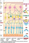
Gap junction proteins in the human retina and their coupling with GJD2(Cx36). Panel (A) shows the gap junction proteins present in the different retinal layers. Panels (B) and (C) show the gap junction coupling that contains GJD2(Cx36). Dot colors define the different gap junction proteins. Numbers represent the species for which the gap junction locations have been described. Gap junctions are detected at the protein level, when additionally detected on RNA (including cDNA) level, color dots are marked with an asterisk. Homotypic GJD2(Cx36)-containing gap junctions are present between dendrites of alpha ganglion cells, between dendrites of AII amacrine cells, between (ON/OFF) cone bipolar cells, between cones, and between rods and cones. For the latter homotypic configuration, most evidence localizes GJD2(Cx36) on the cone side. GJA7(Cx45) is the only gap junction protein forming channels with GJD2(Cx36). Heterotypic channels are present between subtypes of amacrine cells (providing GJD2(Cx36)) and (ON/OFF) cone bipolar cells (providing GJA7(Cx45)). GJA1(Cx43), GJA4(Cx37), and GJA10 (hCx62, mCx57, and pCx60) are the remaining gap junction proteins in the retina, localized between horizontal cells, but these do not colocalize with GJD2(Cx36). In addition to the gap junctions visualized in the figure, GJD2(Cx36) gap junctions have been reported between amacrine cells and alpha ganglion cells. Abbreviations: A, amacrine cell; BC, bipolar cells; C, cone photoreceptor cell; GC, ganglion cell; GCL, ganglion cell layer; H, horizontal cell; ILM, inner limiting membrane; INL, inner nuclear layer; IPL, inner plexiform layer; IS, inner segment; NFL, optic nerve fiber layer; OLM, outer limiting membrane; ONL, outer nuclear layer; OPL, outer plexiform layer; OS, outer segment; R, rod photoreceptor cell; RPE, retinal pigmented epithelium.
Figure 3.
Figure 3.
Gap junction proteins (connexins) in the eye. Color dots define the different gap junction proteins. Numbers represent the species for which the gap junction locations have been described. Gap junctions are detected at the protein level, when additionally detected on RNA (including cDNA) level color dots are marked with an asterisk. GJD2(Cx36) is expressed in the retina exclusively. GJA1(Cx43) is widely expressed throughout the eyes and is identified in all ocular segments.
Figure 4.
Figure 4.
Overview of potential mechanisms for GJD2(Cx36) (marked red) causing myopia. Green arrows indicate a positive (i.e. stimulatory) effect; red arrows a negative (i.e. inhibiting) effect. Dotted lines indicate hypothesized mechanisms not fully advocated yet by the literature but mentioned in the current manuscript as possible mechanisms. Conditions highlighted with black triangles are frequently linked in the literature with decreased myopia/axial elongation, whereas white triangles are associated with increased myopia/axial elongation.

References

    1. Németh J, Tapasztó B, Aclimandos WA, et al. .. Update and guidance on management of myopia. European Society of Ophthalmology in cooperation with International Myopia Institute. Eur J Ophthalmol . Published online March 5, 2021: 1120672121998960. - PMC - PubMed
    1. Haarman AEG, Enthoven CA, Tideman JWL, Tedja MS, Verhoeven VJM, Klaver CCW.. The complications of myopia: A review and meta-analysis. Invest Ophthalmol Vis Sci . 2020; 61(4): 49. - PMC - PubMed
    1. Tideman JWL, Snabel MCC, Tedja MS, et al. .. Association of Axial Length With Risk of Uncorrectable Visual Impairment for Europeans With Myopia. JAMA Ophthalmol. 2016; 134(12): 1355–1363. - PubMed
    1. Verhoeven VJM, Wong KT, Buitendijk GHS, Hofman A, Vingerling JR, Klaver CCW.. Visual consequences of refractive errors in the general population. Ophthalmology. 2015; 122(1): 101–109. - PubMed
    1. Grzybowski A, Kanclerz P, Tsubota K, Lanca C, Saw S-M.. A review on the epidemiology of myopia in school children worldwide. BMC Ophthalmol. 2020; 20(1): 27. - PMC - PubMed

Publication types