Review
. 2022 Mar 11;8(10):eabl9653.
doi: 10.1126/sciadv.abl9653. Epub 2022 Mar 9.

Biomineralization: Integrating mechanism and evolutionary history

Affiliations
Review

Biomineralization: Integrating mechanism and evolutionary history

Pupa U P A Gilbert et al. Sci Adv. .

Abstract

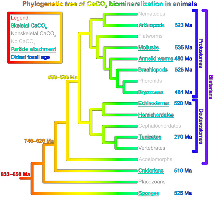
Calcium carbonate (CaCO3) biomineralizing organisms have played major roles in the history of life and the global carbon cycle during the past 541 Ma. Both marine diversification and mass extinctions reflect physiological responses to environmental changes through time. An integrated understanding of carbonate biomineralization is necessary to illuminate this evolutionary record and to understand how modern organisms will respond to 21st century global change. Biomineralization evolved independently but convergently across phyla, suggesting a unity of mechanism that transcends biological differences. In this review, we combine CaCO3 skeleton formation mechanisms with constraints from evolutionary history, omics, and a meta-analysis of isotopic data to develop a plausible model for CaCO3 biomineralization applicable to all phyla. The model provides a framework for understanding the environmental sensitivity of marine calcifiers, past mass extinctions, and resilience in 21st century acidifying oceans. Thus, it frames questions about the past, present, and future of CaCO3 biomineralizing organisms.


Figures

Fig. 1.
Fig. 1.. Phylogenetic distribution of CaCO3 biomineralization in animals.
Besides animals (shown), there are species (not shown) that make CaCO3 skeletons in the foraminiferans, coccolithophorids, green algae, red algae, dinoflagellates, and even a few amoebozoans and brown algae. CaCO3 skeleton–forming animals are shown in turquoise font; dark gray font indicates clades that form nonskeletal CaCO3 biominerals, and light gray font indicates those that do not form CaCO3 at all. Skeletons confirmed to be formed in part by particle attachment (PA) are underlined, and the age of the oldest unambiguous fossil found to date is in blue font. All fossil age estimates have an uncertainty of a few million years. For hemichordates, biomineralized fossils have not yet been identified. Data are from (, , –246). All clades started biomineralizing after they diverged from one another.
Fig. 2.
Fig. 2.. The same amorphous precursors across phyla: Cnidarians, mollusks, and echinoderms.
(A) S. pistillata coral in the Red Sea (photo credit: T.M.). (B) California red abalone Haliotis rufescens (photo credit: P.U.P.A.G.). (C) California purple sea urchin Strongylocentrotus purpuratus (photo credit: P.U.P.A.G.). (D, G, and J) X-ray absorption spectra from nanoscale regions of fresh forming biominerals: S. pistillata skeleton, H. rufescens nacre, and S. purpuratus embryonic spicules. Three distinct spectral line shapes at the Ca L-edge, and thus, three distinct mineral phases or “components” occur in each biomineral: hydrated ACC, anhydrous ACC, and crystalline calcite or aragonite. (E, H, and K) Component maps showing abundant amorphous pixels in the forming parts of each biomineral and submicrometer amorphous particles in nearby cells. (F, I, and L) Color legend for both component spectra (D, G, and J) and component maps (E, H, and K). (M to P) Scanning electron micrographs showing that modern and fossil biominerals show nanoparticulate texture after cryofracturing (M to P), whereas nonbiogenic minerals do not (Q). Insets in (M) to (Q) show photographs of each sample. (M and N) Modern aragonite biominerals: coral skeleton from S. pistillata (M) and nacre from H. rufescens (N). (O) Calcite sea urchin spine from S. purpuratus. (P) Phosphatized Ediacaran Cloudina (550 Ma before present) from Lijiagou, China. (Q) Nonbiogenic aragonite from Sefrou, Morocco. Data are from (, –25). arb. u., arbitrary units.
Fig. 3.
Fig. 3.. Integrated model for CaCO3 biomineralization mechanisms in all marine organisms.
Biomineralization takes place in a privileged space, shaped for the biomineral function, which may be intra- or extracellular, is separated from but partly open to seawater, and is chemically different from seawater (magenta deltas, right). The cell (green and cyan) can be a single cell (e.g., in foraminiferans or coccolithophorids) or part of a layer of cells (e.g., mantle epithelial cells in mollusks), or there may be additional tissue layers (e.g., in sea urchin embryos, spines, teeth, or coral polyps). The privileged space contains an intra- or extracellular calcifying fluid (ECF or ICF; yellow) modified with respect to seawater (31). All acronyms in the model are defined in Table 1. ICF is either endocytosed seawater (step 1a), as observed in foraminiferans but not in corals (61, 247), or ICF is endocytosed ECF (step 1b), as observed in corals (60). In either case, the ICF is actively enriched in Ca and CO3 ions by membrane transporters across all cell and vesicle membranes (purple rectangles), and protons are removed to increase pH and form ACC. In step (2), ACC-H2O solid particles are exocytosed into the ECF. In (3) and (4), particles and ions attach to the biomineral growth front and remain ACC for up to 45 hours (–25, 53). In the last step (5), particles and ions crystallize into crystalline calcium carbonate (CCC; either calcite, aragonite, or vaterite). Both particle and ion attachment (PA + IA) occur at the biomineral growth front. At the bottom, schematic representations of a foraminiferan, a coccolithophorid, three corals, four echinoderms, four brachiopods, and three mollusks show biominerals (colored as in Fig. 4). In all organisms, cells are shown in green and cyan, and black boxes indicate the region where the above model actively deposits biomineral.
Fig. 4.
Fig. 4.. Meta-analysis of the boron, carbon, and oxygen isotopic ratios observed in a variety of taxa.
Each taxon is colored as the corresponding biomineral in the drawings of Fig. 3. (A and B) Inferred pH from δ11B measured in biominerals versus seawater pH, using expected thermodynamic equilibrium for the borate ion at in situ temperature, salinity, and depth (248). The 1:1 line is shown in gray. (C and D) δ13C versus δ18O normalized to the expected values for inorganic calcite or aragonite in equilibrium with the relevant environment. In all panels, data are plotted separately for heterotrophs without symbionts (A and C) and for photosynthesizers and heterotrophs with photosynthesizing symbionts (B and D). In all plots, the data from the other group are displayed in light gray for reference. As indicated by the black symbols in the legend, circles or triangles represent calcite or aragonite, respectively; empty or filled symbols correspond to wild or laboratory-reared organisms. Linear fits to C and O data for each taxon data are plotted with the same taxon color, with dashed or solid lines for calcite or aragonite, respectively (C and D). Brachiopods and mollusks do not show a linear trend. All data in the meta-analysis (~2500 data points) and all the references from which the data were collected are available for download from (241), along with a preprint of this figure, many ancillary figures, and the code for analyzing and plotting them.

References

    1. Lowenstam H. A., Minerals formed by organisms. Science 211, 1126–1131 (1981). - PubMed
    1. H. A. Lowenstam, S. Weiner, On Biomineralization (Oxford Univ. Press on Demand, 1989).
    1. Olszta M. J., Douglas E. P., Gower L. B., Scanning electron microscopic analysis of the mineralization of type I collagen via a polymer-induced liquid-precursor (PILP) process. Calcif. Tissue Int. 72, 583–591 (2003). - PubMed
    1. Mann S., Molecular recognition in biomineralization. Nature 332, 119 (1988).
    1. Li L., Connors M. J., Kolle M., England G. T., Speiser D. I., Xiao X., Aizenberg J., Ortiz C., Multifunctionality of chiton biomineralized armor with an integrated visual system. Science 350, 952–956 (2015). - PubMed