. 2022 Mar 9;14(635):eabj4906.
doi: 10.1126/scitranslmed.abj4906. Epub 2022 Mar 9.

Reducing ischemic kidney injury through application of a synchronization modulation electric field to maintain Na+/K+-ATPase functions

Affiliations

Reducing ischemic kidney injury through application of a synchronization modulation electric field to maintain Na+/K+-ATPase functions

Wei Chen et al. Sci Transl Med. .

Abstract

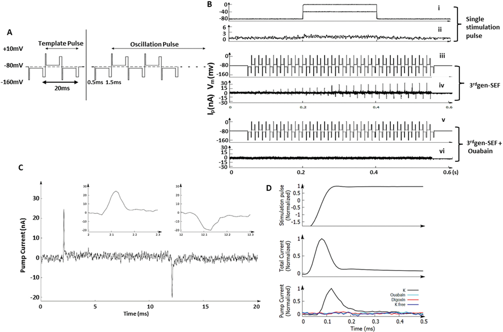
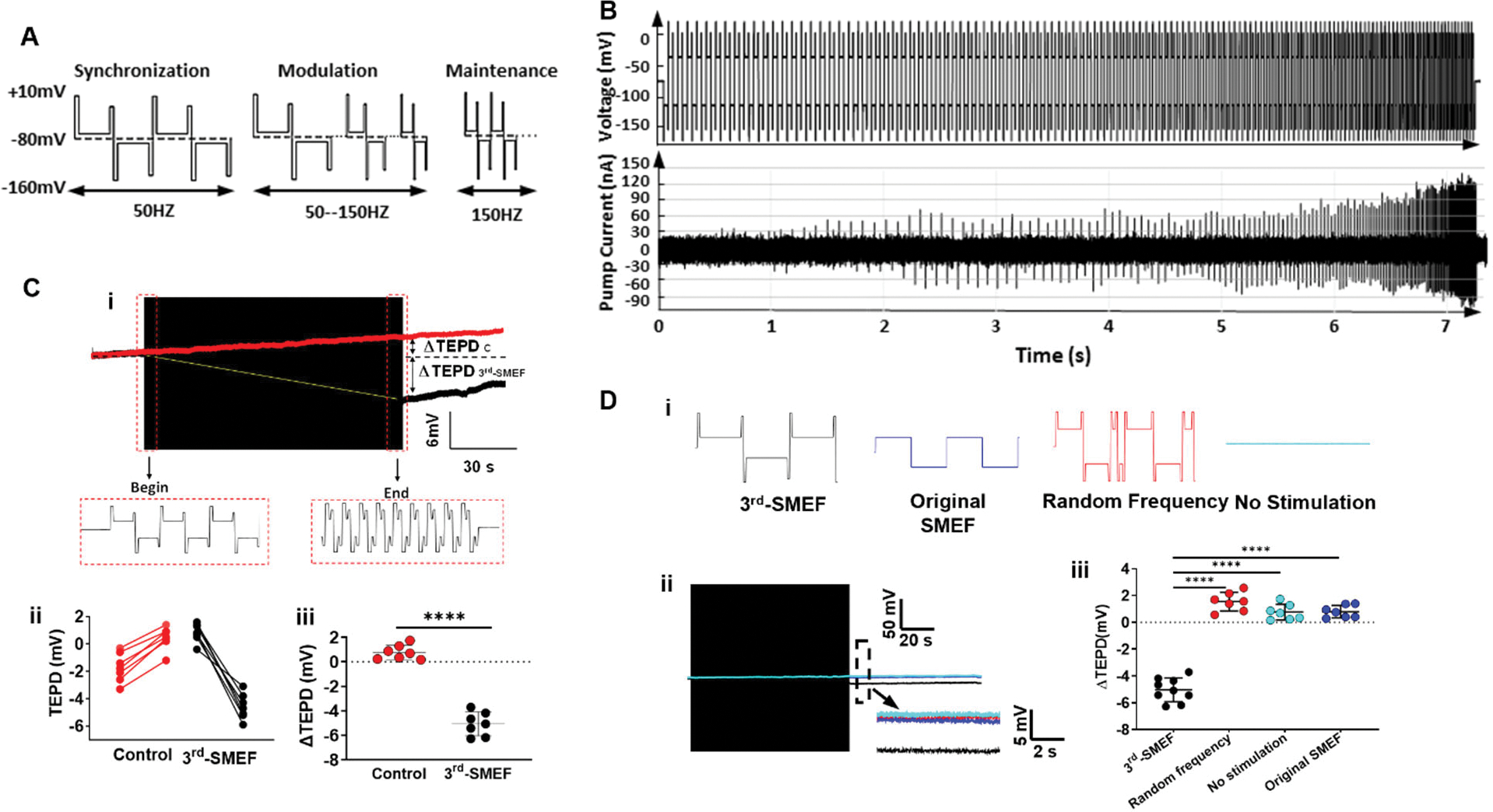
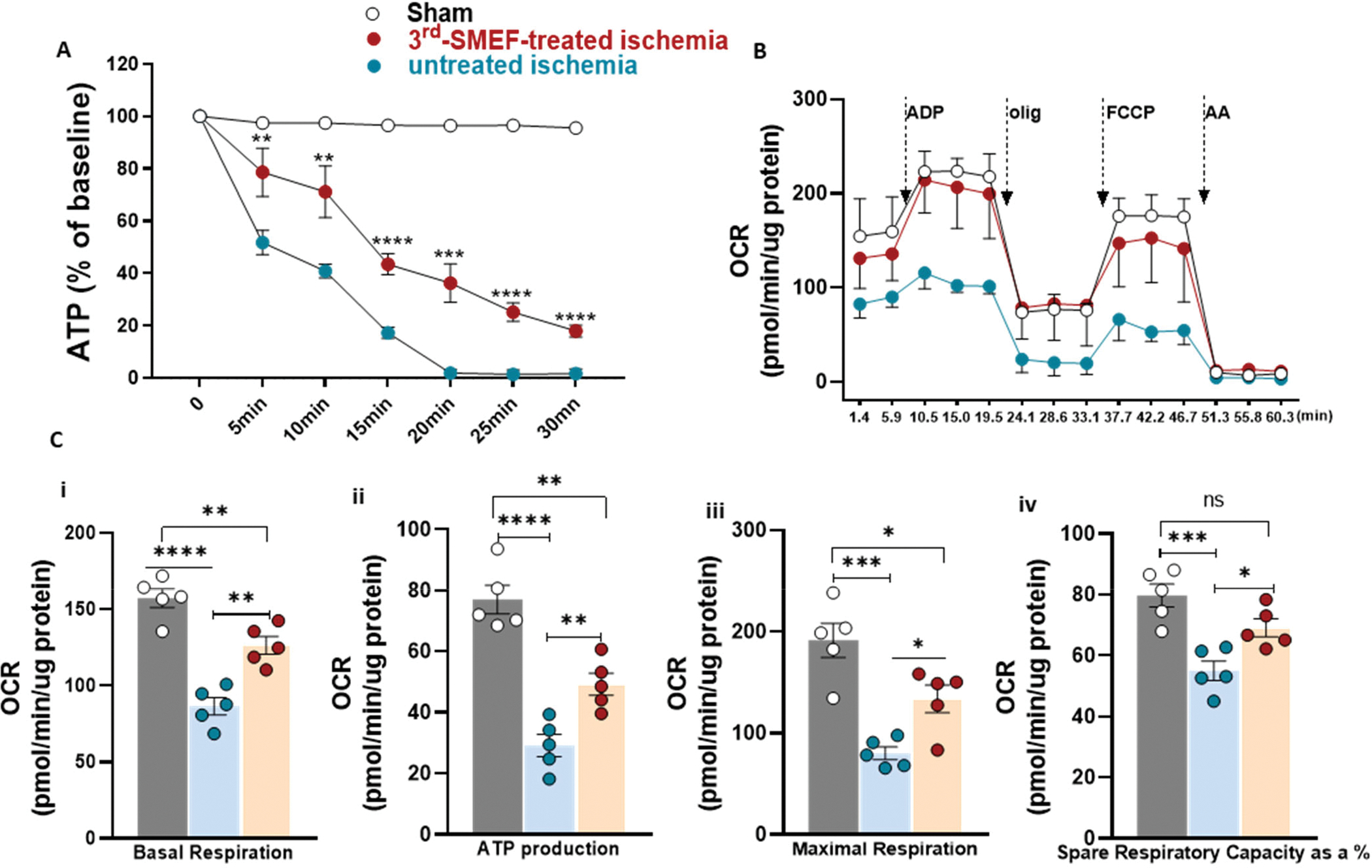
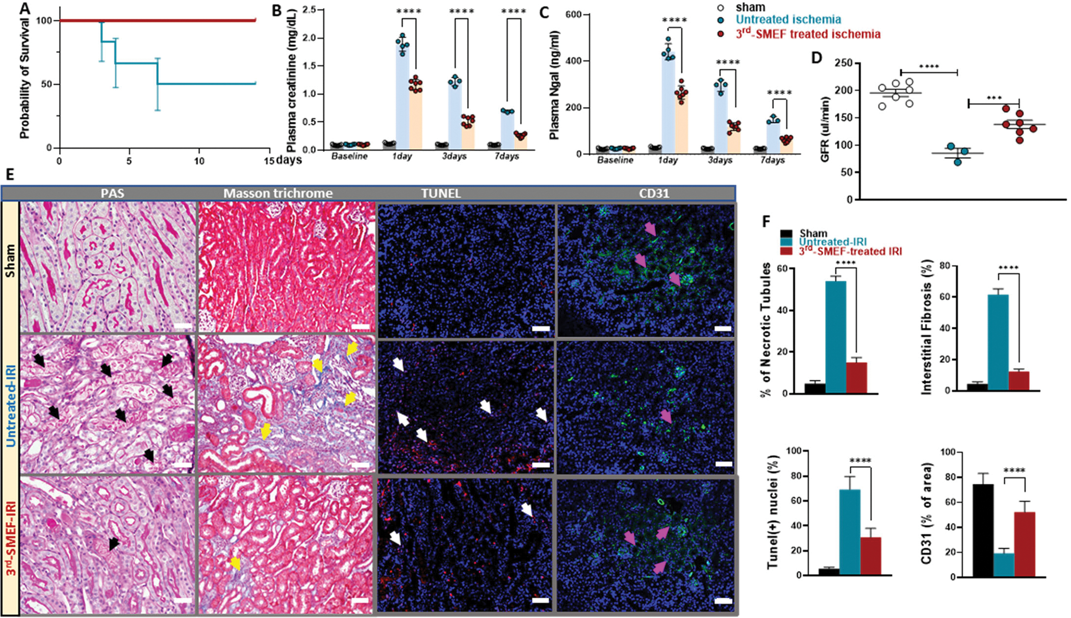
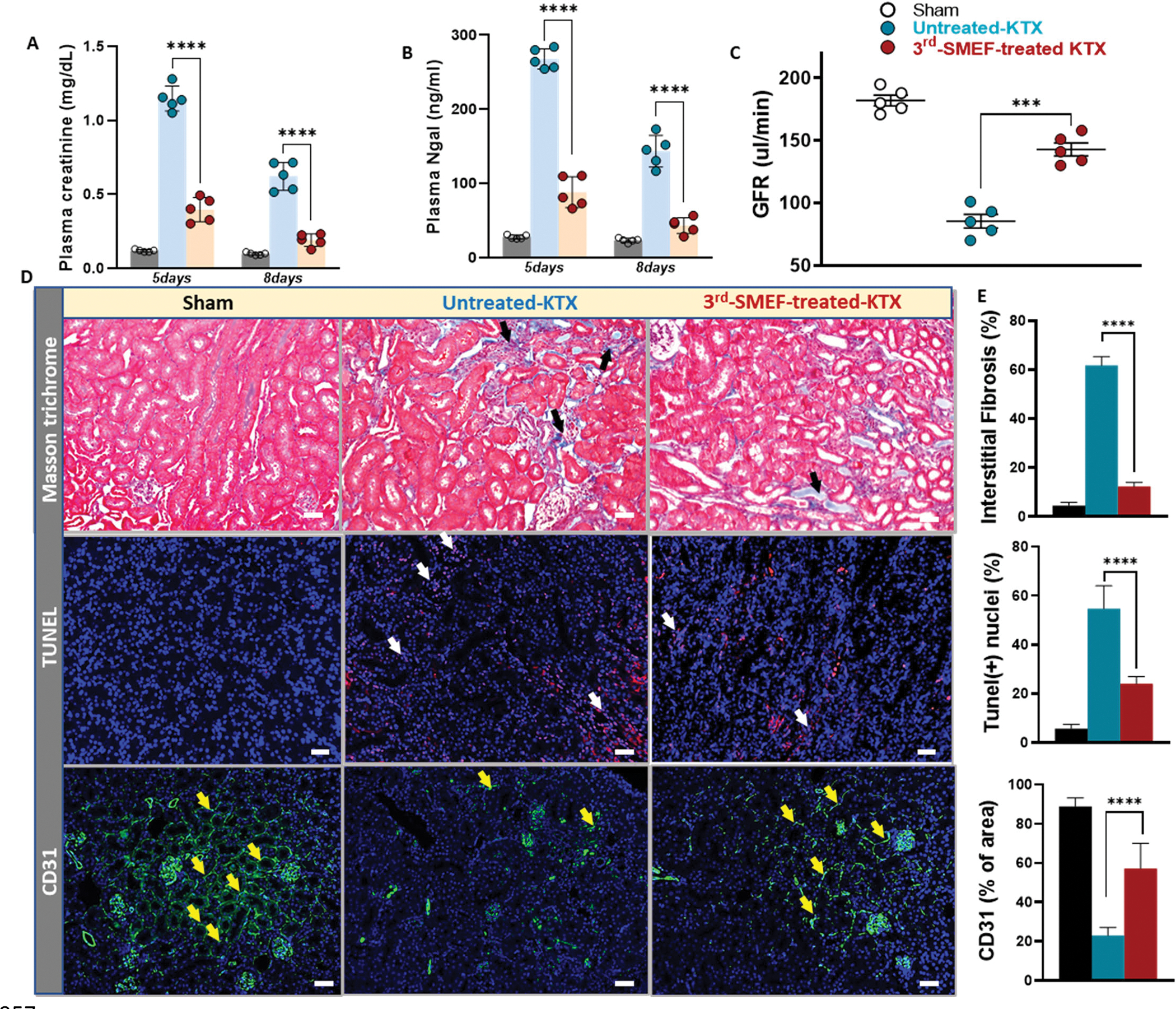
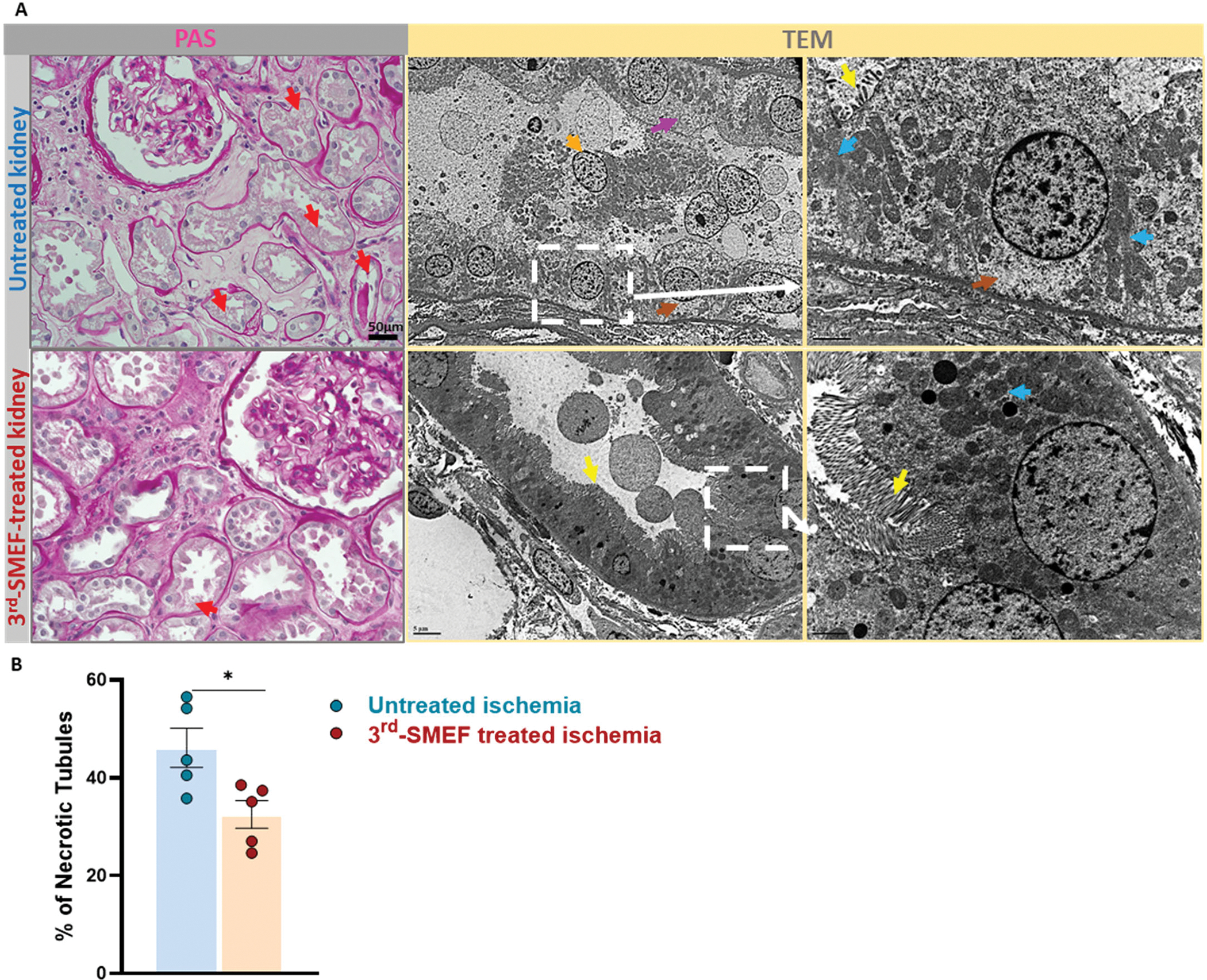
Renal ischemia-reperfusion injury is an important contributor to the development of delayed graft function after transplantation, which is associated with higher rejection rates and poorer long-term outcomes. One of the earliest impairments during ischemia is Na+/K+-ATPase (Na/K pump) dysfunction due to insufficient ATP supply, resulting in subsequent cellular damage. Therefore, strategies that preserve ATP or maintain Na/K pump function may limit the extent of renal injury during ischemia-reperfusion. Here, we applied a synchronization modulation electric field to activate Na/K pumps, thereby maintaining cellular functions under ATP-insufficient conditions. We tested the effectiveness of this technique in two models of ischemic renal injury: an in situ renal ischemia-reperfusion injury model (predominantly warm ischemia) and a kidney transplantation model (predominantly cold ischemia). Application of the synchronization modulation electric field to a renal ischemia-reperfusion injury mouse model preserved Na/K pump activity, thereby reducing kidney injury, as reflected by 40% lower plasma creatinine (1.17 ± 0.03 mg/dl) in the electric field-treated group as compared to the untreated control group (1.89 ± 0.06 mg/dl). In a mouse kidney transplantation model, renal graft function was improved by more than 50% with the application of the synchronization modulation electric field according to glomerular filtration rate measurements (85.40 ± 12.18 μl/min in the untreated group versus 142.80 ± 11.65 μl/min in the electric field-treated group). This technique for preserving Na/K pump function may have therapeutic potential not only for ischemic kidney injury but also for other diseases associated with Na/K pump dysfunction due to inadequate ATP supply.


Conflict of interest statement

Competing Interests: C.W has a patent application pending for “Method of electrogenically controlling pump molecules” (# 8,073,549) and “System and method utilizing electrical energy to fuel and activate ion transporters without consumption of adenosine triphosphate molecules” (#1372.1285 PR). C.W. and L.R. are inventors on a filed patent application (Serial #: 63/162,169): “System and Method Utilizing Electrical Energy to Fuel and Activate Active Ion Transporter without Consumption of Adenosine Triphosphate (ATP) Molecules: Clinical Ischemia Application on Kidney”. All other authors declare that they have no competing interests.

Figures

Figure 1.
Figure 1.. 3rdgen-SEF drives Na/K pump activity in ATP-depleted muscle fibers.
After depleting ATP from the isolated American bullfrog muscle fibers, Na/K pump activity in response to two distinct electrical fields was measured. (A). Waveforms generated by the 3rdgen-SEF. The left panel shows the pre-pulse, whereas the right panel shows the oscillating pulses. The pump current was gained by subtracting the current generated by pre-pulse (a reference) from the current generated by each oscillating pulse. The results are the pump currents responding to this specific oscillating pulse. For the oscillating pulse, each half-pulse consisted of three components: the first overshoot (0.5ms, 90mV), the speeding plateau (8ms, 20mV), and the second overshoot (1.5ms, 70mV). (B). Different electrical fields applied (the traditional single stimulation pulse and the 3rdgen-SEF) and the resultant pump currents. The 3rdgen-SEF was able to generate pump currents in this ATP-depleted milieu (traces iii and iv); these pump currents were eliminated by ouabain, a specific inhibitor of the Na/K pumps (traces v and vi). (C) Representative traces of a pair of synchronized pump currents. The inserts showed the details of Na (left) and K (right) currents with a duration of about 100 μs. (D). Currents measured after reducing the gain of the voltage-clamp. The upper, middle and lower panels depicted the rising-phase of the membrane potential of the first overshot, total transmembrane currents, and the Na/K pump currents, respectively. (In the bottom panel, the black trace was Na/K pump current measured in regular physiological solution; teal trace in solution with ouabain; red trace in solution with Digoxin and blue trace in K-free solution.) The pump and transmembrane capacitance currents have similar durations.
Figure 2.
Figure 2.. The 3rdgen-SMEF maintains the TEPD of ATP-depleted kidneys.
(A). Waveform of all three phases for the 3rdgen-SMEF applied: phase 1 synchronization (left), phase 2 modulation (middle), and phase 3 maintenance (right). (B). Pump currents resulting from this oscillating electrical field. The synchronization frequency was gradually increased in a stepwise pattern (change of 5% to 10% for every 10 to 20 oscillating pulses) up to the target frequency of 150 Hz. The increasing field frequency was accompanied by increases in the magnitude and density of the transient pump currents. (C). The TEPD of PCTs in the isolated Sprague Dawley rat kidneys in the presence (black trace in subpanel i) and absence (red trace in subpanel i) of 3rdgen-SMEF. TEPD was measured with a microelectrode, which was placed in the proximal tubule of the isolated kidneys. The black box was the compressed oscillating electric field of the 3rdgen-SMEF. Panels ii and iii summarized the results from the experiments performed. Significant differences were determined with student t-test. (****P<0.0001, n = 7 kidneys/group) (D). Effects of different electric fields (subpanel i, no stimulation-teal; random frequency-red; original SMEF-blue; and 3rdgen-SMEF-black) on TEPD in PCTs. Detailed TEPD changes right after the application of the different electric fields (black box) were measured in the dash-line box shown as the inserts in D-ii. The results were summarized in D-iii. Results are presented as mean ± SD. Significant differences were determined with one-way ANOVA followed by Dunnett’s multiple comparisons test. (****P<0.0001, n = 7–9 kidneys/group).
Figure 3.
Figure 3.. ATP content and mitochondrial function are preserved with the application of accelerating-3rdgen-SMEF.
(A). ATP content in kidney tissue was measured at different time points of ischemia without reperfusion. Both renal pedicles were clamped, one as untreated control and the other one treated with accelerating-3rdgen-SMEF. Data are presented as mean ± SEM. Two-way ANOVA followed by Tukey multiple comparisons test have been performed (**p<0.01; ***p < 0.001; ****p<0.0001, n = 5 mice/group). (B) Oxygen consumption rate (OCR) traces of isolated mitochondria from C57BL/6J mouse kidneys underwent 15 min of ischemia with or without accelerating-3rdgen-SMEF treatment, expressed as picomoles of O2 per minute, under basal conditions and after the injection of ADP (1mM), oligomycin (2 μM), FCCP (4 μM), and AA+ rotenone (2 μM). Oligo, oligomycin; FCCP, carbonyl cyanide 4-[trifluoromethoxy]phenylhydrazone; AA, antimycin A. (C). Analysis of mitochondrial respiratory parameters obtained from normalized XFe24 graphs (B). Data are presented as mean ± sem. One-way ANOVA followed by Tukey multiple comparisons test have been performed (*p<0.05; **p<0.01; ***p < 0.001; ****p<0.0001, n = 5 kidneys/group).
Figure 4.
Figure 4.. Ischemia reperfusion-induced AKI is attenuated by accelerating-3rdgen-SMEF.
Unilateral kidney ischemia was induced by clamping left renal pedicle for 15 min in C57BL/6J mice with or without application of the accelerating-3rdgen-SMEF, followed by nephrectomy of the counter kidney. Survival rates were monitored for 14 days. Survival curves were analyzed using the log-rank (Mantel–Cox) test (A). Kidney injury was evaluated by plasma creatinine (B) and NGAL (C) measurements at 1-, 3- and 7-days following IR. Two-way ANOVA followed by Tukey multiple comparisons test have been performed (***P < 0.001; ****P<0.0001, n = 3 to 7 mice/group). Kidney function was assessed by GFR measurement in conscious mice at 7 days following IR (D). One-way ANOVA followed by Tukey multiple comparisons test was performed to determine the significance (***P < 0.001; ****P<0.0001, n = 3 to 7 mice/group). Representative morphologic evidence of kidney injury was assessed at 7 days following IR by PAS staining, Masson’s Trichrome staining, TUNEL staining, and CD31 immunofluorescent staining. (black arrows demonstrate necrotic tubules, yellow arrows denote areas of tubulointerstitial fibrosis, white arrows denote apoptosis and pink arrows show peritubular capillary endothelial cell). Scale bars were 50 μm (E). Histological data was quantified with Image-J (F) as described in Supplementary Materials and Methods. Data are presented as mean ± SEM. Student t-test was used to determine the significance (****P<0.0001, n = 3 to 7 kidneys/group).
Figure 5.
Figure 5.. The accelerating-3rdgen-SMEF blunts cold-ischemia induced injury in a murine model of kidney transplantation.
Plasma creatinine (A) and NGAL (B) measurements on day 5 and day 8 following transplant. ****P<0.0001, n = 5 mouse recipients/group, Two-way ANOVA followed by Tukey multiple comparisons test have been performed. (C) GFR measurement in the conscious recipients on day 11 following transplant. Data are presented as mean ± SD. ***P<0.001, Student’s t test comparing untreated group to accelerating-3rdgen-SMEF treated group was performed. (n=5 mouse recipients/group). (D) Representative kidney tissue sections stained with Masson’s Trichrome, TUNEL and immunofluorescent anti-CD31. (black arrows, tubulointerstitial fibrosis; white arrows, apoptosis and yellow arrows, peritubular capillary endothelial cells). (F) Quantification of tubulointerstitial fibrosis area over whole kidney sections, TUNEL-positive cells/nuclei as the percentage (%) of TUNEL and DAPI double-positive cells relative to total cells and CD31-positive area over the kidney section as described in Supplementary Materials and Methods. Data are presented as mean ± SEM. One-way ANOVA followed by Tukey multiple comparisons test have been performed (****P<0.0001, n = 5 kidneys/group).
Figure 6.
Figure 6.. Preliminary evidence suggests that accelerating-3rdgen-SMEF may protect human donor kidneys during cold storage.
Accelerating-3rdgen-SMEF was applied to five pairs of human kidneys during a 24-hour storage period after which morphology was evaluated. (A). The left two panels are representative images of kidney slices stained by PAS. The red arrows indicate the injured tubules (loss of brush borders, cytoplasmic vacuolization and nuclear drop out). Scale bars are 50μm. The right 4 panels are TEM images (yellow arrows, brush border; orange arrows, epithelial cells sloughing into tubular lumen; blue arrows, mitochondria; brown arrows, cytoplasmic vacuolization and pink arrows, apical cytoplasm blebbing). Scale bars are 5μm. (B). The percentage of necrotic tubules based on PAS staining was quantified with Image J as described in Supplementary Materials and Methods. Paired t -test has been performed (*P < 0.05, n = 5 kidneys/group).

Similar articles

Cited by

References

    1. Halloran PF, Aprile MA, Farewell V, Ludwin D, Smith EK, Tsai SY, Bear RA, Cole EH, Fenton SS, Cattran DC, Early function as the principal correlate of graft survival. A multivariate analysis of 200 cadaveric renal transplants treated with a protocol incorporating antilymphocyte globulin and cyclosporine. Transplantation 46, 223–228 (1988). - PubMed
    1. Womer KL, Vella JP, Sayegh MH, Chronic allograft dysfunction: mechanisms and new approaches to therapy. Semin. Nephrol 20, 126–147 (2000). - PubMed
    1. Nehus EJ, Devarajan P, Acute kidney injury: AKI in kidney transplant recipients--here to stay. Nat. Rev Nephrol 8, 198–199 (2012). - PubMed
    1. Daemen MA, de VB, Buurman WA, Apoptosis and inflammation in renal reperfusion injury. Transplantation 73, 1693–1700 (2002). - PubMed
    1. Cooper JE, Wiseman AC, Acute kidney injury in kidney transplantation. Curr. Opin. Nephrol. Hypertens 22, 698–703 (2013). - PubMed

Publication types

Substances