

The effect of N6-methyladenosine (m6A) factors on the development of acute respiratory distress syndrome in the mouse model
- PMID: 35263199
- PMCID: PMC8973778
- DOI: 10.1080/21655979.2022.2049473
The effect of N6-methyladenosine (m6A) factors on the development of acute respiratory distress syndrome in the mouse model
Abstract
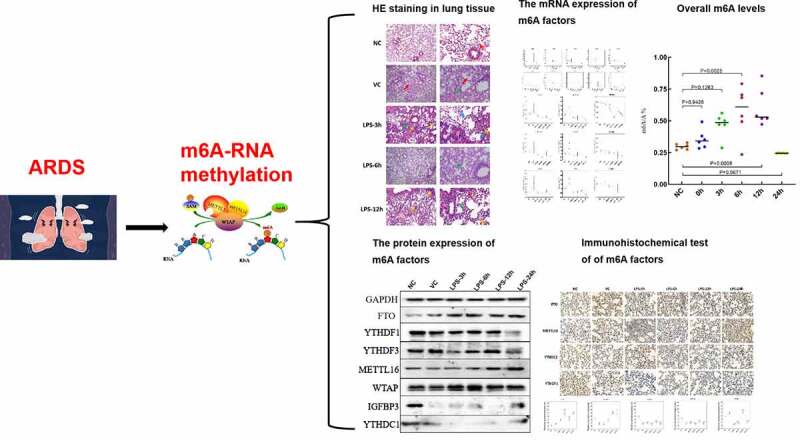
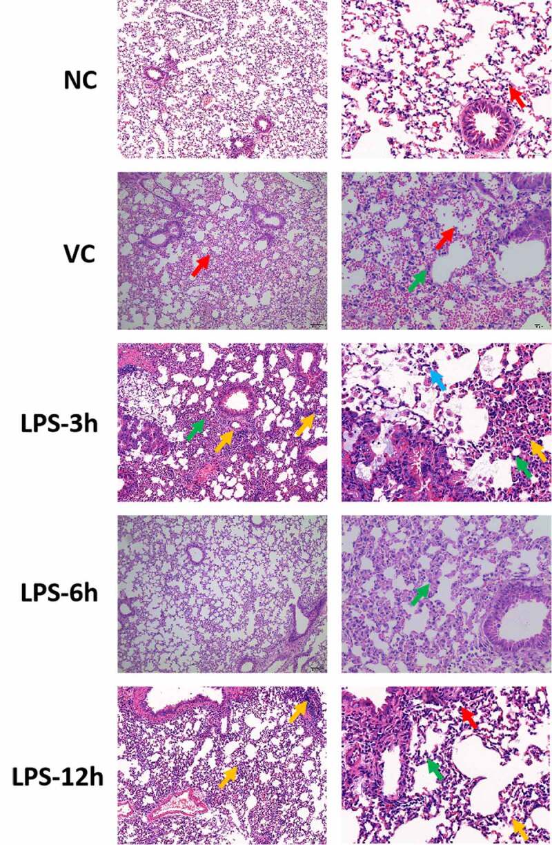
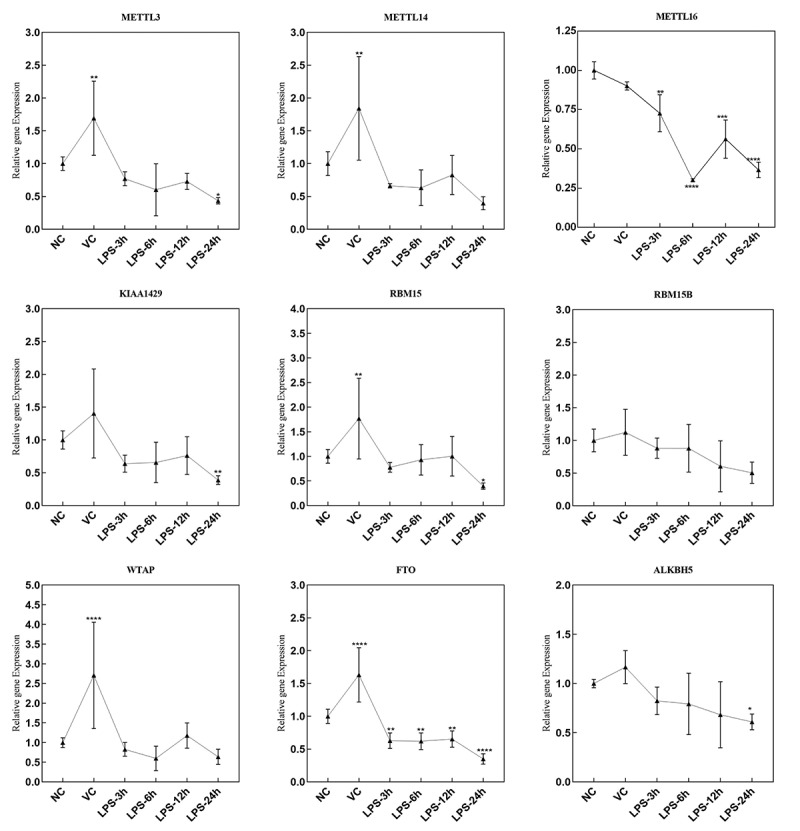
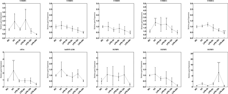
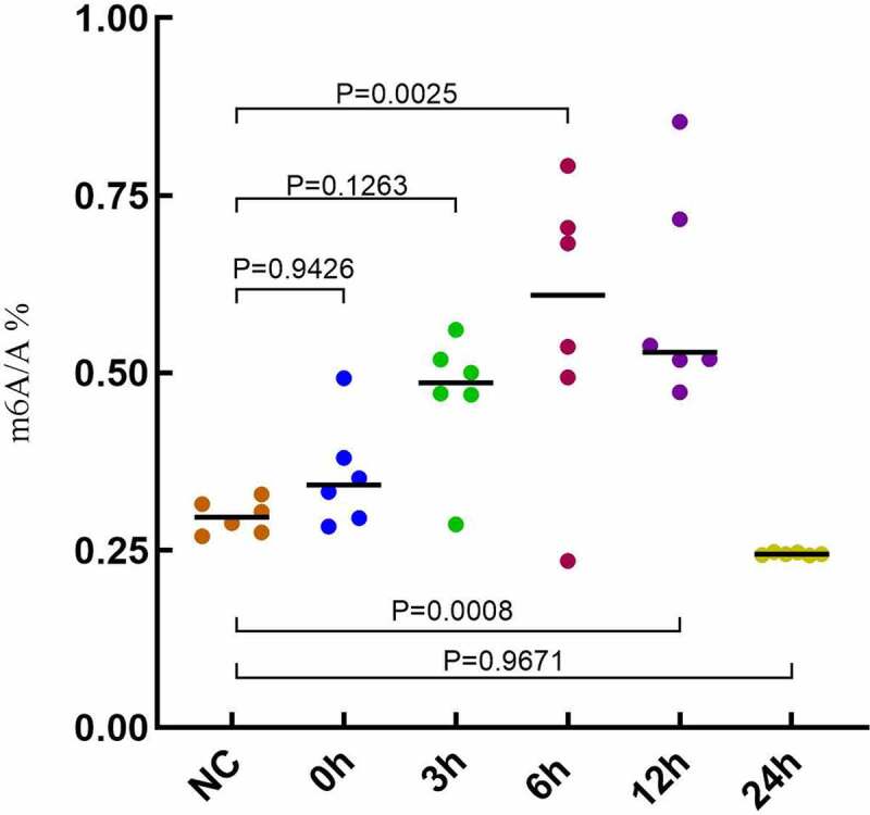
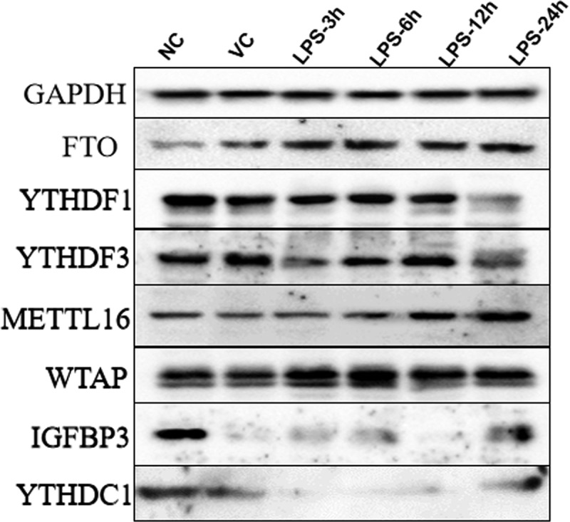
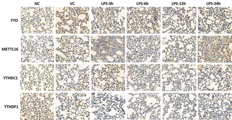
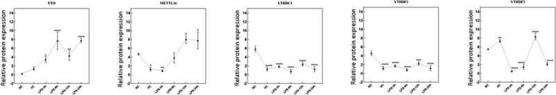
Acute respiratory distress syndrome (ARDS) can cause loss of alveolar-capillary membrane integrity and life-threatening immune responses. The underlying molecular mechanisms of ARDS remain unclear. N6-methyladenosine (m6A)-RNA modification plays an important part in many biological processes. However, it is not clear whether ARDS alters RNA methylation in lung tissue. We tried to investigate the changes of m6A-RNA methylation in lung tissues of lipopolysaccharide (LPS)-induced ARDS mice. Lung tissue samples were collected to detect the expression of m6A factors through hematoxylin and eosin (HE) staining, quantitative reverse transcriptase-polymerase chain reaction (qRT-PCR), immunohistochemical analysis and western blot. The overall m6A levels in lung tissue of ARDS in mouse were detected by UPLC-UV-MS. HE staining showed that the degree of the inflammatory response was more severe in the LPS-3 h group. The mRNA expression of YTHDF1, YTHDC1 and IGFBP3 was remarkably up-regulated at, respectively, 6, 6 and 12 h after LPS treatment. The mRNA expression of METTL16, FTO, METTL3, KIAA1429, RBM15, ALKBH5, YTHDF2, YTHDF3, YTHDC2 and IGFBP2 was significantly down-regulated at 24 h after LPS treatment. The protein expression of METTL16 and FTO increased, YTHDC1, IGFBP3 YTHDF1 and YTHDF3 showed a down-regulation trend after LPS induction. Overall m6A-RNA methylation levels were significantly increased at 6 h after LPS induction. In ARDS mice, LPS-induced m6A methylation may be involved in the expression regulation of inflammatory factors and may play important roles in the occurrence and development of lung tissue. It is suggested that m6A modification may be a promising therapeutic target for ARDS.
Keywords: Acute respiratory distress syndrome; RNA methylation; inflammatory response; lung; m6A.
Conflict of interest statement
No potential conflict of interest was reported by the author(s).
Figures
References
-
- Bellani G, Laffey JG, Pham T, et al. Epidemiology, Patterns of Care, and Mortality for Patients With Acute Respiratory Distress Syndrome in Intensive Care Units in 50 Countries. Jama. 2016;315(8):788–800. - PubMed
-
- Vincent JL, Sakr Y, Ranieri VM.. Epidemiology and outcome of acute respiratory failure in intensive care unit patients. Crit Care Med. 2003;31(Supplement):S296–9. - PubMed
-
- Tomashefski JF Jr. Pulmonary pathology of acute respiratory distress syndrome. Clin Chest Med. 2000;21(3):435–466. - PubMed
-
- Ranieri VM, Rubenfeld GD, Thompson BT, et al. Acute respiratory distress syndrome: the Berlin Definition. Jama. 2012;307(23):2526–2533. - PubMed
MeSH terms
Substances
LinkOut - more resources
Full Text Sources
Other Literature Sources
Miscellaneous