. 2021 Jul-Sep;62(3):713-721.
doi: 10.47162/RJME.62.3.08.

Hypertension induces compensatory left ventricular hypertrophy by a mechanism involving gap junction lateralization and overexpression of CD36, PKC and MMP-2

Affiliations

Hypertension induces compensatory left ventricular hypertrophy by a mechanism involving gap junction lateralization and overexpression of CD36, PKC and MMP-2

Mădălina Dumitrescu et al. Rom J Morphol Embryol. 2021 Jul-Sep.

Abstract

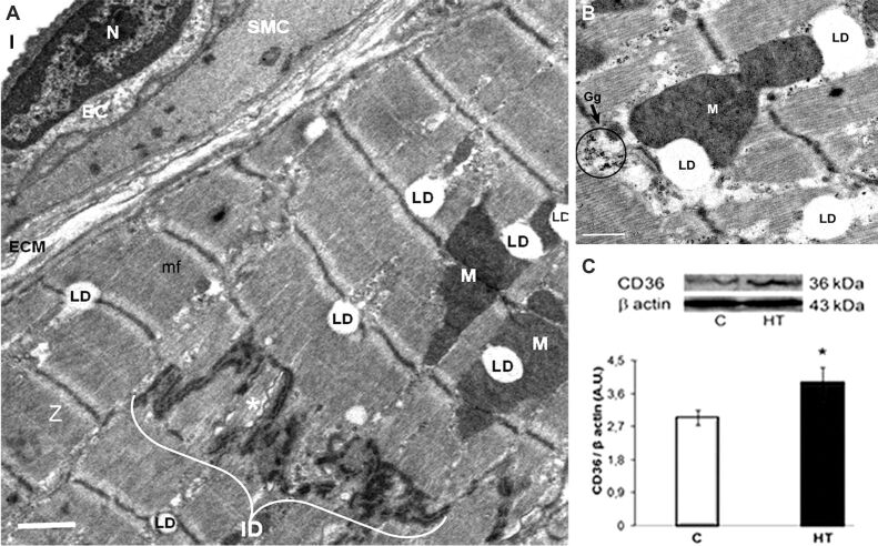
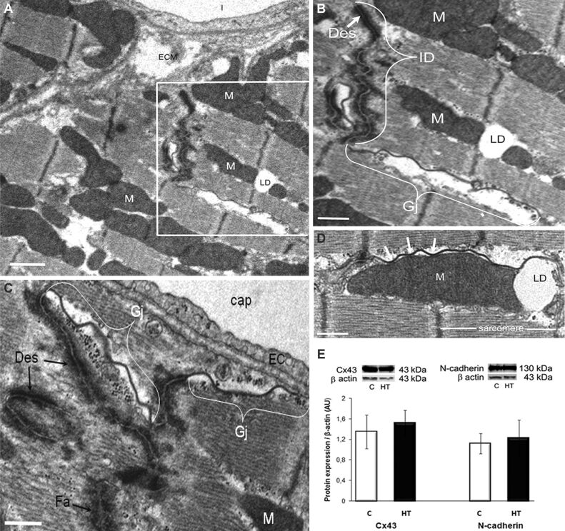
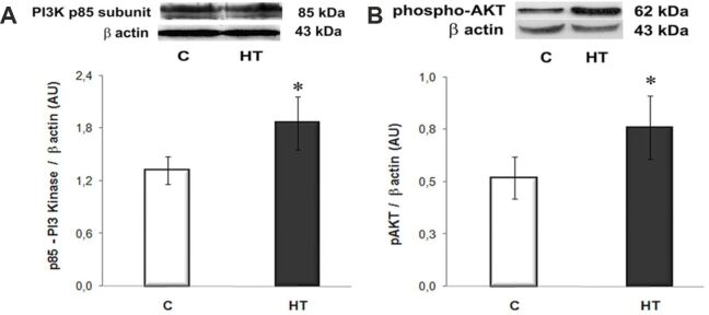
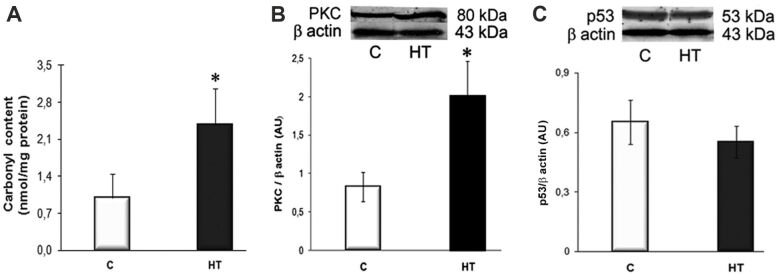
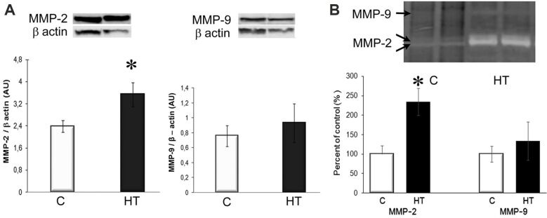
Hypertension-induced left ventricular hypertrophy evolves initially as an adaptive response meant to minimize ventricular wall stress. The mechanisms involved in the preservation of the cardiac function during the "compensatory" phase of the left ventricular hypertrophy are still unclear. Therefore, we aimed at uncovering fine changes that aid the heart to cope with the increased stress in hypertension. Male golden Syrian hamsters were given NG-nitro-L-arginine methyl ester (L-NAME) for 16 weeks, and they became hypertensive (HT), developing left ventricular hypertrophy with no impaired contractility or fibrosis. As compared to age-matched control hamsters, the hypertrophied left ventricles in L-NAME-induced HT hamsters exhibited the following structural and molecular changes: (i) accumulation of lipid droplets (LDs) within cardiomyocytes and relocation of gap junctions to the lateral membrane of cardiomyocytes or close to mitochondria (revealed by electron microscopy); (ii) overexpression of the cluster of differentiation 36 (CD36) fatty acid transporter, protein kinase C (PKC), and matrix metalloproteinase-2 (MMP-2), enhanced activation of the phosphoinositide 3-kinase (PI3K)/protein kinase B (AKT) pathway, and unchanged expression of the connexin 43 (Cx43) and N-cadherin junctional proteins (assessed by Western blot); (iii) increased protein carbonyl content, assessed with a 2,4-Dinitrophenylhydrazine (DNPH)-based spectrophotometric assay, indicative of an enhanced reactive oxygen species (ROS) production; and (iv) augmented MMP-2 activity (determined by gelatin zymography). These changes may participate in an orchestrated adaptive hypertrophic growth response that helps to maintain cardiac performance, in HT hamsters. Together, these findings could provide support for designing future strategies meant to prevent the transition from compensatory left ventricular hypertrophy to decompensated heart failure.


Conflict of interest statement

The authors declare that there is no conflict of interests.

Figures

Figure 1
Figure 1
LV of a HT hamster with compensatory myocardial hypertrophy: (A and B) TEM revealing the apparent normal structure of a cardiomyocyte with regular arrangement of mf and Z. Note in (A) the abundance of LD, often at the level of Z, associated with M or adjacent to an ID that encloses a long, ribbon-shaped Gj (asterisk), and in (B) the proximity between M, LD and Gg. Scale bar: (A) 1 μm; (B) 0.5 μm; (C) CD36 protein expression in the LV homogenate is significantly increased compared to controls [C]. Representative immunoblots (upper panel) and the densitometric analysis of the bands expressed as AU are shown. The intensities of the bands were quantified with TotalLab Quant software. β-Actin served as protein loading control. n: 3–4 hamsters in each group. *p<0.05 vs. [C] group. AU: Arbitrary units; CD36: Cluster of differentiation 36; EC: Endothelial cell; Gg: Glycogen granules; Gj; Gap junction; HT: Hypertensive; ID: Intercalated disc; l: Capillary lumen; LD: Lipid droplets; LV: Left ventricle; M: Mitochondria; mf: Myofibrils; N: Nucleus; SMC: Smooth muscle cell; TEM: Transmission electron microscopy; Z: Z-lines
Figure 2
Figure 2
The architecture of the IDs between cardiomyocytes in the LV of a HT hamster with compensatory cardiac hypertrophy is slightly modified, whereas the expression of Cx43 and N-cadherin junctional proteins remains unchanged, as compared to controls [C]: (A and B) Electron micrographs highlighting an ID, and Gj redistribution to the lateral membrane of cardiomyocyte (lateralization). An enlarged view of the boxed area in (A) is shown in (B). Scale bar: (A and B) 1 μm; (C and D) Electron micrographs representative for lateralized Gjs that associate with electron-dense structures with the morphology of Des and Fa in (C), or in close connection with an interfibrillar M (white arrows) and its adjoining LD in (D). Scale bar: (C and D) 0.5 μm; (E) Representative immunoblots and densitometry of the bands (AU) revealing no differences in the expression of Cx43 and N-cadherin in the LV homogenates in HT and [C] hamsters. β-Actin was used as protein loading control. n: 3–4 hamsters in each group. AU: Arbitrary units; cap: Capillary; Cx43: Connexin 43; Des: Desmosomes; EC: Endothelial cell; Fa: Fascia adherens; Gj; Gap junction; HT: Hypertensive; ID: Intercalated disc; LD: Lipid droplet; LV: Left ventricle; M: Mitochondria; mf: Myofibrils
Figure 3
Figure 3
The expression levels of PI3K (p85 subunit) (A) and phospho-AKT (Ser473) (B) are significantly increased in the LVs of HT hamsters compared to controls [C] and assessed by Western blot. Representative immunoblots and the densitometric analysis (AU) are displayed in the upper and lower panels, respectively. β-Actin was used as protein loading control. n: 3–4 hamsters in each group. *p<0.05 vs. [C] group. AKT: Protein kinase B; AU: Arbitrary units; HT: Hypertensive; LV: Left ventricle; PI3K: Phosphoinositide 3-kinase
Figure 4
Figure 4
Assessment of the ROS production in the LVs of HT hamsters by determining the protein carbonyl content and the expression levels of redox-sensitive PKC and p53: (A) Protein carbonyl content, assessed with a DNPH-based spectrophotometric assay, is significantly higher in homogenates of HT hamsters than in control [C] ones; (B and C) As compared to [C], the protein expression of PKC is significantly increased, whereas the expression of p53 is unchanged in HT hamsters. Representative Western blots and quantification of the bands’ intensities (AU) are shown in the upper and lower panels, respectively. β-Actin was used as protein loading control. n: 3–4 hamsters in each group. *p<0.05 vs. [C] group. AU: Arbitrary units; DNPH: 2,4-Dinitrophenylhydrazine; HT: Hypertensive; LV: Left ventricle; PKC: Protein kinase C; ROS: Reactive oxygen species
Figure 5
Figure 5
MMP-2 protein expression and gelatinolytic activity significantly increased in the hypertrophic LV of HT hamsters: (A) MMP-2 and MMP-9 protein expression. Representative immunoblots (top) and the densitometric analysis (bottom) of the bands intensities, expressed as AU; (B) Gelatin zymography indicating an increased proteolytic activity of MMP-2 in HT hamsters. β-Actin was used as protein loading control. n: 3–4 hamsters in each group. *p<0.05 vs. [C] group. AU: Arbitrary units; [C]: Control; HT: Hypertensive; LV: Left ventricle; MMP: Matrix metalloproteinase

References

    1. Saheera S, Krishnamurthy P. Cardiovascular changes associated with hypertensive heart disease and aging. Cell Transplant. 2020;29:963689720920830–963689720920830. - PMC - PubMed
    1. Habets DDJ, Coumans WA, Voshol PJ, den Boer MAM, Febbraio M, Bonen A, Glatz JFC, Luiken JJFP. AMPK-mediated increase in myocardial long-chain fatty acid uptake critically depends on sarcolemmal CD36. Biochem Biophys Res Commun. 2007;355(1):204–210. - PubMed
    1. Heier C, Haemmerle G. Fat in the heart: the enzymatic machinery regulating cardiac triacylglycerol metabolism. Biochim Biophys Acta. 2016;1861(10):1500–1512. - PubMed
    1. Abumrad NA, Goldberg IJ. CD36 actions in the heart: lipids, calcium, inflammation, repair and more. Biochim Biophys Acta. 2016;1861(10):1442–1449. - PMC - PubMed
    1. Simon JN, Duglan D, Casadei B, Carnicer R. Nitric oxide synthase regulation of cardiac excitation-contraction coupling in health and disease. J Mol Cell Cardiol. 2014;73:80–91. - PubMed

MeSH terms

Substances