

Intranodal palisaded myofibroblastoma expressing DOG1: focusing on the potential diagnostic pitfalls
- PMID: 35263416
- PMCID: PMC9019625
- DOI: 10.47162/RJME.62.3.25
Intranodal palisaded myofibroblastoma expressing DOG1: focusing on the potential diagnostic pitfalls
Abstract
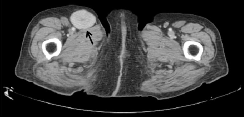
Intranodal palisaded myofibroblastoma (IPM) is a rare, benign mesenchymal neoplasm of the lymph nodes with a broad differential diagnosis. We report a case of an 82-year-old woman presenting with a slow growing, right inguinal mass. The tumor arose as a circumscribed neoplasm inside a lymph node and consisted of bland spindle cells with nuclear palisading and intervening areas of amianthoid-like fibers among interstitial hemorrhage and hemosiderin-laden histiocytes in the stroma, typical histomorphological characteristics of IPM. Immunohistochemically, the neoplastic cells were positive for vimentin, smooth muscle actin (SMA), β-catenin, cyclin D1 and discovered on gastrointestinal stromal tumor (GIST) 1 (DOG1) immunostainings. A literature review and differential diagnosis of IPM are discussed. To the best of our knowledge, this is the first case of DOG1 immunoexpression in a case of IPM.
Conflict of interest statement
The authors declare no potential conflict of interests with respect to the research, authorship, and/or publication of this article.
Figures


Similar articles
-
Intranodal palisaded myofibroblastoma - a rare case report and literature review.APMIS. 2016 Oct;124(10):905-10. doi: 10.1111/apm.12580. Epub 2016 Aug 8. APMIS. 2016. PMID: 27500890 Review.
-
Cytopathological findings of intranodal palisaded myofibroblastoma: Case report and review of the literature.Diagn Cytopathol. 2023 Aug;51(8):E248-E254. doi: 10.1002/dc.25172. Epub 2023 May 27. Diagn Cytopathol. 2023. PMID: 37243568 Review.
-
Intranodal palisaded myofibroblastoma: a case report and an update on etiopathogenesis and differential diagnosis.J Cancer Res Ther. 2013 Apr-Jun;9(2):295-8. doi: 10.4103/0973-1482.113395. J Cancer Res Ther. 2013. PMID: 23771380
-
Intranodal palisaded myofibroblastoma (intranodal hemorrhagic spindle cell tumor with amianthoid fibers): a case report and literature review.Diagn Pathol. 2010 Feb 9;5:12. doi: 10.1186/1746-1596-5-12. Diagn Pathol. 2010. PMID: 20181136 Free PMC article. Review.
-
Fine-Needle Aspiration Cytology of Intranodal Palisaded Myofibroblastoma of the Inguinal Lymph Node.Acta Cytol. 2016;60(1):89-92. doi: 10.1159/000445162. Epub 2016 Mar 31. Acta Cytol. 2016. PMID: 27027816
References
-
- Weiss SW, Gnepp DR, Bratthauer GL. Palisaded myofibroblastoma. A benign mesenchymal tumor of lymph node. Am J Surg Pathol. 1989;13(5):341–346. - PubMed
-
- Suster S, Rosai J. Intranodal hemorrhagic spindle-cell tumor with "amianthoid" fibers. Report of six cases of a distinctive mesenchymal neoplasm of the inguinal region that simulates Kaposi’s sarcoma. Am J Surg Pathol. 1989;13(5):347–357. - PubMed
-
- Lee JY, Abell E, Shevechik GJ. Solitary spindle cell tumor with myoid differentiation of the lymph node. Arch Pathol Lab Med. 1989;113(5):547–550. - PubMed
-
- Karabulut YY, Kara T, Berkeşoğlu M. Intranodal palisaded myofibroblastoma - a rare case report and literature review. APMIS. 2016;124(10):905–910. - PubMed
-
- Fletcher CDM, Stirling RW. Intranodal myofibroblastoma presenting in the submandibular region: evidence of a broader clinical and histological spectrum. Histopathology. 1990;16(3):287–293. - PubMed
Publication types
MeSH terms
Substances
LinkOut - more resources
Full Text Sources
Medical
Research Materials
