Review
. 2022 Jun 20;23(12):e202100689.
doi: 10.1002/cbic.202100689. Epub 2022 Mar 19.

Strategies to Reduce the On-Target Platelet Toxicity of Bcl-xL Inhibitors: PROTACs, SNIPERs and Prodrug-Based Approaches

Affiliations
Review

Strategies to Reduce the On-Target Platelet Toxicity of Bcl-xL Inhibitors: PROTACs, SNIPERs and Prodrug-Based Approaches

Arvind Negi et al. Chembiochem. .

Abstract

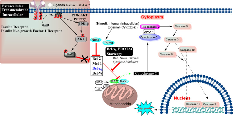
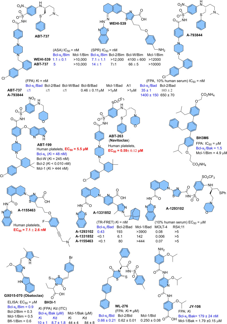
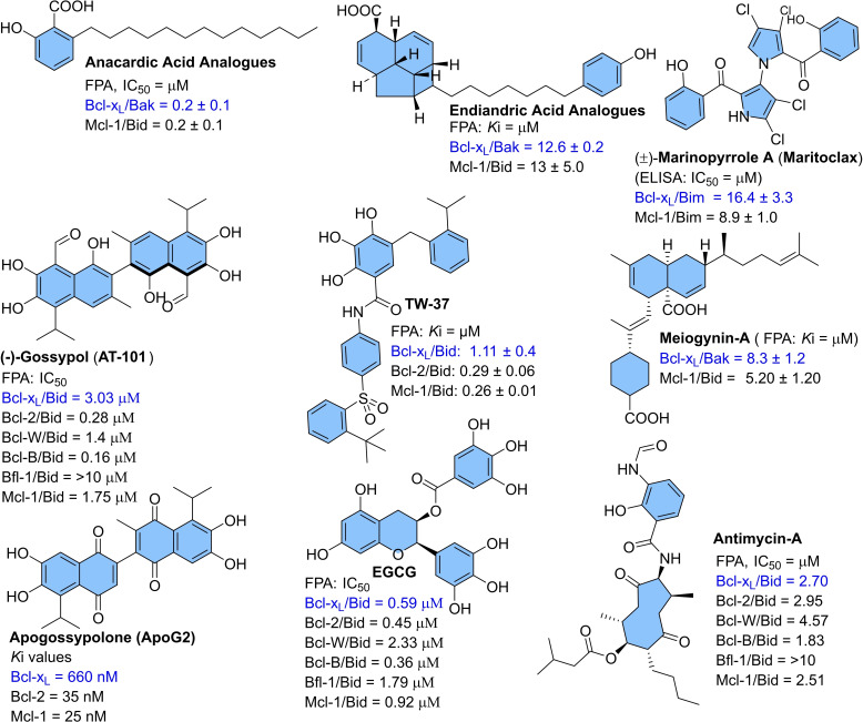
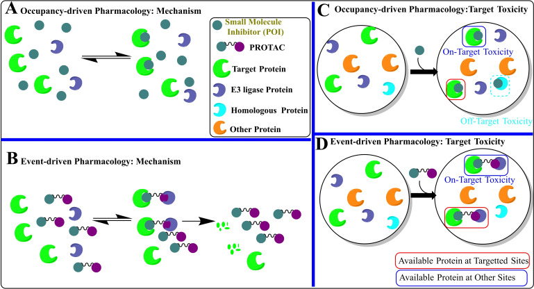
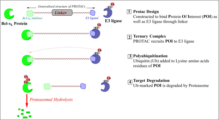
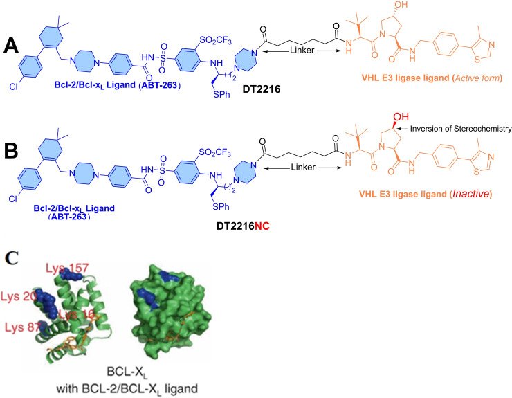
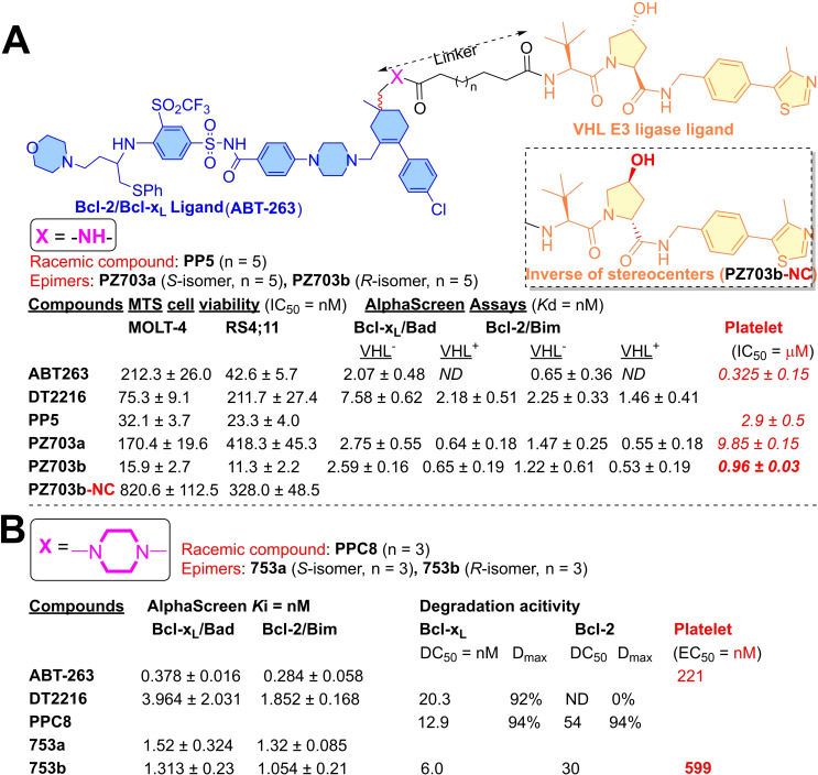
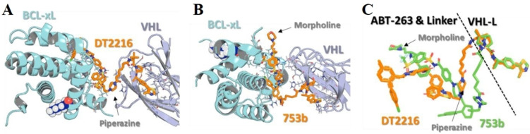
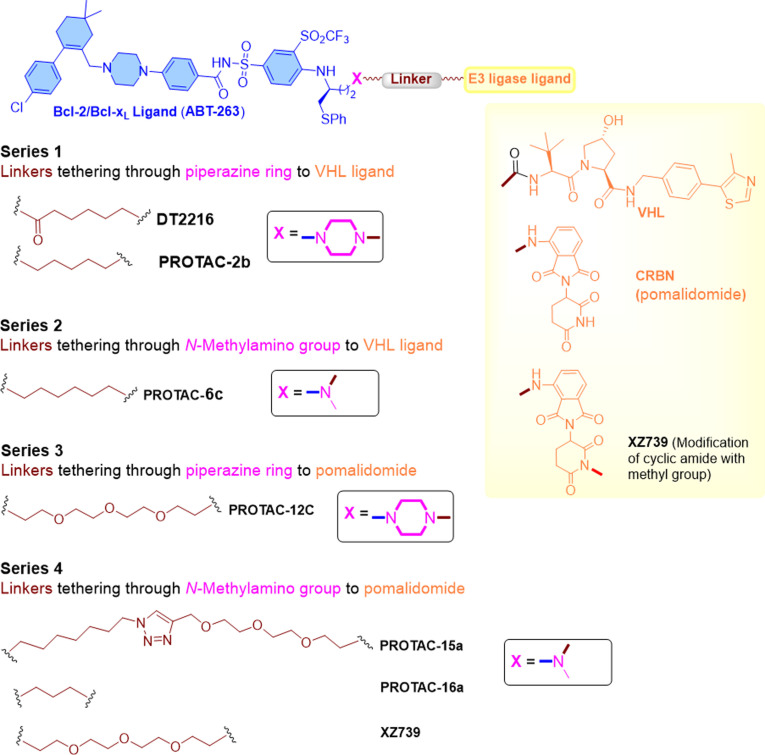
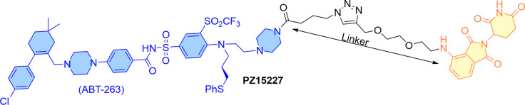
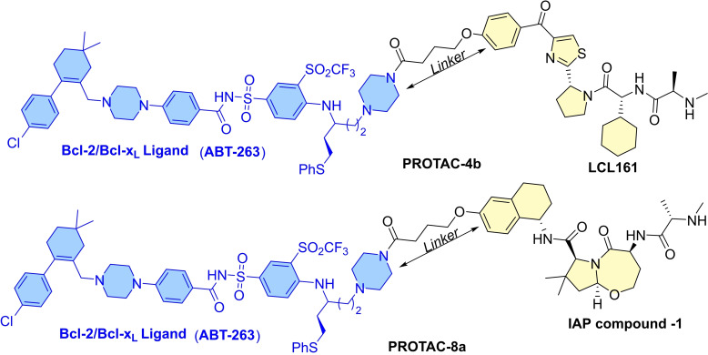
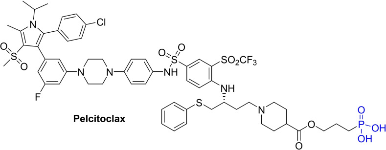
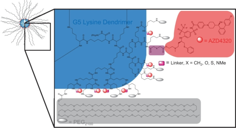
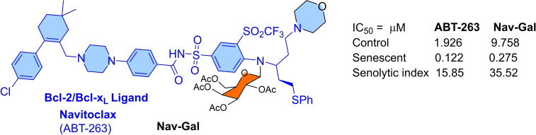
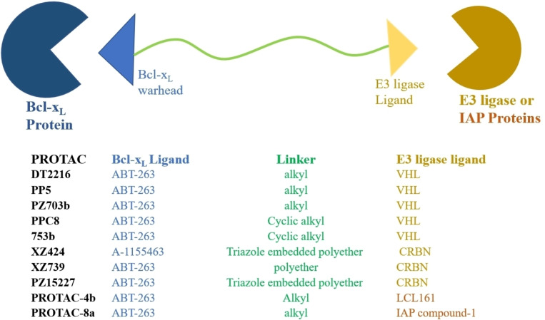
Apoptosis is a highly regulated cellular process. Aberration in apoptosis is a common characteristic of various disorders. Therefore, proteins involved in apoptosis are prime targets in multiple therapies. Bcl-xL is an antiapoptotic protein. Compared to other antiapoptotic proteins, the expression of Bcl-xL is common in solid tumors and, to an extent, in some leukemias and lymphomas. The overexpression of Bcl-xL is also linked to survival and chemoresistance in cancer and senescent cells. Therefore, Bcl-xL is a promising anticancer and senolytic target. Various nanomolar range Bcl-xL inhibitors have been developed. ABT-263 was successfully identified as a Bcl-xL /Bcl-2 dual inhibitor. But it failed in the clinical trial (phase-II) because of its on-target platelet toxicity, which also implies an essential role of Bcl-xL protein in the survival of human platelets. Classical Bcl-xL inhibitor designs utilize occupancy-driven pharmacology with typical shortcomings (such as dose-dependent off-target and on-target platelet toxicities). Hence, event-driven pharmacology-based approaches, such as proteolysis targeting chimeras (PROTACs) and SNIPERs (specific non-genetic IAP-based protein erasers) have been developed. The development of Bcl-xL based PROTACs was expected, as 600 E3-ligases are available in humans, while some (such as cereblon (CRBN), von Hippel-Lindau (VHL)) are relatively less expressed in platelets. Therefore, E3 ligase ligand-based Bcl-xL PROTACs (CRBN: XZ424, XZ739; VHL: DT2216, PZ703b, 753b) showed a significant improvement in platelet therapeutic index than their parent molecules (ABT-263: DT2216, PZ703b, 753b, XZ739, PZ15227; A1155463: XZ424). Other than their distinctive pharmacology, PROTACs are molecularly large, which limits their cell permeability and plays a role in improving their cell selectivity. We also discuss prodrug-based approaches, such as antibody-drug conjugates (ABBV-155), phosphate prodrugs (APG-1252), dendrimer conjugate (AZD0466), and glycosylated conjugates (Nav-Gal). Studies of in-vitro, in-vivo, structure-activity relationships, biophysical characterization, and status of preclinical/clinical inhibitors derived from these strategies are also discussed in the review.

Keywords: Bcl-xL; Bcl-xL inhibitors; PROTACs; SNIPERs; drug conjugates; on-target toxicity; platelet toxicity.


Conflict of interest statement

The authors declare no conflict of interest.

Figures

Figure 1
Figure 1
Apoptosis‐inducing proteins catalyze the MOMP process to release the cytochrome‐c. The cytochrome‐c forms an apoptosome complex with adaptor Apaf‐1 and caspase‐9 in the cytosol, and ultimately activates the caspase‐9 to initiate caspase‐cascade cellular apoptosis. The antiapoptotic proteins (Bcl‐2, Mcl‐1, and Bcl‐xL) inhibit the cytochrome‐c release, whereas Bax, Bak, Bid and other apoptosis‐inducing proteins, promote the release of cytochrome‐c from mitochondria. The inhibitors of Bcl‐2/Mcl‐1/Bcl‐xL prevent PPIs of Bcl‐2/Mcl‐1/Bcl‐xL with apoptosis‐inducing proteins (BH3 helix of Bid, Bim, Bad, Puma, Bmf, and Noxa), which increase the cellular level of BAX/BAK, their availability for oligomerization, which eventually leads to the release of cytochrome‐c.[ 1 , 2 ] The Insulin‐like growth factor receptor and insulin receptor tyrosine kinase initiate the phosphorylation which activates the intracellular accessary signaling cascade via the PI3 K‐AKT pathway, resulting in activating the BAD into its phosphorylated form, which then modifies the mitochondrial functions in senescent cells.
Figure 2
Figure 2
Synthetically developed inhibitors of Bcl‐xL protein with associated platelet toxicities: ABT‐199, ABT‐263, BH3I1, WL‐276,[15] Obatoclax, WEHI‐539, A‐793844, ABT‐737,[ 7 , 17 ] A‐1293102, A‐1331852, A‐1155463,; Oligomers: p‐terphenyl derivative (BH3‐M6),[ 17 , 19 ] oligoamide‐foldamer (JY‐1106 (IC50: Bcl‐xL/Bak=394±54 nM, Mcl‐1/Bak=10.21±0.83 μM[21])). ELISA: enzyme‐linked immunosorbent assay.
Figure 3
Figure 3
Naturally derived preferential or dual Bcl‐xL/Bcl‐2/Mcl‐1 inhibitors: Anacardic acids, Endiandric acids, Marinopyrroles, Polyphenols (Gossypol, ApoG2, EGCG, TW‐37),[ 25 , 26 ] Meiogynins, and antimycin‐A. However, most of them were not studied for their on‐target platelet toxicity.
Figure 4
Figure 4
(A) Occupancy‐driven pharmacology target toxicity: This type of pharmacology is mainly shown by classical inhibitors (green color spheres) which inhibit or modify the signaling of a protein of interest (POI) (green crescent). However, the success of a complete inhibition is proportional to the time‐dependent bound concentration of SMI to the POI, therefore, these approaches require highly potent affinities of SMIs towards the POI. (B) Representation of event‐driven pharmacology: A bifunctional molecule (dumbbell‐shaped) subsequently binds to the target protein (green crescent) and E3 ligase protein (violet crescent). The binding with E3 ligase (violet crescent) induces the POI hydrolysis, e. g. protein degradation approaches. (C) Occupancy‐driven pharmacology target toxicity: Off‐target toxicity, as well as on‐target toxicity, are typical with SMIs as a virtue of their smaller sizes. Their smaller sizes and high potency, not only allow them to target the POI in other non‐relevant cells (on‐target toxicity) but also inherited them with a flaw to adopt non‐selective entropy binding conformation with homologous (orange crescent) as well as non‐homologous proteins (cyan crescent). (D) Occupancy‐driven pharmacology target toxicity: These strategies mainly use large structures, which reduces their cross‐membrane permeation and the degree of entropy‐based non‐selective conformations for homologous as well as non‐homologous proteins, and therefore relatively less prone to the off‐target toxicities. However, the wider cellular availability of target protein leads to minimal‐to‐moderate on‐target toxicity from these approaches.
Figure 5
Figure 5
Schematic representation and mechanism involved in PROTACs targeted protein degradation. The PROTACs inhibition is usually measured in DC50 (half‐maximal degradation concentration) and Dmax (maximum degradation).
Figure 6
Figure 6
(A) Chemical structure of DT2216 for Bcl‐xL degradation containing ABT‐263 (left‐hand side) and VHL E3 ligase ligand (right‐hand side). (B) Chemical structure of DT2216NC for Bcl‐xL degradation containing ABT‐263 (left‐hand side) and hydroxylated version of VHL E3 ligase ligand (right‐hand side), which doesn't bind to the VHL. (C) Showcasing the surface lysine residues in Bcl‐xL (Left side) and Bcl‐2 (Right side), reproduced with permission from Khan et al. Copyright 2019 Nature Publishing Group.
Figure 7
Figure 7
(A) PROTAC analogs derivatized ABT‐263 and VHL E3‐ligase ligand. VHL=without VHL; VHL+=with VHL. (B) PROTAC analogs derivatized ABT‐263 and VHL E3‐ligase ligand.
Figure 8
Figure 8
Ternary complexes of DT2216 (A), 753b (B), and superpose of DT2216/753 b (C). Reproduced with permission from Lv et al. Copyright 2021 Nature Publishing Group.
Figure 9
Figure 9
Chemical structure of A1155463 based PROTAC with CRBN E3 ligase.
Figure 10
Figure 10
Bcl‐xL degraders: Investigation of linker chemical space and tethering points to the warheads.
Figure 11
Figure 11
PZ15227: ABT‐263 warhead (Bcl‐XL) that recruits CRBN E3 ligase.
Figure 12
Figure 12
Chemical structure of PROTAC‐4b and PROTAC‐8a that recruits IAPs (LCL161 and IAP compound 1) for Bcl‐XL degradation.
Figure 13
Figure 13
Chemical structure of Phosphate prodrug of Bcl‐xL inhibitors (APG‐1252).
Figure 14
Figure 14
Representation of AZD4320‐dendrimer conjugate: Each dendrimer presenting 32 PEG2100 terminals (in grey color), 32 AZD4320 (in red color) linker regions (marked in pink color), where if X=CH2 for SPL‐8931, X=S for SPL‐8932 and X=O for SPL‐8933. Reproduced with permission from Patterson et al Copyright 2021 Nature.
Figure 15
Figure 15
ABT‐263‐derived Nav‐Gal structure.
Figure 16
Figure 16
Summarized structures of Bcl‐xL PROTACs and SNIPERs.

Similar articles

Cited by

References

    1. Negi A., Murphy P. V., Eur. J. Med. Chem. 2021, 210, 113038. - PubMed
    1. Wu H., Medeiros L. J., Young K. H., Blood 2018, 32, 8–28. - PubMed
    1. Negi A., Ramarao P., Kumar R., Mini-Rev. Med. Chem. 2013, 13, 653–681. - PubMed
    1. Silke J., Meier P., Cold Spring Harbor Perspect. Biol. 2013, 5, 1–19. - PMC - PubMed
    1. Vogler M., Adv. Med. 2014, 2014, 943648. - PMC - PubMed