. 2022 Mar 8;38(10):110482.
doi: 10.1016/j.celrep.2022.110482.

Induction of broadly reactive influenza antibodies increases susceptibility to autoimmunity

Affiliations

Induction of broadly reactive influenza antibodies increases susceptibility to autoimmunity

Jocelyn G Labombarde et al. Cell Rep. .

Abstract

Infection and vaccination repeatedly expose individuals to antigens that are conserved between influenza virus subtypes. Nevertheless, antibodies recognizing variable influenza epitopes greatly outnumber antibodies reactive against conserved epitopes. Elucidating factors contributing to the paucity of broadly reactive influenza antibodies remains a major obstacle for developing a universal influenza vaccine. Here, we report that inducing broadly reactive influenza antibodies increases autoreactive antibodies in humans and mice and exacerbates disease in four distinct models of autoimmune disease. Importantly, transferring broadly reactive influenza antibodies augments disease in the presence of inflammation or autoimmune susceptibility. Further, broadly reactive influenza antibodies spontaneously arise in mice with defects in B cell tolerance. Together, these data suggest that self-tolerance mechanisms limit the prevalence of broadly reactive influenza antibodies, which can exacerbate disease in the context of additional risk factors.

Keywords: antibody; autoimmunity; influenza; universal influenza vaccine.


Conflict of interest statement

Declaration of interests P.G.T. has consulted for Illumina, 10X, Immunoscape, and Cyotagents. P.G.T. and J.C.C. filed patents related to the treatment of severe respiratory infections (not based on research in this paper).

Figures

Figure 1.
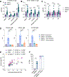
Figure 1.. Induction of broadly reactive influenza antibodies is accompanied by elevated levels of autoreactive antibodies
(A) C57BL/6 mice were given HKx31 and treated daily with rapamycin or PBS. Serum taken 14 days after infection was analyzed for reactivity to 128 autoantigens via protein arrays. Antigens with a statistically significant increase in reactivity to IgM antibodies in rapamycin-treated mice are depicted. Data are representative of two independent experiments of four to five mice per group per experiment. (B and C) Signal intensities from the autoantigen array of groups of antigens associated with (B) arthritis and (C) lupus. (D–G) Sera from mice 14 days after HKx31 exposure was tested by ELISA for reactivity to (D) collagen II (IgM), (E) histone H4 (IgM), (F) histone H1 (IgM), and (G) Smith antigen (IgM). (H) Sera was taken from mice 90 days after HKx31 and tested for IgM reactivity against collagen, whole histones, GM1 + GD1a, histone H1, and histone H4. (I–O) Sera from mice 14 days after HKx31 exposure was tested by ELISA for reactivity to (I) insulin (IgM), (J) LPS (IgM), (K) dsDNA (IgM), (L) nuclear antigens (IgM), (M) KLH (IgM), (N) GM1 + GD1a (IgM), and (O) GM1 (IgG). (P) Sera from mice 14 days after administration of HKx31, MCMV, SARS-CoV-2 RBD/CFA, or OVA/CFA was analyzed by ELISA for GM1/GD1a IgM antibodies. ELISAs are representative of two to seven independent experiments with at least four mice per group. Data are represented as mean ± SEM. *p < 0.05; **p < 0.01; ***p < 0.001, ****p < 0.0001 determined by Mann-Whitney test.
Figure 2.
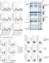
Figure 2.. Antibodies that bind influenza also bind self-proteins
(A) Serum taken from mice 14 days after HKx31 and daily rapamycin or PBS was analyzed by ELISA for IgM binding to histones after adsorption to HKx31, histones, or insulin. (B and C) IgG (B) and IgM (C) monoclonal antibodies were generated from mice given HKx31 and rapamycin (red text) or PBS (blue text) and tested by ELISA for reactivity to influenza antigens HKx31 (H3N2), Vn1203 (H5N1), and PR8 (H1N1) and the indicated self-proteins. Optical densities (ODs) for each antibody and antigen are plotted. (D and E) IgG (D) and IgM (E) monoclonal antibodies were tested by ELISA to rH3 and rH5. (F and G) Reactivity to HKx31 whole virus was evaluated by western blot with (F) IgG and (G) IgM monoclonal antibodies. (H and I) Serum taken 14 days after HKx31 was analyzed by ELISA for IgM antibodies specific for (H) PI(4)P or (I) cardiolipin. (J and K) IgG and IgM monoclonal antibodies were tested for binding to lipids with lipid strips. Lipid strips depicted in Figure S3 were scored for reactivity on a scale of 0–3. Data are representative of two to four experiments (A, H, I, data are mean ± SEM). *p < 0.05; **p < 0.01; ***p < 0.001, ****p < 0.0001 as determined by Mann-Whitney test.
Figure 3.
Figure 3.. Defects in B cell tolerance lead to spontaneous generation of broadly reactive influenza antibodies
(A and B) Serum from uninfected (A) 9-week-old or (B) 28- to 31-week-old NZBWF1 or C57BL/6 mice was analyzed by ELISA for IgM antibodies specific for HKx31, PR8, Vn1203, and histones + GM1. (C) Serum from uninfected 6- to 11-week-old C57BL/6, NZBWF1, and NOD mice were analyzed by ELISA for IgM antibodies specific for rH1, rH3, and rH5 proteins. (D) Serum from uninfected NZBWF1 mice was analyzed for IgM binding to histones, HKx31, and Vn1203 after adsorption to HKx31 or histones. (E) Linear regression of histones/ganglioside IgM versus HKx31, PR8, and Vn1203 IgM in serum from 28- to 31-week-old NZBWF1 mice. (F) Serum from 18-week-old, uninfected, and HKx31-infected (9 weeks after infection) NZBWF1 and C57BL/6 mice was analyzed in a hemagglutination inhibition assay. Data are mean ± SEM and are representative of (A–C) two, (D) four, or (F) three independent experiments and analyzed by (A, B, and F) Mann-Whitney test or (C and D) Kruskal-Wallis test with Dunn’s multiple comparison test.
Figure 4.
Figure 4.. Mice with increased levels of broadly reactive influenza antibodies are more susceptible to autoimmune disease
(A and B) C57BL/6 mice were (A) treated daily with rapamycin or PBS or (B) given HKx31and rapamycin or PBS for 14 days, allowed to rest for 2 weeks, immunized with native MBP in CFA, and scored daily for disease. (C and D) C57BL/6 mice were given HKx31, treated with rapamycin or PBS for 21 days, and immunized with P0 peptide in CFA 2 weeks later. To monitor for disease, activity was measured (C) in an open field test or (D) by measuring stride length 75 days after disease induction. (E) C57BL/6 mice were given HKx31, treated with rapamycin or PBS for 20 days, allowed to rest for 6 days, and infected with C. jejuni. Control mice were treated with rapamycin or PBS without HKx31 and infected with C. jejuni. Activity was measured in an open field test. (F–J) Nine-week-old NZBWF1 mice were given HKx31 and rapamycin or PBS for 28 days. Serum was analyzed by ELISA for IgM antibodies specific for (H) HKx31, (G) PR8, (H) Vn1203, or (J) whole histones. Statistical significance between HKx31 + PBS and HK31 + RAP is indicated by an asterisk and between RAP only and HK31 + RAP by a hashtag. (I) Serum from NZBWF1 mice 10 days after HKx31 was analyzed by ELISA for IgM antibodies specific rH3, rH1, and rH5. (K and L) Serum from NZBWF1 mice 28–38 days after HKx31 was tested for IgM antibodies specific for (K) dsDNA, (L) GM1 + GD1a, and histones. (M) The amount of protein in the urine was measured 113 days after HKx31. All data show mean ± SEM and are representative of at least two experiments and were analyzed by (A–C) two-way ANOVA, (D, I, and K–M) Mann-Whitney test, or (E–H and J) mixed-effects model with Tukey’s multiple corrections.
Figure 5.
Figure 5.. Transfer of broadly reactive monoclonal antibodies increases susceptibility to autoimmune disease
(A) C57BL/6 mice received either broadly reactive or HKx31-specific (single reactive) IgM and IgG monoclonal antibodies. Kidneys were harvested 24 h later and stained for IgM and IgG. (B) Area of IgM and IgG staining in kidney sections was calculated as a proportion of area encompassing individual glomeruli. Data are mean ± SEM and representative of two independent experiments evaluated by Mann-Whitney test. (C and D) Six- to eight-week-old Balb/c mice were given two injections of broadly reactive or HKx31-specific IgG monoclonal antibodies with or without poly I:C, which was given three times per week. Proteinuria was measured weekly, and the incidence of mice with 65 mg/dL urine protein or greater is depicted. (E) NOD mice received two to three injections of broadly reactive or HKx31-specific IgM and IgG monoclonal antibodies. Blood glucose levels were monitored weekly. Incidence of (C and D) proteinuria and (E) diabetes was compared via log rank (Mantel-Cox) test.
Figure 6.
Figure 6.. Influenza infection in humans is associated with increased anti-MAG and -ganglioside antibodies
(A) Plasma from individuals infected with influenza between 2009 and 2014 and uninfected controls was analyzed by ELISA for reactivity to gangliosides and MAG. The normalized ODs were compared via non-parametric, rank-based MANOVA to test for statistical variations of normalized ODs as effects of infection status and year. The overall effect was significant (p = 0.003), and asterisks indicate significance of post hoc Dunnett’s tests comparing infection years with the influenza-negative condition. (B) Reactivity to each ganglioside was plotted with binned microneutralization log10 titers. Non-parametric, rank-based MANOVA was used to test for statistical significance. Overall effect was significant (p < 0.001), and asterisks indicate significance of post hoc Tukey’s pairwise comparisons. (C) Monoclonal antibodies isolated from humans were tested for reactivity to MAG, gangliosides, ANA, and phospholipids via ELISA, and normalized ODs are plotted in the heatmap. (D–F) Normalized ODs of monoclonal antibody reactivity to MAG, ANAs, and phospholipids were plotted based on (D) reactivity to influenza subtypes, (E) location of HA binding, or (F) previously characterized polyreactivity and were tested by multiple Mann-Whitney tests. Mean ± SEM is depicted.

References

    1. Ahmed SS, Schur PH, MacDonald NE, and Steinman L (2014). Narcolepsy, 2009 A(H1N1) pandemic influenza, and pandemic influenza vaccinations: what is known and unknown about the neurological disorder, the role for autoimmunity, and vaccine adjuvants. J. Autoimmun. 50, 1–11. - PubMed
    1. Altman MO, Bennink JR, Yewdell JW, and Herrin BR (2015). Lamprey VLRB response to influenza virus supports universal rules of immunogenicity and antigenicity. Elife 4, e07467. - PMC - PubMed
    1. Amanat F, Stadlbauer D, Strohmeier S, Nguyen THO, Chromikova V, McMahon M, Jiang K, Arunkumar GA, Jurczyszak D, Polanco J, et al. (2020). A serological assay to detect SARS-CoV-2 seroconversion in humans. Nat. Med. 26, 1033–1036. - PMC - PubMed
    1. Anderson MS, and Bluestone JA (2005). The NOD mouse: a model of immune dysregulation. Annu. Rev. Immunol. 23, 447–485. - PubMed
    1. Andrews SF, Huang Y, Kaur K, Popova LI, Ho IY, Pauli NT, Henry Dunand CJ, Taylor WM, Lim S, Huang M, et al. (2015). Immune history profoundly affects broadly protective B cell responses to influenza. Sci. Transl. Med. 7, 316ra192. - PMC - PubMed

Publication types