Comparative Study
. 2022 Mar 9;17(1):151.
doi: 10.1186/s13018-022-03039-y.

A biomechanical comparison between cement packing combined with extra fixation and three-dimensional printed strut-type prosthetic reconstruction for giant cell tumor of bone in distal femur

Affiliations
Comparative Study

A biomechanical comparison between cement packing combined with extra fixation and three-dimensional printed strut-type prosthetic reconstruction for giant cell tumor of bone in distal femur

Xin Hu et al. J Orthop Surg Res. .

Abstract

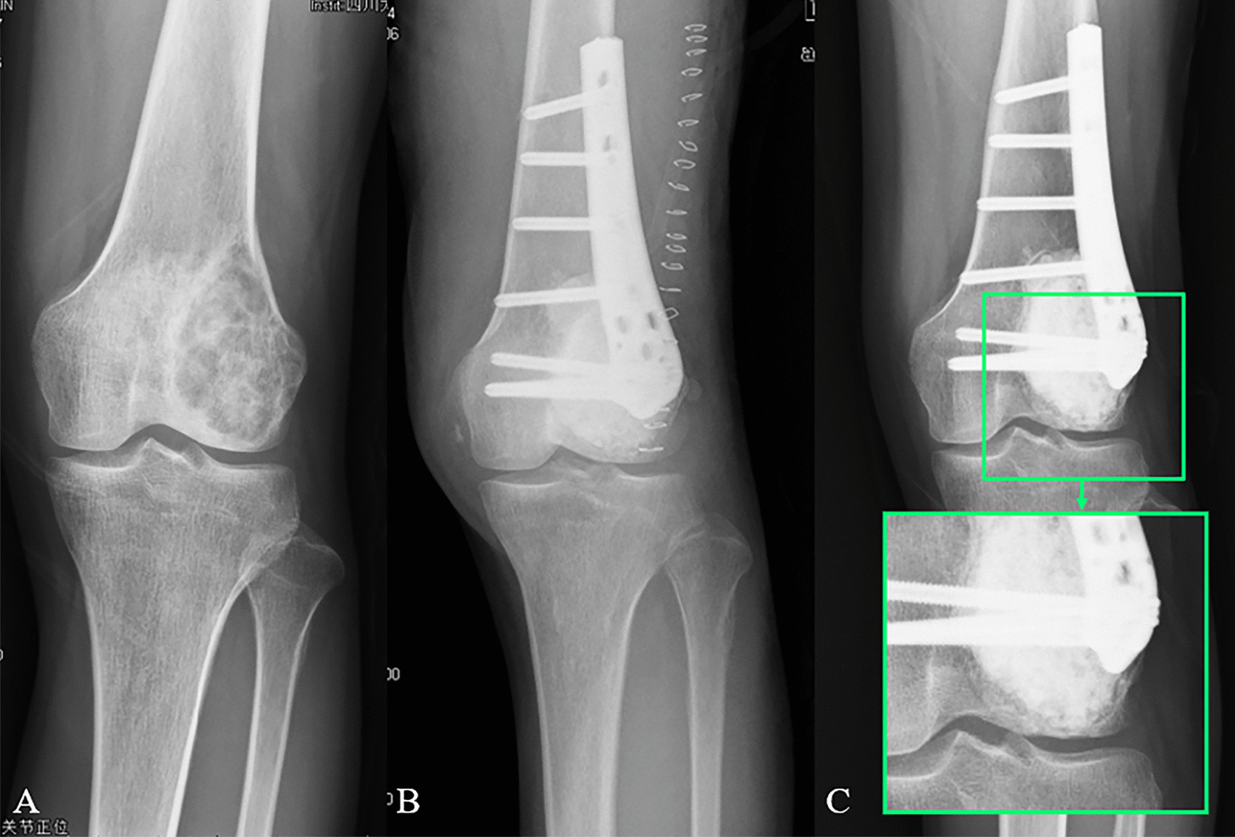
Background: The most common reconstruction method for bone defects caused by giant cell tumor of bone (GCTB) is cement packing combined with subchondral bone grafting and extra fixation. However, this method has several limitations involving bone cement and bone graft, which may lead to poor prognosis and joint function. A titanium-based 3D-printed strut-type prosthesis, featured with excellent biocompatibility and osseointegration ability, was developed for this bone defect in our institution. The goal of this study is to comparatively analyze the biomechanical performance of reconstruction methods aimed at the identification of better operative strategy.

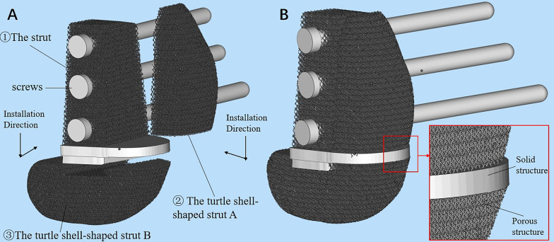
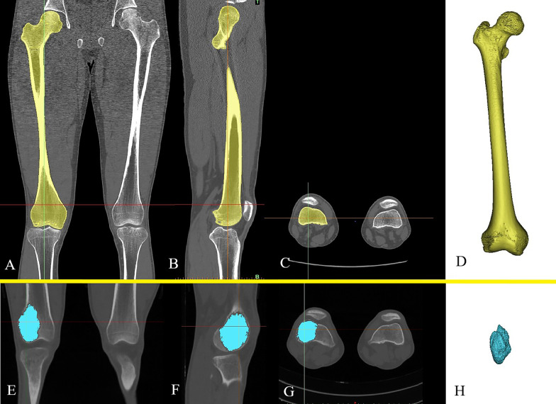
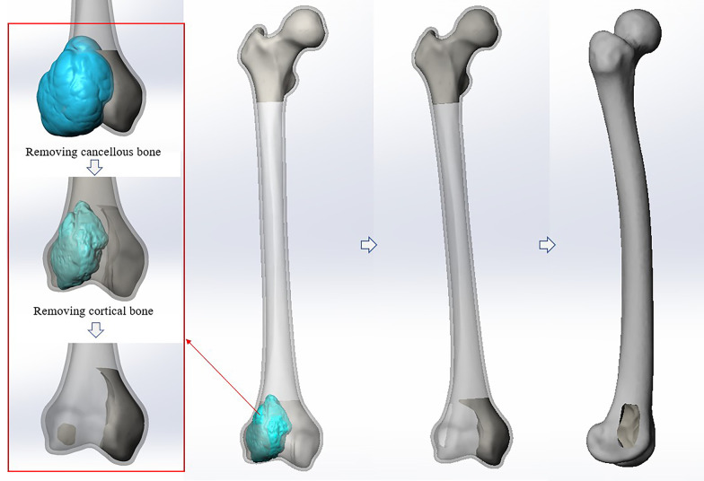
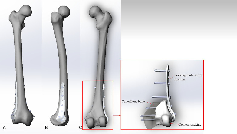
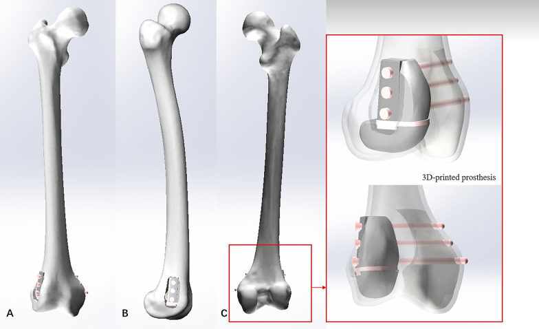
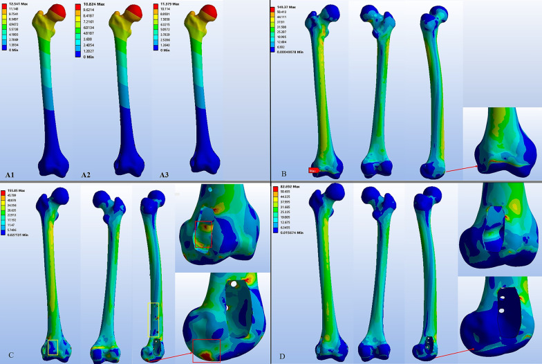
Methods: Four different 3D finite element models were created. Model #1: Normal femur; Model #2: Femur with tumorous cavity bone defects in the distal femur; Model #3: Cavity bone defects reconstructed by cement packing combined with subchondral bone grafting and extra fixation; Model #4: Cavity bone defects reconstructed by 3D-printed strut-type prosthesis combined with subchondral bone grafting. The femoral muscle multiple forces were applied to analyze the mechanical difference among these models by finite element analysis.

Results: Optimal stress and displacement distribution were observed in the normal femur. Both reconstruction methods could provide good initial stability and mechanical support. Stress distributed unevenly on the femur repaired by cement packing combined with subchondral bone grafting and extra fixation, and obvious stress concentration was found around the articular surface of this femur. However, the femur repaired by 3D-printed strut-type prosthetic reconstruction showed better performance both in displacement and stress distribution, particularly in terms of the protection of articular surface and subchondral bone.

Conclusions: 3D-printed strut-type prosthesis is outstanding in precise shape matching and better osseointegration. Compared to cement packing and extra fixation, it can provide the almost same support and fixation stiffness, but better biomechanical performance and protection of subchondral bone and articular cartilage. Therefore, 3D-printed strut-type prosthetic reconstruction combined with subchondral bone grafting may be evaluated as an alternative for the treatment of GCTBs in distal femur.

Keywords: 3D-printed prosthesis; Bone cement; Distal femur; Finite element analysis; Giant cell tumor.


Conflict of interest statement

The authors declare that they have no competing interests or personal relationships that could have appeared to influence the work reported in this paper.

Figures

Fig. 1
Fig. 1
Diagram of the novel design of 3D-printed strut-type prosthesis: A the detached prosthesis, B the assembled prosthesis
Fig. 2
Fig. 2
The reconstruction of the normal femur model and the tumor model: A Coronal, B sagittal, and C axial CT images of the normal femur. D The normal femur model. E Coronal, F sagittal, and G axial CT images of the femur with GCTB. H The tumor model
Fig. 3
Fig. 3
Diagram of the femur model with tumorous bone defects. The surgical procedures, creating cortical window and intralesional extended curettage, had been simulated by executing modules of Offsetting polygons and Boolean Operation in Solidworks 2016
Fig. 4
Fig. 4
Diagram of the cement packing combined with fixation reconstruction model: A Front view, B Side view, and C Back view
Fig. 5
Fig. 5
Diagram of the 3D-printed strut-type prosthetic reconstruction model: A Front view, B Side view, and C Back view
Fig. 6
Fig. 6
The hip joint-femur muscle multiple force was applied to these femur models, and the distal condyle articular surface was fixed
Fig. 7
Fig. 7
Postoperative T-SMART showed osseointegration: A a AP view of a 29 years olf male patient with GCTB. B Extended curettage, subchondral bone grafting and 3D-printed strut-type prosthetic reconstruction were performed. C T-SMART in postoperative day 1 showed interfacial gap between bone and implant (green box). D T-SMART taken at 2 years after surgery showed that excellent osseointegration
Fig. 8
Fig. 8
Preoperative and postoperative X-ray evaluations: A a AP view of a 29 years old male patient with GCTB. B Extended curettage, cement packing, subchondral bone grafting and plate-screws fixation were performed. C A sclerotic rim occurred (green box), and an interfacial gap between bone and cement can be observed
Fig. 9
Fig. 9
The displacement and stress distribution of femurs: A13 The displacement of femur in Model #1, #3, and #4. B The stress distribution of the normal femur. C The stress distribution of femur in Model #3, stress shielding (yellow box) and stress concentration (red box) occurred. D The stress distribution of femur in Model #4
Fig. 10
Fig. 10
The displacement and stress distribution of implants: A, B The stress and displacement distribution of the bone cement, high stress concentration and displacement occurred at the bottom of the cement near the articular surface. C, D The stress and displacement distribution of the locking plate and screws. E, F The stress and displacement distribution of the 3D-printed strut-type prosthesis

Similar articles

Cited by

References

    1. Campanacci M, Baldini N, Boriani S, Sudanese A. Giant-cell tumor of bone. J Bone Jt Surg-Am. 1987;69A(1):106–114. doi: 10.2106/00004623-198769010-00018. - DOI - PubMed
    1. Sobti A, Agrawal P, Agarwala S, Agarwal M. Giant cell tumor of bone—an overview. Arch Bone Jt Surg-ABJS. 2016;4(1):2–9. - PMC - PubMed
    1. Niu XH, Zhang Q, Hao L, Ding Y, Li Y, Xu HR, Liu WF. Giant cell tumor of the extremity retrospective analysis of 621 Chinese patients from one institution. J Bone Jt Surg-Am. 2012;94A(5):461–467. doi: 10.2106/JBJS.J.01922. - DOI - PubMed
    1. Montgomery C, Couch C, Emory CL, Nicholas R. Giant cell tumor of bone: review of current literature, evaluation, and treatment options. J Knee Surg. 2019;32(4):331–336. doi: 10.1055/s-0038-1675815. - DOI - PubMed
    1. Vaishya R, Pokhrel A, Agarwal AK, Vijay V. Current status of bone cementing and bone grafting for giant cell tumour of bone: a systemic review. Ann R Coll Surg Engl. 2019;101(2):79–85. doi: 10.1308/rcsann.2019.0004. - DOI - PMC - PubMed

Publication types