. 2022 Mar 9;20(1):122.
doi: 10.1186/s12951-022-01332-w.

Neuron secrete exosomes containing miR-9-5p to promote polarization of M1 microglia in depression

Affiliations

Neuron secrete exosomes containing miR-9-5p to promote polarization of M1 microglia in depression

Xian Xian et al. J Nanobiotechnology. .

Abstract

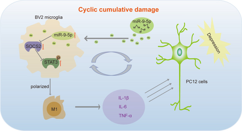
Background: Neuroinflammation is an important component mechanism in the development of depression. Exosomal transfer of MDD-associated microRNAs (miRNAs) from neurons to microglia might exacerbate neuronal cell inflammatory injury.

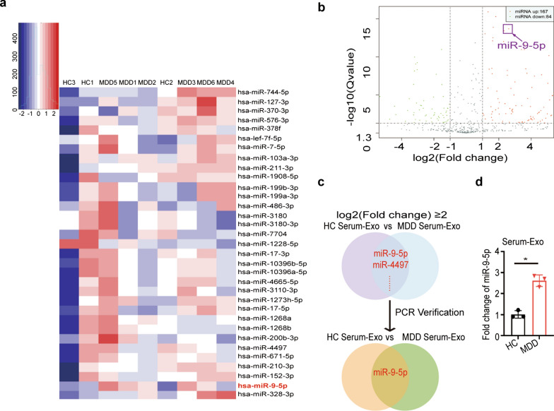
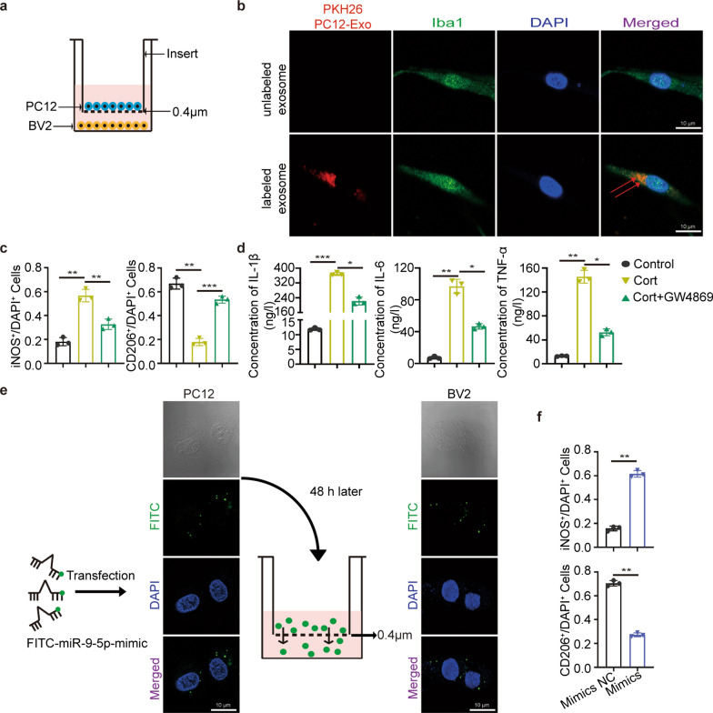
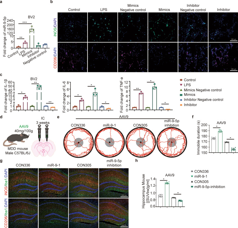
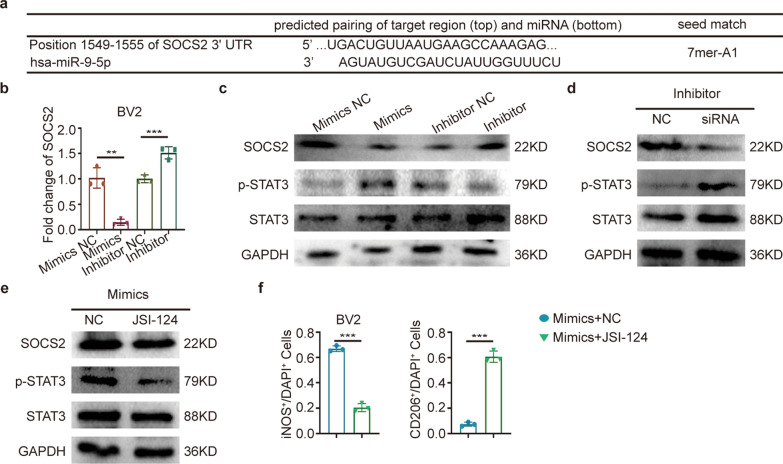
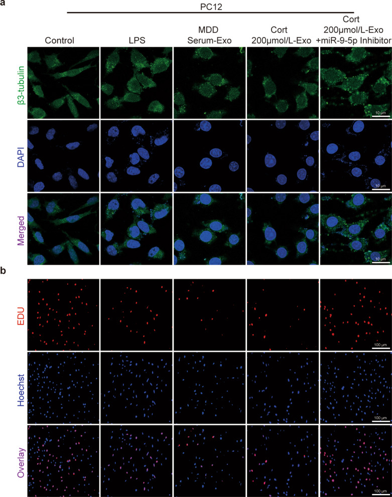
Results: By sequence identification, we found significantly higher miR-9-5p expression levels in serum exosomes from MDD patients than healthy control (HC) subjects. Then, in cultured cell model, we observed that BV2 microglial cells internalized PC12 neuron cell-derived exosomes while successfully transferring miR-9-5p. MiR-9-5p promoted M1 polarization in microglia and led to over releasing of proinflammatory cytokines, such as interleukin-1β (IL-1β), interleukin-6 (IL-6) and tumor necrosis factor-alpha (TNF-α), which exacerbated neurological damage. Furthermore, we identified suppressor of cytokine signaling 2 (SOCS2) as a direct target of miR-9-5p. Overexpression of miR-9-5p suppressed SOCS2 expression and reactivated SOCS2-repressed Janus kinase (JAK)/signal transducer and activator of transcription 3 (STAT3) pathways. Consistently, we confirmed that adeno-associated virus (AAV)-mediated overexpression of miR-9-5p polarized microglia toward the M1 phenotype and exacerbated depressive symptoms in chronic unpredictable mild stress (CUMS) mouse mode.

Conclusion: MiR-9-5p was transferred from neurons to microglia in an exosomal way, leading to M1 polarization of microglia and further neuronal injury. The expression and secretion of miR-9-5p might be novel therapeutic targets for MDD.

Keywords: Depression; Exosomes; Microglial polarization; Neuroinflammation; miR-9-5p.


Conflict of interest statement

The authors declare that they have no competing interests.

Figures

Fig. 1
Fig. 1
Serum-derived exosomes from depression patients induced microglia M1 polarization. Serum exosomes isolation, identification and uptake. ac Exosomal characterization was profiled with electron microscopy, western blotting and NTA. d Schematic diagram of serum or serum exosomes co-culture with BV2 cells. e Internalization of PKH-26-labeled exosomes was analyzed in BV2 cells cocultured with serum exosomes. Scale bar = 10 µm. f iNOS+ and CD206+ staining for microglia in indicated four groups. Scale bar = 100 µm. n = 3. g Related fold change of IL-1β, IL-6 and TNF-α mRNA in microglia treated as described above. Their relative expression levels were measured by 2−ΔΔCt. h Flow chart of the animal experiment. i Representative tracks depicting mice in the Control-Exo and Cort-Exo groups during OFT. The red lines represent animal move path. The black dot represents the center of the arena. The black circle represents the outer boundary of the arena. The gray circle represents the boundary of the center arena area (see “Materials and methods”section). n = 6. j Immobile duration of mice in the two groups was recorded. k (Left) after exosomes injection, C57BL/6 J mice were sacrificed for immunofluorescence staining in the hippocampus of brain tissue. Scale bar = 100 µm. (Right) the proportions of M1 and M2 polarized microglia are shown in the statistical plots. l (Left) the representative PET/CT images. (Right) accumulation of [18F]DPA-714 in the hippocampus
Fig. 2
Fig. 2
miRNA microarray analysis and qRT-PCR validation for serum exosomes derived from depression patients. a Partial heatmap of differentially expressed serum exosomal miRNAs between MDD patients and HC subjects. b Volcano maps showed that there were 167 miRNAs with higher expression and 84 miRNAs with lower expression in the serum exosomes of MDD patients compared to HC subjects. c Venn diagram showed that miR-9-5p was a miRNA with high expression in the serum exosomes of MDD patients. d Real-time qRT-PCR validated miR-9-5p expression in the serum exosomes of MDD patients
Fig. 3
Fig. 3
Depression-cells-derived exosomes containing miR-9-5p were engulfed by microglia and promoted the polarization of microglia. a Schematic diagram of PC12/BV2 cell co-culture system. b Internalization of PKH-26-labeled exosomes derived from PC12 cells was analyzed in BV2 cells. Scale bar = 10 µm. c Quantification of iNOS+ and CD206+ microglia in the three groups. d Extracelluler inflammatory cytokines IL-6, IL-1β and TNF-α levels in indicated three groups. e PC12 cells transfected with the FITC-miR-9-5p mimic (green fluorescence) were plated in the upper chamber and coincubated with BV2 cells in the lower chamber in a coculture system with a 0.4-µm pore membrane. Green fluorescence was detected in the PC12 recipient cells under the fluorescence microscope. Scale bar = 10 µm. f Quantification of iNOS+ and CD206+ staining in microglia in the two groups
Fig. 4
Fig. 4
miR-9-5p promoted M1 polarization of microglia in vitro. a Related miR-9-5p level in the different groups. b iNOS+ and CD206+ staining for microglia under various treatments. Scale bar = 100 µm. n = 3. c Related fold change of IL-1β, IL-6 and TNF-α mRNA. Their relative expression levels were measured by 2−ΔΔCt. d Schematic diagram of adenovirus stereotactic injection into mouse brain. e, f Representative tracks and immobile duration of mice in different groups. n = 6. g Immunofluorescence for brain tissues in indicated groups. Scale bar = 100 µm. n = 6. h [18F]DPA-714 uptake in uptake hippocampus
Fig. 5
Fig. 5
Exosomal miR-9-5p activated STAT3 pathway through suppressing SOCS2. a Sequence alignment of miR-9-5p and SOCS2 3′UTR sequences. b Real-time qRT-PCR confirmed the effects of miR-9-5p mimics and inhibitor on SOCS2. c Protein levels of SOCS2, p-STAT3 and STAT3 in the BV2 cells treated with mimics and inhibitor. n = 3. d SiRNA reversed the effect of inhibitor to SOCS2 and p-STAT3. n = 3. e, f Western blot analysis of SOCS2, p-STAT3 and STAT3 expression and quantification of iNOS+ and CD206+ staining in microglia treated with mimics and JSI-124. GAPDH served as the loading control. n = 3
Fig. 6
Fig. 6
Polarized M1 cells contributed to damaging PC12 cells. To further study the changes in PC12 cells cocultured with M1 microglia, β3-tubulin and EdU immunofluorescence staining were performed to measure synaptic growth and cell proliferation. a Total neurite length of PC12 cells after different treatments was imaged by β3-tubulin staining. Scale bar = 10 µm. n = 3. b PC12 cell proliferation after co-culture was detected by EdU staining. Scale bar = 100 µm. n = 3
Fig. 7
Fig. 7
The schematic representation of the interaction between PC12 cells and BV2 microglia in depression. The schematic representation of cyclic cumulative damage: first, the depressed PC12 cells generated exosomes with high miR-9-5p contents. BV2 microglia phagocytosed these exosomes, in which enriched miR-9-5p, suppressed SOCS2 expression and activated JAK/STAT3 pathways, leading to polarization into M1 type. Meanwhile, proinflammatory factors including IL-β, IL-6 and TNF-α were secreted by M1 microglia. These proinflammatory factors further accelerated PC12 cell damage

References

    1. Jo WK, Zhang Y, Emrich HM, Dietrich DE. Glia in the cytokine-mediated onset of depression: fine tuning the immune response. Front Cell Neurosci. 2015;9:268. - PMC - PubMed
    1. Daly EJ, Singh JB, Fedgchin M, Cooper K, Lim P, Shelton RC, Thase ME, Winokur A, Van Nueten L, Manji H, et al. Efficacy and safety of intranasal esketamine adjunctive to oral antidepressant therapy in treatment-resistant depression: a randomized clinical trial. JAMA Psychiatry. 2018;75(2):139–148. - PMC - PubMed
    1. Dwivedi Y. Emerging role of microRNAs in major depressive disorder: diagnosis and therapeutic implications. Dialogues Clin Neurosci. 2014;16(1):43–61. - PMC - PubMed
    1. Haapakoski R, Ebmeier KP, Alenius H, Kivimaki M. Innate and adaptive immunity in the development of depression: an update on current knowledge and technological advances. Prog Neuropsychopharmacol Biol Psychiatry. 2016;66:63–72. - PMC - PubMed
    1. Brites D, Fernandes A. Neuroinflammation and depression: microglia activation, extracellular microvesicles and microRNA dysregulation. Front Cell Neurosci. 2015;9:476. - PMC - PubMed

LinkOut - more resources