. 2022 Jun;33(6):1105-1119.
doi: 10.1681/ASN.2021060843. Epub 2022 Mar 9.

Intestinal Bacterial Translocation Contributes to Diabetic Kidney Disease

Affiliations

Intestinal Bacterial Translocation Contributes to Diabetic Kidney Disease

Hoang Thuy Linh et al. J Am Soc Nephrol. 2022 Jun.

Abstract

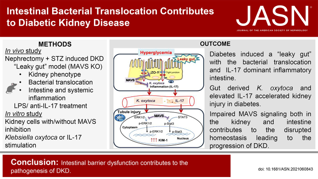
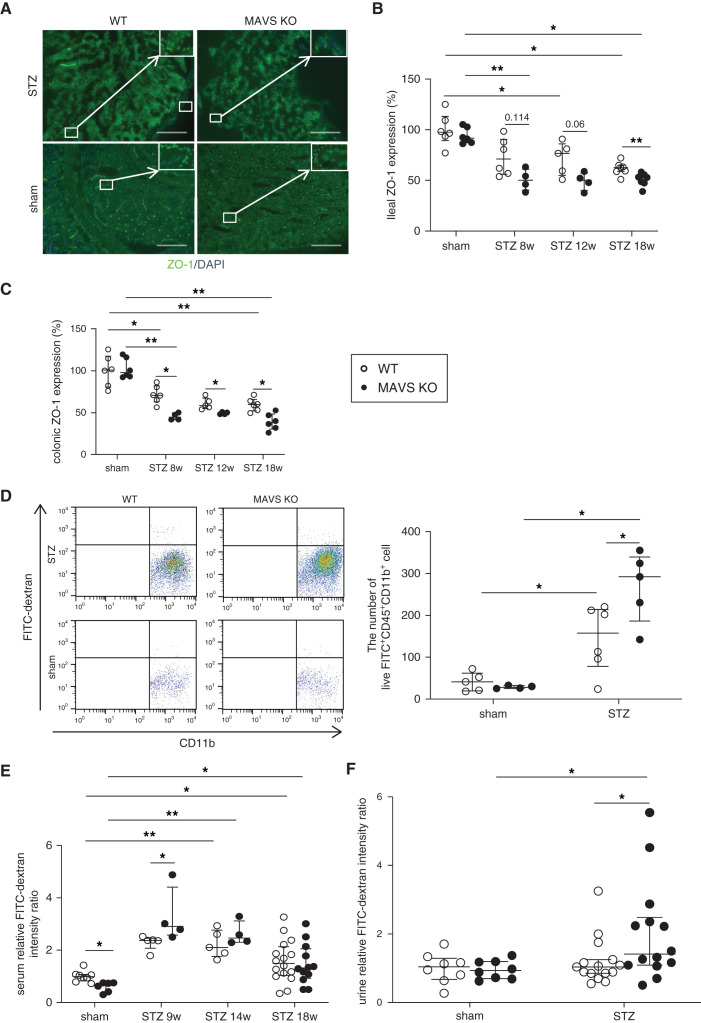
Background: In recent years, many studies have focused on the intestinal environment to elucidate pathogenesis of various diseases, including kidney diseases. Impairment of the intestinal barrier function, the "leaky gut," reportedly contributes to pathologic processes in some disorders. Mitochondrial antiviral signaling protein (MAVS), a component of innate immunity, maintains intestinal integrity. The effects of disrupted intestinal homeostasis associated with MAVS signaling in diabetic kidney disease remains unclear.

Methods: To evaluate the contribution of intestinal barrier impairment to kidney injury under diabetic conditions, we induced diabetic kidney disease in wild-type and MAVS knockout mice through unilateral nephrectomy and streptozotocin treatment. We then assessed effects on the kidney, intestinal injuries, and bacterial translocation.

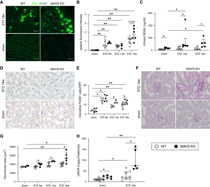
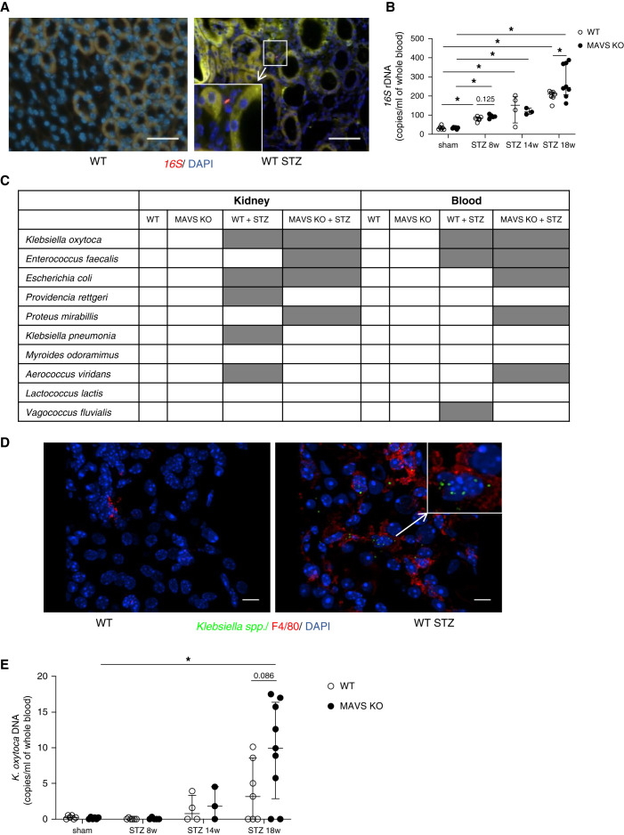
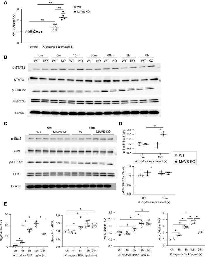
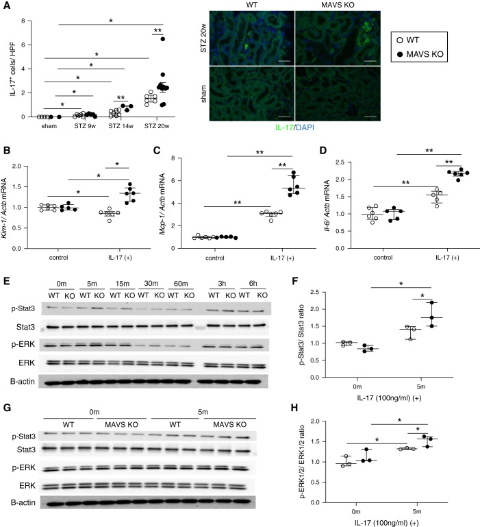
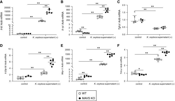
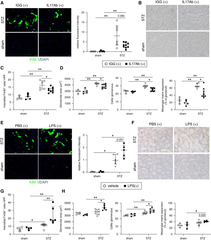
Results: MAVS knockout diabetic mice showed more severe glomerular and tubular injuries compared with wild-type diabetic mice. Owing to impaired intestinal integrity, the presence of intestine-derived Klebsiella oxytoca and elevated IL-17 were detected in the circulation and kidneys of diabetic mice, especially in diabetic MAVS knockout mice. Stimulation of tubular epithelial cells with K. oxytoca activated MAVS pathways and the phosphorylation of Stat3 and ERK1/2, leading to the production of kidney injury molecule-1 (KIM-1). Nevertheless, MAVS inhibition induced inflammation in the intestinal epithelial cells and KIM-1 production in tubular epithelial cells under K. oxytoca supernatant or IL-17 stimulation. Treatment with neutralizing anti-IL-17 antibody treatment had renoprotective effects. In contrast, LPS administration accelerated kidney injury in the murine diabetic kidney disease model.

Conclusions: Impaired MAVS signaling both in the kidney and intestine contributes to the disrupted homeostasis, leading to diabetic kidney disease progression. Controlling intestinal homeostasis may offer a novel therapeutic approach for this condition.

Keywords: bacterial translocation; diabetes; diabetic kidney disease; gut-kidney axis; inflammation; microbiota.


Figures

None
Graphical abstract
Figure 1.
Figure 1.
MAVS deficiency promotes kidney injury in diabetic condition. (A and B) Immunofluorescence analysis of KIM-1, a marker for tubule injury (magnification, ×200; scale bar, 50 μm). (C) NGAL concentration in the urine. (D and E) Representative kidney histology showing interstitial F4/80 macrophage infiltration (magnification, ×400; scale bar, 50 μm). (F and G) Increase in the glomerular area (magnification, ×400; scale bar, 100 μm). (H) Urinary albumin excretion (UACR) is normalized to creatinine level. Data are shown as median±IQR; *P<0.05, **P<0.01.
Figure 2.
Figure 2.
Detection of intestinal bacteria including K. oxytoca in the blood and kidney of diabetic mice. (A) Bacterial 16S in kidney sections by FISH experiment. (B) 16S rDNA quantification in the blood. (C) Bacterial strains in the kidney and blood culture. (D) Detection of Klebsiella spp. in the kidney of diabetic mice using immunofluorescence staining. (E) K. oxytoca DNA quantification in the blood. Data are shown as median±IQR; *P<0.05.
Figure 3.
Figure 3.
Hyperglycemia induces the formation of a “leaky gut.” (A) Ileal ZO-1 staining (magnification, ×200; scale bar, 50 μm) and relative quantification of (B) an ileal section and (C) a colon section. (D) Mice were fasted for 4 hours and gavaged with FITC-dextran 4 kDa. Flow cytometry showed the number of live FITC+CD11b+CD45+ cells in the kidney at 18 weeks after STZ injection. FITC intensity was measured in (E) serum and (F) urine. Data are shown as median±IQR; *P<0.05, **P<0.01.
Figure 4.
Figure 4.
Hyperglycemia induced an inflammatory state in the intestine. Rorγt/Foxp3 mRNA expression normalized with β-actin in (A) the lamina propria, (B) Peyer’s patch, and (C) mesenteric lymph nodes at 18 weeks after STZ injection. Il-17 mRNA expression over 18 weeks in (D) the ileal epithelium and (E) Peyer’s patch. (F) IL-17 staining and (G) quantification in Peyer’s patch (magnification, ×200; scale bar, 50 μm) at 18 weeks of diabetes. (H) IL-17 levels in the serum. Data are shown as median±IQR; *P<0.05, **P<0.01.
Figure 5.
Figure 5.
Effect of the K. oxytoca supernatant on primary TECs. (A) Kim-1 mRNA expression after treatment with the K. oxytoca supernatant (1:20) for 24 hours. (B) Western blot analysis of phospho-Stat3, total Stat3, phospho-ERK1/2, and total ERK1/2 in WT and MAVS KO TECs after K. oxytoca supernatant stimulation (1:20) at different time points. (C and D) Western blot analysis and densitometric quantification of phospho-Stat3, total Stat3, phospho-ERK1/2, and total ERK1/2 in WT and MAVS KO TECs after K. oxytoca supernatant stimulation (1:20) at the indicated time. (E) Upregulation of Rig-I, Mavs, Traf-6, and Kim-1 mRNA was shown under K. oxytoca RNA transfection. Data are shown as median±IQR; *P<0.05, **P<0.01.
Figure 6.
Figure 6.
Change in IL-17 levels in diabetic mice and the effect of IL-17 on primary TECs. (A) IL-17 staining (magnification, ×400; scale bar, 100 μm) and quantification in the kidney. Levels of (B) Kim-1, (C) Mcp-1, and (D) Il-6 mRNA under IL-17 stimulation (100 ng/ml) for 24 hours. (E) Western blot analysis of phospho-Stat3, total Stat3, phospho-ERK1/2, and total ERK1/2 in WT and MAVS KO TECs after IL-17 stimulation (100 ng/ml) at different time points. (F–H) Western blot analysis and densitometric quantification of phospho-Stat3, total Stat3, phospho-ERK1/2, and total ERK1/2 in WT and MAVS KO TECs after IL-17 stimulation (100 ng/ml) at the indicated time. Data are shown as median±IQR; *P<0.05, **P<0.01.
Figure 7.
Figure 7.
Effects of K. oxytoca supernatant to BMDMs. Proinflammatory cytokines, namely (A) Il-6, (B) Il-1β, (C) Tgf-β, (D) Il-23p19, (E) Mcp-1, and (F) Tnf-α were expressed under the K. oxytoca supernatant (1:100) stimulation for 24 hours. Data are shown as median±IQR; *P<0.05, **P<0.01.
Figure 8.
Figure 8.
Neutralizing IL-17 antibody treatment ameliorates DKD whereas the administration of LPS to WT diabetic mice aggravates kidney injury. (A) Immunofluorescence analysis of KIM-1 (magnification, ×200; scale bar, 50 μm), (B and C) representative kidney histology showing interstitial F4/80 macrophage infiltration (magnification, 400×; scale bar, 50 μm), and (D) pathologic glomerular analysis (glomerular area, hypercellularity, and mesangial matrix expansion) in IL-17 antibody-treated WT diabetic mice. (E) Immunofluorescence analysis of KIM-1 (magnification, ×200; scale bar, 50 μm), (F and G) representative kidney histology showing interstitial F4/80 macrophage infiltration (magnification, ×400; scale bar, 50 μm), and (H) pathologic glomerular analysis (glomerular area, hypercellularity, and mesangial matrix expansion) in LPS-treated WT diabetic mice. Data are shown as median±IQR; *P<0.05, **P<0.01.

Comment in

References

    1. Locatelli F, Pozzoni P, Del Vecchio L: Renal replacement therapy in patients with diabetes and end-stage renal disease. J Am Soc Nephrol 15[Suppl 1]: S25–S29, 2004 - PubMed
    1. Schieber AM, Lee YM, Chang MW, Leblanc M, Collins B, Downes M, et al. : Disease tolerance mediated by microbiome E. coli involves inflammasome and IGF-1 signaling. Science 350: 558–563, 2015 - PMC - PubMed
    1. Zhu S, Jiang Y, Xu K, Cui M, Ye W, Zhao G, et al. : The progress of gut microbiome research related to brain disorders. J Neuroinflammation 17: 25, 2020 - PMC - PubMed
    1. Kikuchi K, Saigusa D, Kanemitsu Y, Matsumoto Y, Thanai P, Suzuki N, et al. : Gut microbiome-derived phenyl sulfate contributes to albuminuria in diabetic kidney disease. Nat Commun 10: 1835, 2019 - PMC - PubMed
    1. Muskiet MH, Smits MM, Morsink LM, Diamant M: The gut-renal axis: Do incretin-based agents confer renoprotection in diabetes? Nat Rev Nephrol 10: 88–103, 2014 - PubMed

Publication types