. 2022 Mar 9;13(3):220.
doi: 10.1038/s41419-022-04605-2.

Bone marrow-independent adventitial macrophage progenitor cells contribute to angiogenesis

Affiliations

Bone marrow-independent adventitial macrophage progenitor cells contribute to angiogenesis

Florian Kleefeldt et al. Cell Death Dis. .

Abstract

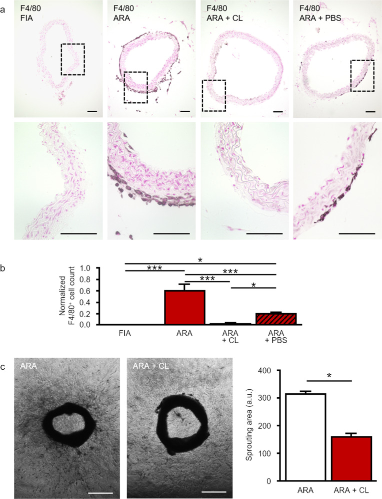
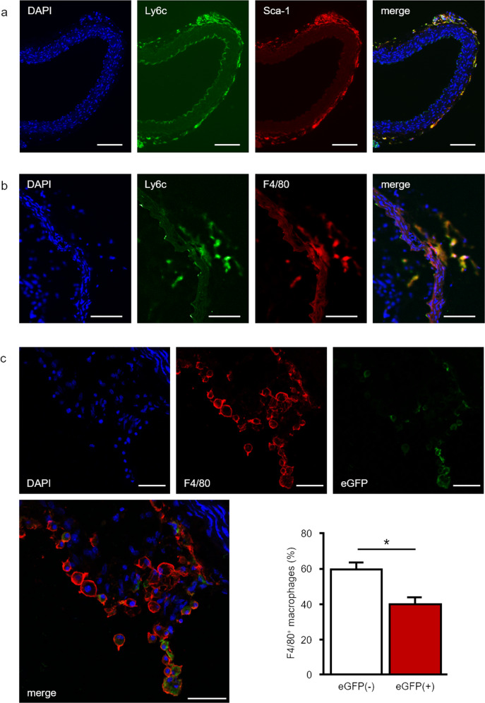
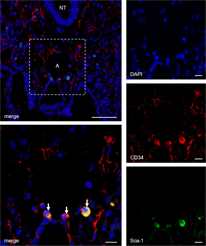
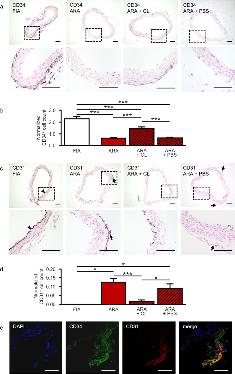
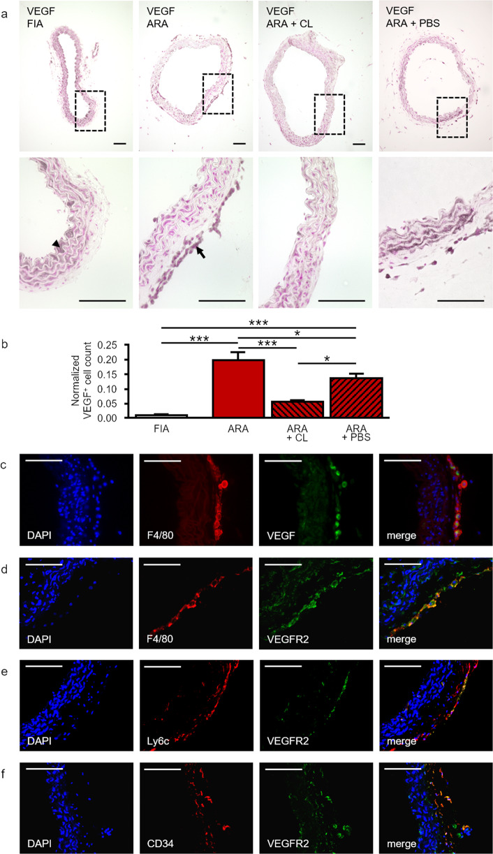
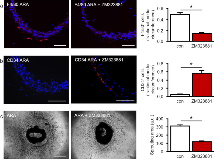
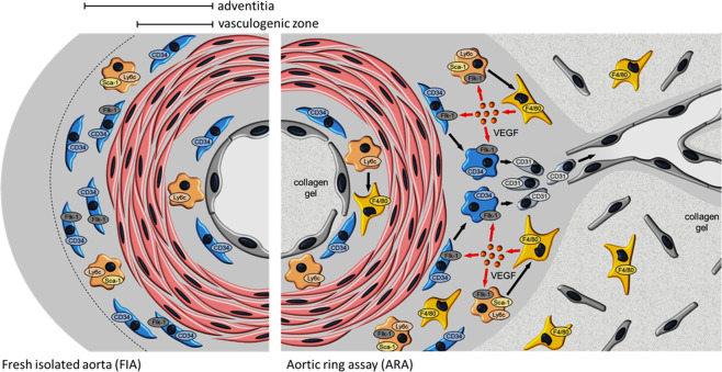
Pathological angiogenesis promotes tumor growth, metastasis, and atherosclerotic plaque rupture. Macrophages are key players in these processes. However, whether these macrophages differentiate from bone marrow-derived monocytes or from local vascular wall-resident stem and progenitor cells (VW-SCs) is an unresolved issue of angiogenesis. To answer this question, we analyzed vascular sprouting and alterations in aortic cell populations in mouse aortic ring assays (ARA). ARA culture leads to the generation of large numbers of macrophages, especially within the aortic adventitia. Using immunohistochemical fate-mapping and genetic in vivo-labeling approaches we show that 60% of these macrophages differentiate from bone marrow-independent Ly6c+/Sca-1+ adventitial progenitor cells. Analysis of the NCX-/- mouse model that genetically lacks embryonic circulation and yolk sac perfusion indicates that at least some of those progenitor cells arise yolk sac-independent. Macrophages represent the main source of VEGF in ARA that vice versa promotes the generation of additional macrophages thereby creating a pro-angiogenetic feedforward loop. Additionally, macrophage-derived VEGF activates CD34+ progenitor cells within the adventitial vasculogenic zone to differentiate into CD31+ endothelial cells. Consequently, depletion of macrophages and VEGFR2 antagonism drastically reduce vascular sprouting activity in ARA. In summary, we show that angiogenic activation induces differentiation of macrophages from bone marrow-derived as well as from bone marrow-independent VW-SCs. The latter ones are at least partially yolk sac-independent, too. Those VW-SC-derived macrophages critically contribute to angiogenesis, making them an attractive target to interfere with pathological angiogenesis in cancer and atherosclerosis as well as with regenerative angiogenesis in ischemic cardiovascular disorders.


Conflict of interest statement

The authors declare no competing interests.

Figures

Fig. 1
Fig. 1. Generation of F4/80+ macrophages in ARA.
a Cross-sections of murine aortae were immunostained for the marker of mature macrophages F4/80. In FIA, hardly any F4/80+ macrophage could be found. After ARA a few F4/80+ macrophages appeared within the intima. However, within the adventitia, the number of F4/80+ macrophages increased substantially. Compared to control, cultured aortic rings treated with clodronate-containing liposomes displayed almost no F4/80 immunostaining due to efficient depletion of F4/80+ macrophages within the adventitia. b Statistical analysis of the number of adventitial F4/80+ macrophages with and without liposome treatment. Application of PBS-containing liposomes had a less pronounced effect compared to clodronate-containing liposomes. Cell counts were normalized to total media area (n = 7–10). c Phase-contrast images and quantification of sprouting area of cultured aortic rings with or without clodronate treatment. Depletion of macrophages by clodronate-containing liposomes reduced total cellular sprouting as well as capillary-like tube formation (n = 23–31). *P < 0.05; ***P < 0.001. Scale bars: in a 100 µm, in c 500 µm. ARA aortic ring assay, ARA + CL ARA with clodronate-containing liposomes, ARA + PBS ARA with PBS-containing liposomes.
Fig. 2
Fig. 2. The majority of macrophages in ARA originates from bone marrow-independent progenitors.
a Cross-sections of freshly isolated murine aortae were analyzed by immunofluorescence for the presence of Ly6c+ and Sca-1+ progenitor cells. Within the adventitia, a substantial part of Ly6c+ cells co-expressed Sca-1 distinguishing them from Ly6c+ monocytes that derived from the bone marrow and circulate in peripheral blood. b After ARA, cross-sections of murine aortae were analyzed by immunofluorescence for the presence of Ly6c+/F4/80+ cells. The majority of F4/80+ macrophages also expressed Ly6c suggesting their derivation from Ly6c+ progenitor cells. c Confocal image of a cross-section of a cultured aortic ring from the FlkSwitch mouse. These sections were screened for F4/80+ cells (red immunofluorescence) and eGFP+ cells (bone marrow-derived). Quantitative analysis of these sections (n = 6) confirmed that 40% of F4/80 macrophages originate from bone marrow-derived cells (F4/80+/eGFP+) whereas 60% are generated from bone marrow-independent progenitors (F4/80+/GFP-). Scale bars: in a, b 100 µm, in c 50 µm.
Fig. 3
Fig. 3. Aorta-associated progenitor cells may partially be yolk sac-independent.
Confocal image of a transversal section from an NCX1−/− mouse embryo (E9.5) immunostained for CD34 and Sca-1. The presence of CD34+/Sca-1+ cells surrounding the aortic region of these embryos that lack a functional vascular network to the yolk sac indicates a yolk sac-independent origin of these progenitor cells. Scale bars: lower magnification 50 µm, higher magnification 10 µm; NT neural tube, A aorta.
Fig. 4
Fig. 4. Macrophage depletion prevents activation of the vasculogenic zone and generation of CD31+ endothelial cells in ARA.
a Cross-sections of murine aortae were immunostained for the progenitor cell marker CD34. In FIA, a large number of CD34+ progenitor cells was present within the vasculogenic zone of the adventitia. By performing ARA, these cells were activated within their adventitial stem cell niche and differentiated thereby losing their CD34 immunoreactivity. Depletion of macrophages preserved the number of CD34+ cells within the adventitia during ARA. b Statistical analysis of the number of adventitial CD34+ cells with and without liposome treatment. Cell counts were normalized to the total media area (n = 6–12). c Cross-sections of murine aortae were immunostained for the endothelial cell marker CD31. In FIA, CD31+ cells were present within the intima as expected (arrowhead), but were almost completely absent from the adventitia. After ARA, a substantial number of CD31+ cells appeared within the adventitia (arrows). Their generation was largely prevented by the depletion of macrophages. d Statistical analysis of the number of adventitial CD31+ cells with and without liposome treatment. Cell counts were normalized to total media area (n = 3–10). e Cross-section of a cultured aortic ring at day 3 immunostained for CD34 and CD31. CD34+/CD31+ cells represent a transitional stage in the differentiation of CD34+ adventitial progenitors towards CD31+ endothelial cells. *P < 0.05; ***P < 0.001. Scale bars: in a and c 100 µm, in e 50 µm. FIA fresh isolated aorta, ARA aortic ring assay, ARA + CL ARA with clodronate-containing liposomes, ARA + PBS ARA with PBS-containing liposomes.
Fig. 5
Fig. 5. Expression of VEGF/ VEGFR2 in macrophages and CD34+ progenitor cells in ARA.
a Cross-sections of murine aortae were immunostained for the pro-angiogenic factor VEGF. In FIA, VEGF is predominantly expressed by intimal cells (arrowhead) and cells within the media. In ARA the number of VEGF+ cells increased substantially especially within the adventitia (arrow). This increase was largely prevented by the depletion of macrophages. b Statistical analysis of the number of adventitial VEGF+ cells with and without liposome treatment. Cell counts were normalized to total media area (n = 6–8). c Cross-sections of cultured aortic rings were analyzed by confocal co-immunofluorescence for the presence of VEGF+ and F4/80+ cells, respectively. Almost all adventitial cells that were highly positive for VEGF co-express F4/80. d Additionally, F4/80+ cells in cross-sections of cultured aortic rings co-express VEGFR2. e, f Similarly, in cross-sections of FIA Ly6c+ and CD34+ progenitor cells show at least partially co-expression of VEGFR2 by confocal immunofluorescence microscopy. *P < 0.05; ***P < 0.001. Scale bars: 100 µm. FIA fresh isolated aorta, ARA aortic ring assay, ARA + CL ARA with clodronate-containing liposomes, ARA + PBS ARA with PBS-containing liposomes.
Fig. 6
Fig. 6. Macrophage-derived VEGF activates the vasculogenic zone and promotes macrophage generation.
a After ARA, a substantial number of F4/80+ macrophages was generated whereas hardly any macrophage was detected after ARA in the presence of the VEGFR2 antagonist ZM323881 (1 µM). For quantification, the fraction of media circumference that is covered by F4/80+ cells was analyzed (n ≥ 5). b Furthermore, the application of ZM323881 (1 µM) resulted in the preservation of CD34+ cells within the adventitial vasculogenic zone. For quantification, the fraction of media circumference that is covered by CD34+ cells was analyzed (n ≥ 4). c Consequently, cellular sprouting, as well as capillary-like tube formation in ARA, were reduced in the presence of ZM323881 (1 µM; n = 31). *P < 0.05. Scale bars: 100 µm, phase contrast 500 µm. ARA aortic ring assay.
Fig. 7
Fig. 7. Interplay of adventitial cells in angiogenic activation.
In ARA the majority of macrophages arise from resident bone marrow-independent and at least partially yolk sac-independent Ly6c+/Sca-1+ adventitial progenitor cells. VEGF released by macrophages on the one hand induces angiogenetic sprouting by stimulating the differentiation of CD34+ progenitor cells within the adventitial vasculogenic zone into CD31+ endothelial cells. On the other hand, macrophage-derived VEGF potentiates the generation of macrophages via VEGF/VEGFR2 signaling in terms of a feedforward loop thereby maintaining the angiogenic process.

References

    1. Filipowska J, Tomaszewski KA, Niedźwiedzki Ł, Walocha JA, Niedźwiedzki T. The role of vasculature in bone development, regeneration and proper systemic functioning. Angiogenesis. 2017;20:291–302. - PMC - PubMed
    1. Carmeliet P. Angiogenesis in life, disease and medicine. Nature. 2005;438:932–6. - PubMed
    1. Gariano RF, Gardner TW. Retinal angiogenesis in development and disease. Nature. 2005;438:960–6. - PubMed
    1. Subhani S, Vavilala DT, Mukherji M. HIF inhibitors for ischemic retinopathies and cancers: options beyond anti-VEGF therapies. Angiogenesis. 2016;19:257–73. - PubMed
    1. Wang S, Li X, Parra M, Verdin E, Bassel-Duby R, Olson EN. Control of endothelial cell proliferation and migration by VEGF signaling to histone deacetylase 7. Proc Natl Acad Sci USA. 2008;105:7738–43. - PMC - PubMed

Publication types

Substances