. 2022 May;27(5):2502-2513.
doi: 10.1038/s41380-022-01496-9. Epub 2022 Mar 9.

Central amygdala corticotropin-releasing factor neurons promote hyponeophagia but do not control alcohol drinking in mice

Affiliations

Central amygdala corticotropin-releasing factor neurons promote hyponeophagia but do not control alcohol drinking in mice

Max Kreifeldt et al. Mol Psychiatry. 2022 May.

Abstract

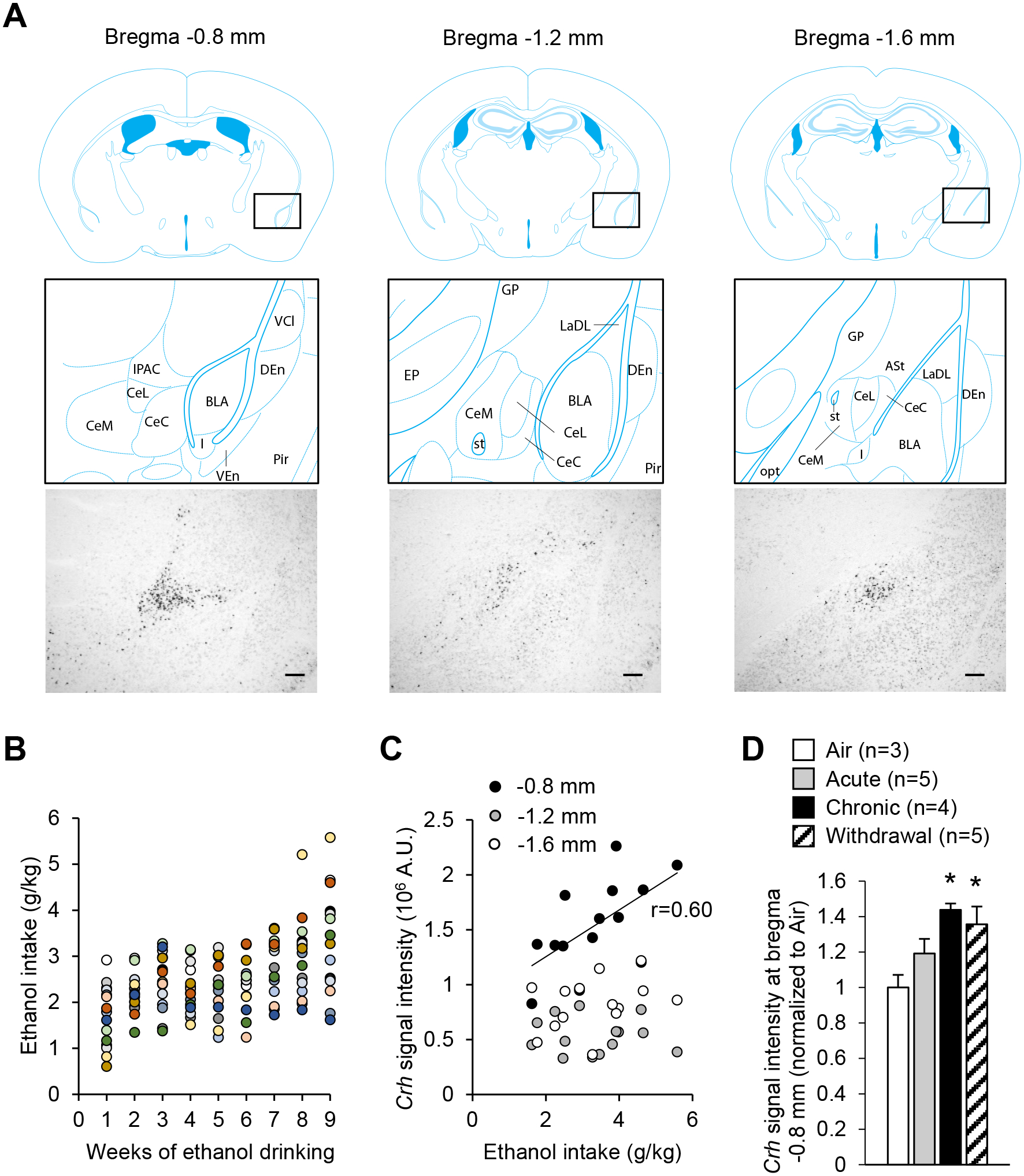
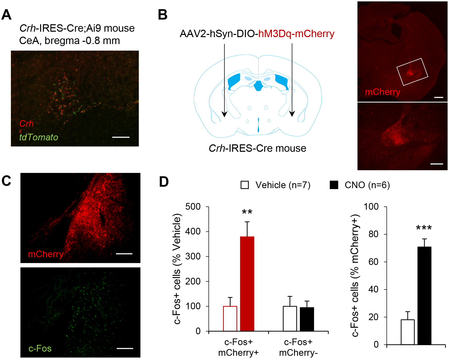
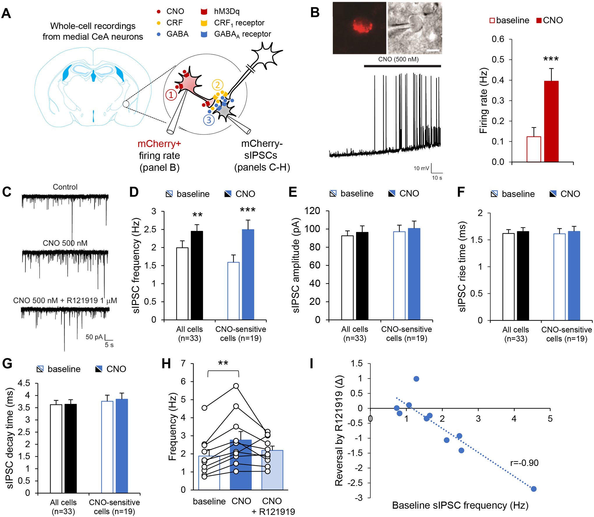
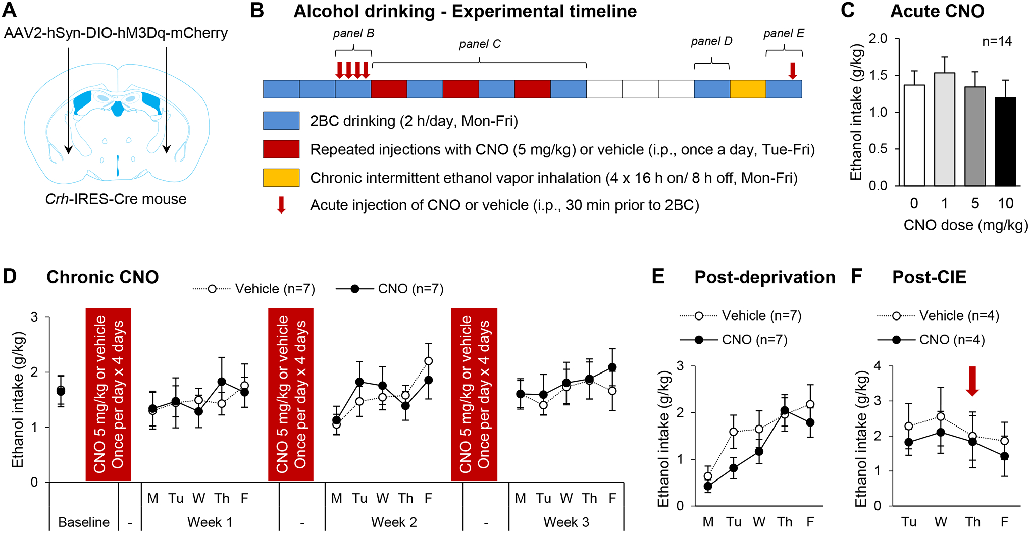
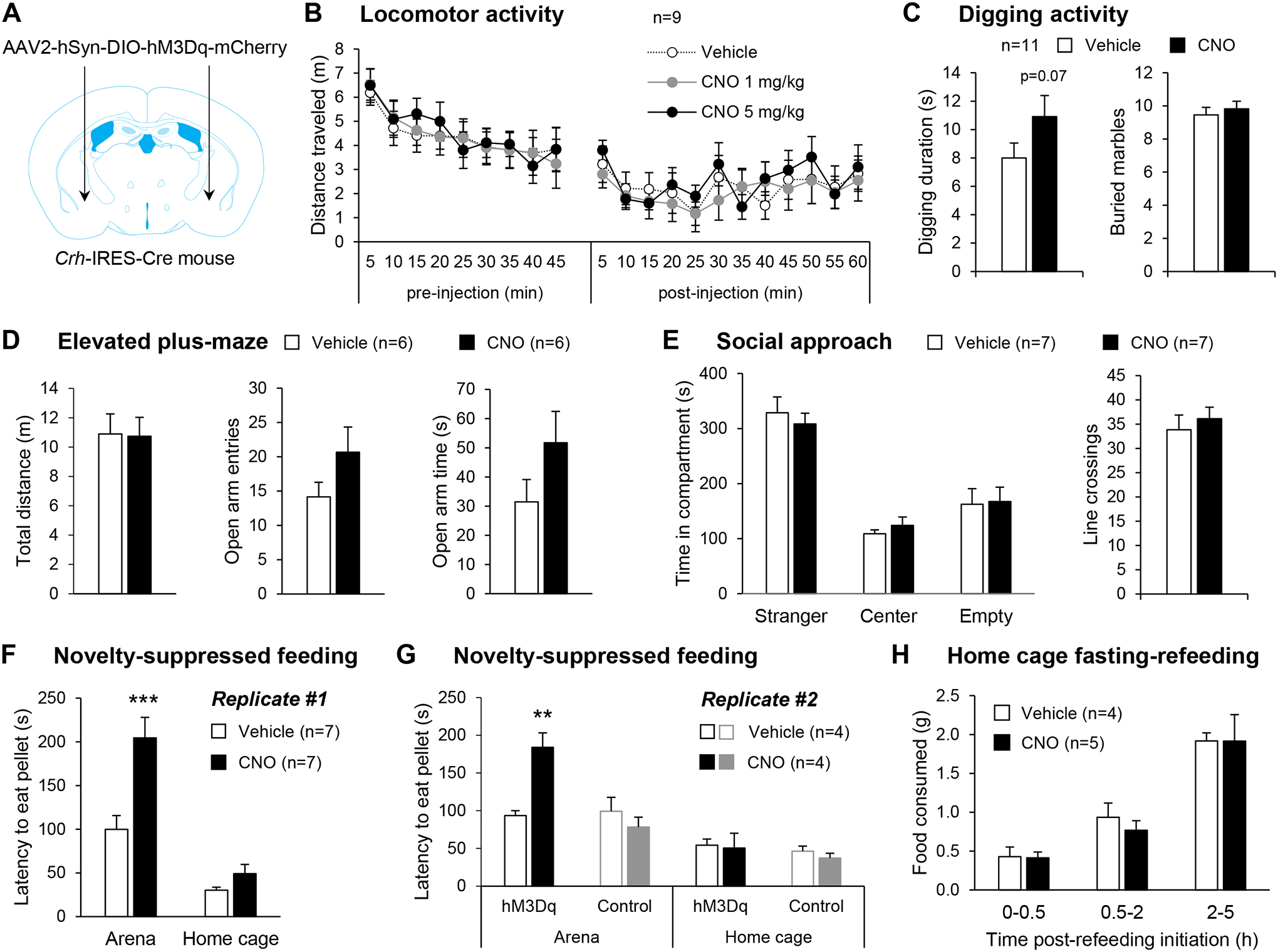
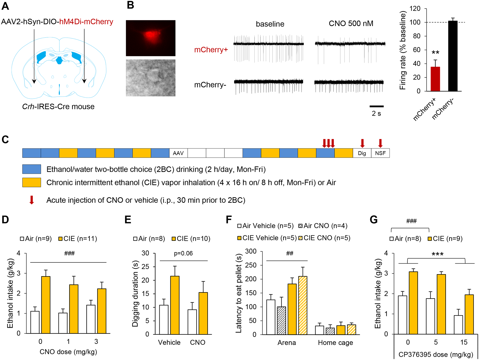
Corticotropin-releasing factor (CRF) signaling in the central nucleus of the amygdala (CeA) plays a critical role in rodent models of excessive alcohol drinking. However, the source of CRF acting in the CeA during alcohol withdrawal remains to be identified. In the present study, we hypothesized that CeA CRF interneurons may represent a behaviorally relevant source of CRF to the CeA increasing motivation for alcohol via negative reinforcement. We first observed that Crh mRNA expression in the anterior part of the mouse CeA correlates positively with alcohol intake in C57BL/6J males with a history of chronic binge drinking followed by abstinence and increases upon exposure to chronic intermittent ethanol (CIE) vapor inhalation. We then found that chemogenetic activation of CeA CRF neurons in Crh-IRES-Cre mouse brain slices increases gamma-aminobutyric acid (GABA) release in the medial CeA, in part via CRF1 receptor activation. While chemogenetic stimulation exacerbated novelty-induced feeding suppression (NSF) in alcohol-naïve mice, thereby mimicking the effect of withdrawal from CIE, it had no effect on voluntary alcohol consumption, following either acute or chronic manipulation. Furthermore, chemogenetic inhibition of CeA CRF neurons did not affect alcohol consumption or NSF in chronic alcohol drinkers exposed to air or CIE. Altogether, these findings indicate that CeA CRF neurons produce local release of GABA and CRF and promote hyponeophagia in naïve mice, but do not drive alcohol intake escalation or negative affect in CIE-withdrawn mice. The latter result contrasts with previous findings in rats and demonstrates species specificity of CRF circuit engagement in alcohol dependence.


Conflict of interest statement

Disclosure

The authors have no conflicts of interest to declare.

Figures

Figure 1.
Figure 1.. Crh mRNA levels in the anterior CeA increase upon chronic intermittent alcohol exposure.
A. Representative images of Crh mRNA distribution at three antero-posterior levels of the mouse CeA (scale bars = 200 μm) and corresponding brain atlas diagrams highlighting a V-shaped cluster of CRF neurons at the junction of the anterior CeL and IPAC (left panels), and scattered CRF neurons at more posterior levels of the CeL (middle and right panels). Brain atlas diagrams were reproduced from. BLA, basolateral amygdaloid nucleus; CeC, capsular part of the CeA; CeL, lateral division of the CeA; CeM, medial division of the CeA; DEn, dorsal endopiriform claustrum; EP, entopeduncular nucleus; IPAC, interstitial nucleus of the posterior limb of the anterior commissure; GP, globus pallidus; I, intercalated nuclei of the amygdala; LaDL, lateral amygdaloid nucleus, dorsolateral part; opt, optic tract; Pir, piriform cortex; st, stria terminalis; VCl, ventral part of claustrum; VEn, ventral endopiriform claustrum. B. Average weekly ethanol intake in a cohort of mice subjected to 2-h two-bottle choice (ethanol 15% v:v vs water) sessions five days per week for nine weeks. Each color represents an individual mouse. C. Crh chromogenic in situ hybridization signal density at three antero-posterior levels of the CeA as a function of ethanol intake during the last week. There was a significant correlation at the most anterior level (≈ bregma −0.8 mm, p<0.05), but not at more posterior levels. D. Crh chromogenic in situ hybridization signal density at the most anterior level of the CeA in mice exposed to a single 16-h bout of alcohol vapor inhalation (Acute), eight bouts (Chronic), or eight bouts followed by three days of withdrawal (Withdrawal). Data are shown as mean ± s.e.m. of Crh signal normalized to Air controls. Data were analyzed by one-way ANOVA; *, p<0.05 vs. Air, Dunnett’s posthoc test.
Figure 2.
Figure 2.. Validation of Cre activity and chemogenetic stimulation in CeA CRF neurons of Crh-IRES-Cre mice.
A. Distribution of Crh (red) and tdTomato (green) mRNAs in the CeA of Crh-IRES-Cre;Ai9 mice, as visualized by double fluorescent in situ hybridization (scale bar = 100 μm). B. mCherry immunolabeling recapitulates the V-shaped pattern of Crh expression in the CeA of Crh-IRES-Cre mice injected with a Cre-dependent AAV vector encoding hM3Dq-mCherry. The area framed in the top picture (scale bar = 500 μm) is shown at higher magnification in the bottom picture (scale bar = 100 μm). C. Double immunostaining of mCherry (red) and c-Fos (green) was used to evaluate neuronal activation in the CeA following i.p. injection of vehicle (n=7) or CNO 5 mg/kg (n=6) in Crh-IRES-Cre mice injected with AAV2-hSyn-DIO-hM3Dq-mCherry in the CeA (scale bars = 100 μm). D. Chemogenetic stimulation of CeA CRF neurons increased c-Fos expression selectively in mCherry-positive CeA neurons. Data are shown as mean ± s.e.m. of the number of c-Fos+ cells expressed as percentage of Vehicle values (left graph) or percentage of mCherry+ cells (right graph). Data were analyzed by unpaired t-test: **, p<0.01; ***, p<0.001.
Figure 3.
Figure 3.. Chemogenetic stimulation of CeA CRF neurons increases GABA release onto medial CeA neurons in a CRF1-dependent manner.
A. Crh-IRES-Cre mice were injected in the anterior CeA with a Cre-dependent AAV vector encoding hM3Dq-mCherry. Whole-cell recordings were then obtained from mCherry+ neurons (panel B) or mCherry− neurons (panels C-I) to test the hypothesis that chemogenetic stimulation of CeA CRF neurons (red, ①) triggers the release of GABA (blue) and CRF (yellow) in the medial CeA (②), which may in turn stimulate GABA release (③) onto non-CRF neurons (grey) via the activation of CRF1 receptors located on intrinsic or extrinsic GABAergic presynaptic terminals. B. Firing rates were recorded from CeA mCherry+ neurons. Top: red fluorescence and differential interference contrast images of patched CeA neuron. Bottom: representative current-clamp trace before and during CNO application (500 nM). Right: Firing rates are shown as mean ± s.e.m. (n=8 neurons). Data were analyzed by paired t-test: ***, p<0.001. C-I. Spontaneous inhibitory postsynaptic currents (sIPSCs) were recorded from medial CeA mCherry− neurons. C. Representative traces before and during CNO application (500 nM) followed by subsequent R121919 co-application (1 μM). CNO increased sIPSC frequency (D), but did not affect sIPSC amplitude (E), rise time (F) or decay time (G). Data are shown as mean ± s.e.m. for the whole set of recorded neurons (n=33, black bars), as well as for the subset of neurons whose sIPSC frequency was significantly increased by CNO (n=19, blue bars). Data were analyzed by paired t-test: **, p<0.01; ***, p<0.001. H-I. In a subset of CNO-sensitive cells (n=10), the CRF1 antagonist R121919 was co-applied following CNO alone. H. sIPSC frequencies are shown as mean ± s.e.m. and were analyzed by one-way ANOVA; **, p<0.01, Tukey posthoc test. I. The effect of R121919 (expressed as sIPSC frequency difference before and after addition of R121919) was correlated with baseline sIPSC frequency; p<0.001.
Figure 4.
Figure 4.. Chemogenetic stimulation of CeA CRF neurons does not affect alcohol drinking.
A. Crh-IRES-Cre male mice were injected with a Cre-dependent hM3Dq-encoding vector in the anterior CeA and were tested for voluntary ethanol intake under different experimental conditions. B. Experimental timeline. Each box represents one week, the color code indicates the experimental procedure conducted during that week. Two-bottle choice (2BC) drinking sessions were conducted Mon-Fri, starting at the beginning of the dark phase and lasting 2 h (blue boxes). Mice were given ten baselining sessions prior to testing the acute effect of CNO (0, 1, 5 and 10 mg/kg, i.p., 30-min pretreatment) according to a within-subject Latin-square design over four consecutive days (red arrows; data shown in panel C). An additional 2BC session without pretreatment was conducted and mice were then split in two groups exhibiting equivalent baseline ethanol intake, which were repeatedly injected with either CNO (5 mg/kg) or vehicle. Weeks of CNO or vehicle administration (once per day, Tue-Fri; red boxes) were alternated with weeks of 2BC drinking sessions (Mon-Fri, as described above; blue boxes; data shown in panel D) for a total of 3 rounds. Mice were then given a 3-week ethanol deprivation period (white boxes), after which 2BC sessions were resumed for a week (blue box; data shown in panel E). Next, the mice were exposed to four cycles of chronic intermittent ethanol exposure (16-h ethanol vapor inhalation followed by 8-h air inhalation, Mon-Fri; yellow box). The mice were then returned to their home cages and 2BC sessions resumed four days later (Tue-Fri; blue box; data shown in panel F). On the third session (Thu), CNO (5 mg/kg) or vehicle was administered 30 min prior to the session (red arrow). C-F. Ethanol intake is expressed in g ethanol per kg body weight in 2-h session. Data are shown as mean ± s.e.m. Number of mice per group is shown in the legend of each graph.
Figure 5.
Figure 5.. Chemogenetic stimulation of CeA CRF neurons exacerbates hyponeophagia without altering other affective responses nor appetite.
A. Crh-IRES-Cre male mice were injected with a Cre-dependent hM3Dq-encoding vector in the anterior CeA. B. Locomotor activity 45 min prior and 60 min following i.p. injection of vehicle or CNO. C-H. Mice were tested in assays probing affective behavior 30 min after i.p. injection of saline or CNO (5 mg/kg). C. CNO tended to increase digging duration but did not affect marble burying. D. CNO did not affect open arm exploration in the elevated plus maze. E. CNO did not affect the preference for social interaction. F-G. CNO increased the latency to start feeding in an anxiogenic arena, but not in the home cage. This effect of CNO was replicated in an independent cohort and was not observed in control mice with no hM3Dq expression. **, p<0.01; ***, p<0.001, Tukey posthoc test. H. CNO did not alter the amount of food consumed following 24-h food deprivation. Data are shown as mean ± s.e.m. Number of mice per group is shown in the legend of each graph.
Figure 6.
Figure 6.. Chemogenetic inhibition of CeA CRF neurons does not reverse ethanol intake escalation or affective disturbance in alcohol-dependent mice.
A-F. Crh-IRES-Cre male mice were injected with a Cre-dependent hM4Di-encoding vector in the anterior CeA. B. Firing rates were recorded from CeA mCherry+ and mCherry− neurons. Left: red fluorescence and differential interference contrast images of patched CeA neuron. Middle: representative traces before and during CNO application (500 nM). Right: CNO-induced change in firing rate (mCherry+, n=5; mCherry-, n=4). **, p<0.01, one-sample t-test. C. Experimental timeline for behavioral testing. Each box represents one week. AAV designates the timepoint at which the viral vector was injected, mice recovered for 3 weeks before resuming alcohol drinking sessions. The digging test (Dig) was performed 7 and 10 days after last vapor exposure (within-subject design). The novelty-suppressed feeding (NSF) test was conducted 14 days after last vapor exposure (between-subject design). D-F. Exposure to CIE increased ethanol intake (D), digging activity (E), and hyponeophagia (F). Acute CNO administration had no significant effect on these measures. Data were analyzed by two-way ANOVA (main effect of CIE: ##, p<0.01; ###, p<0.001). G. C57BL/6J mice subjected to the CIE-2BC procedure were injected with the CRF1 antagonist CP376395 30 min prior to 2BC. CP376395 reduced ethanol intake in both Air and CIE mice. Data were analyzed by two-way repeated-measures ANOVA (###, p<0.001, main effect of CIE; ***, p<0.001, Dunnett’s posthoc test). Data are shown as mean ± s.e.m. Number of mice per group is shown in the legend of each graph.

Similar articles

Cited by

References

    1. National Survey on Drug Use and Health: Substance Abuse and Mental Health Services Administration; 2015. - PubMed
    1. Collaborators GBDA. Alcohol use and burden for 195 countries and territories, 1990–2016: a systematic analysis for the Global Burden of Disease Study 2016. Lancet 2018; 392(10152): 1015–1035. - PMC - PubMed
    1. Koob GF, Le Moal M. Addiction and the brain antireward system. Annual review of psychology 2008; 59: 29–53. - PubMed
    1. Becker HC, Lopez MF. Increased ethanol drinking after repeated chronic ethanol exposure and withdrawal experience in C57BL/6 mice. Alcohol Clin Exp Res 2004; 28(12): 1829–1838. - PubMed
    1. Rhodes JS, Best K, Belknap JK, Finn DA, Crabbe JC. Evaluation of a simple model of ethanol drinking to intoxication in C57BL/6J mice. Physiology & behavior 2005; 84(1): 53–63. - PubMed

Publication types

Substances