. 2022 Mar;603(7901):509-514.
doi: 10.1038/s41586-022-04487-6. Epub 2022 Mar 9.

Bacterial ribosome collision sensing by a MutS DNA repair ATPase paralogue

Affiliations

Bacterial ribosome collision sensing by a MutS DNA repair ATPase paralogue

Federico Cerullo et al. Nature. 2022 Mar.

Abstract

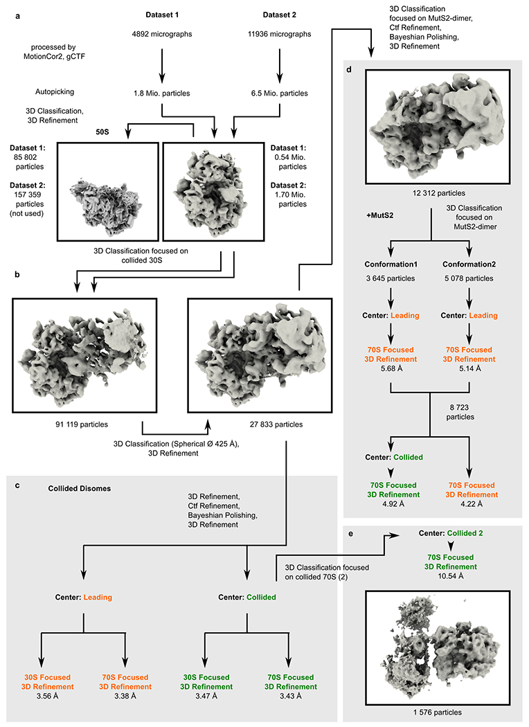
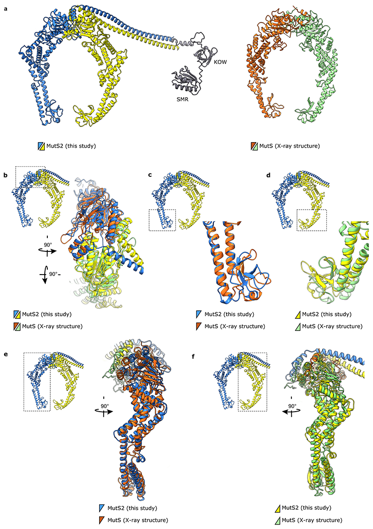
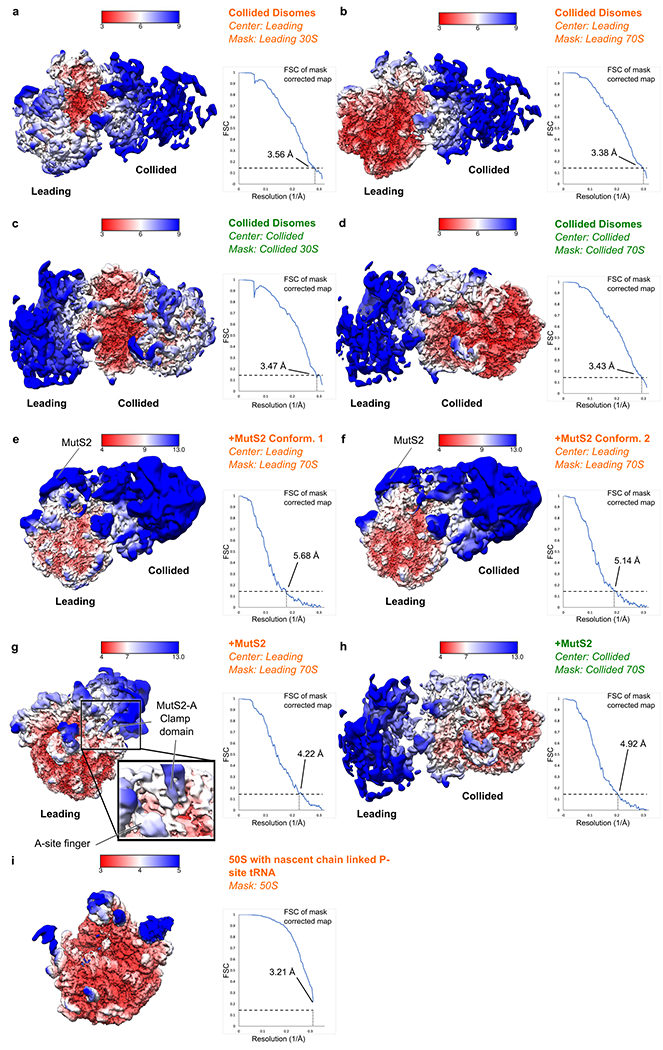
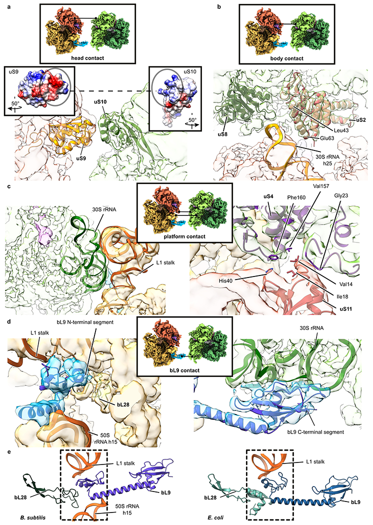
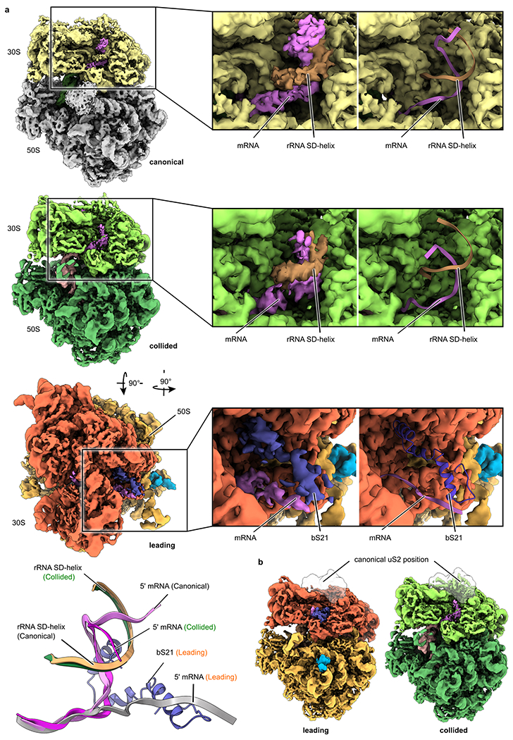
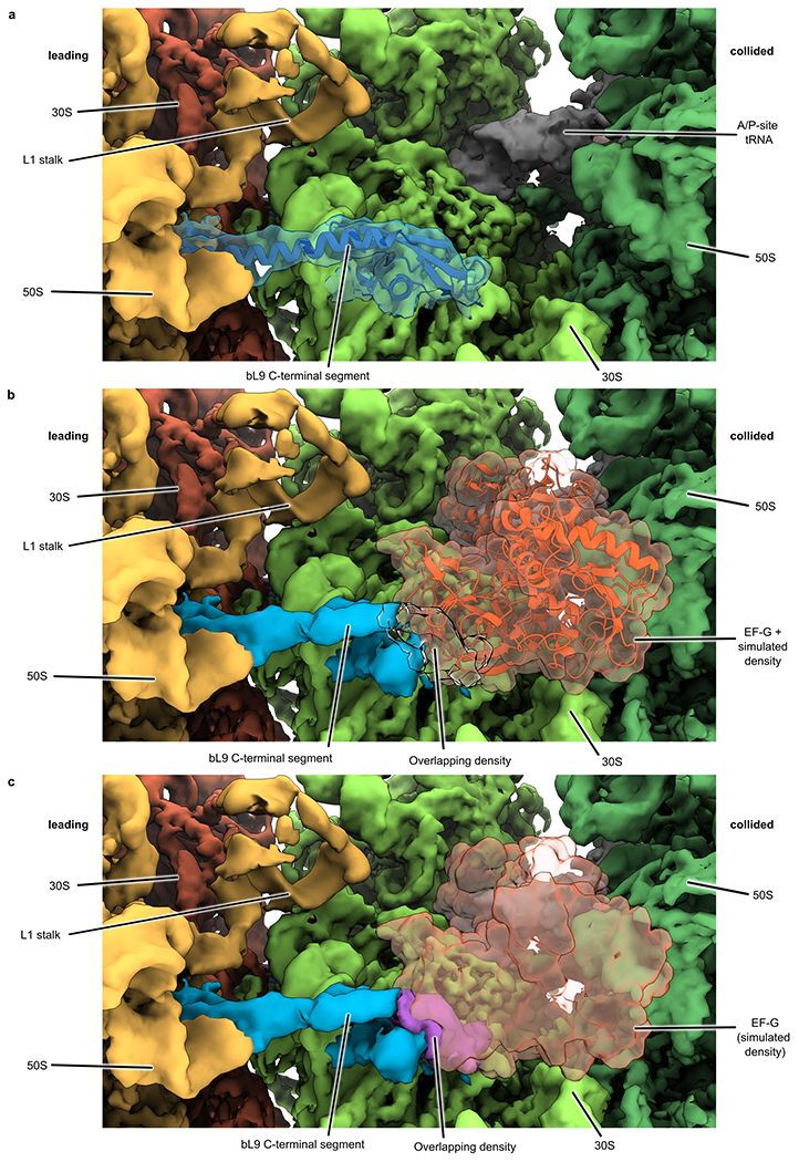
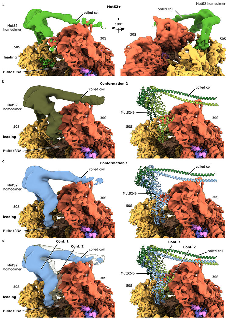
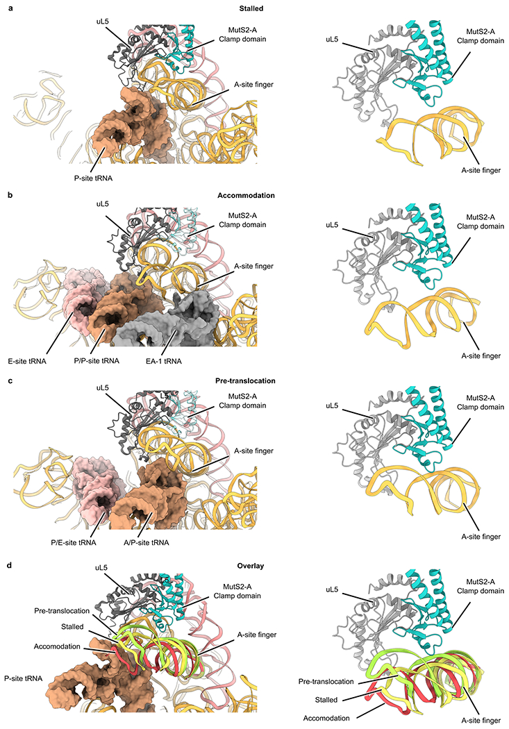
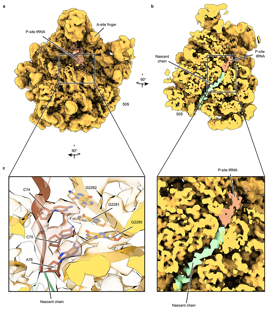
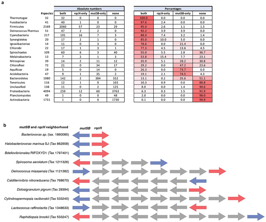
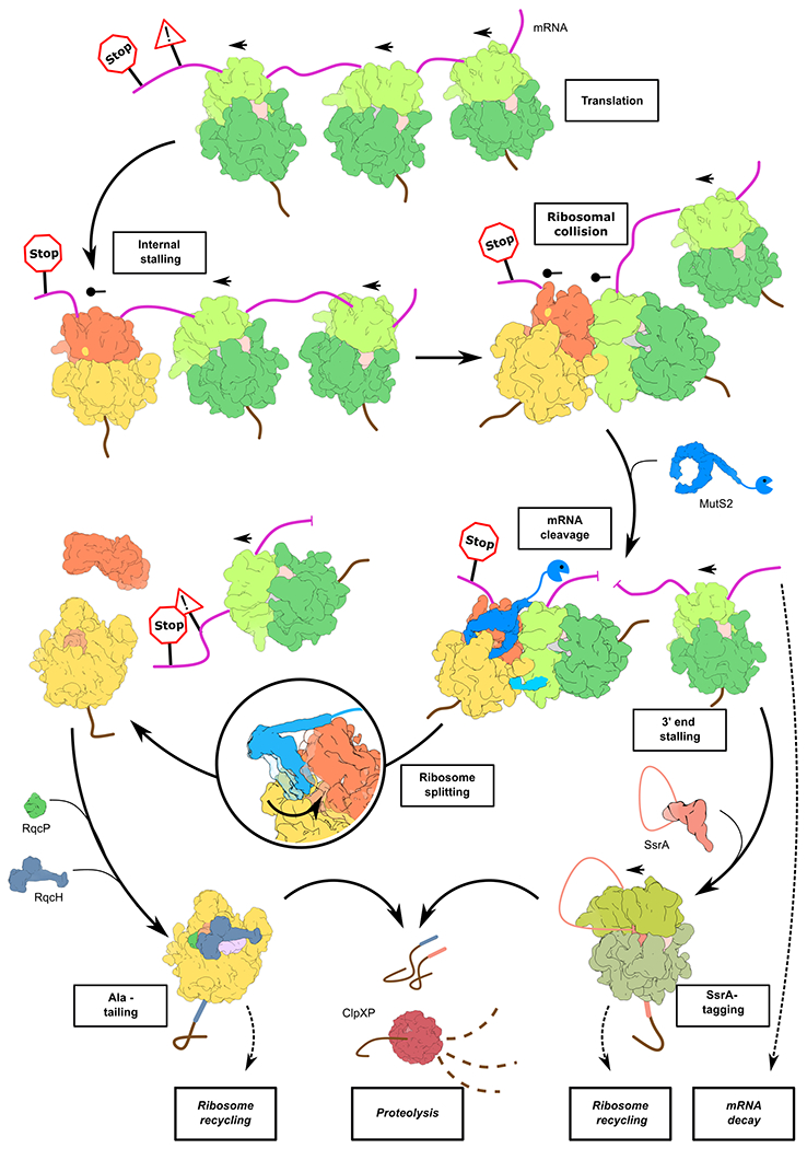
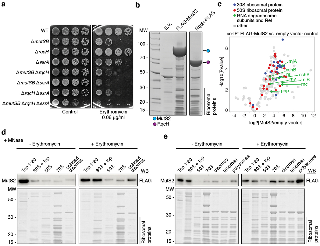
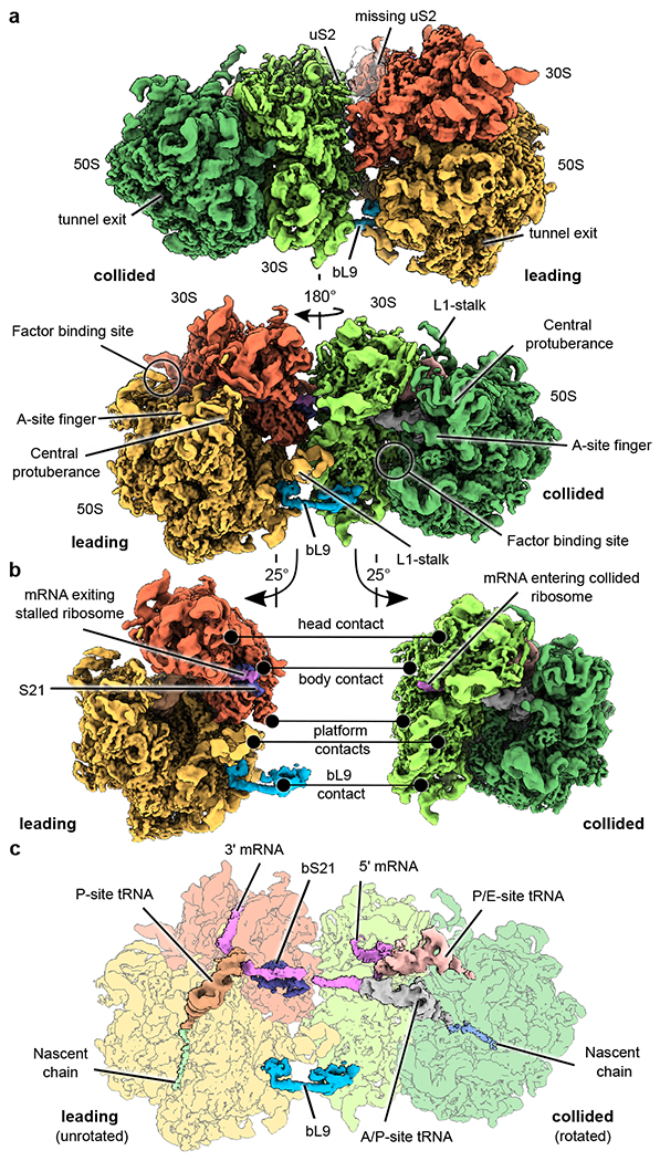
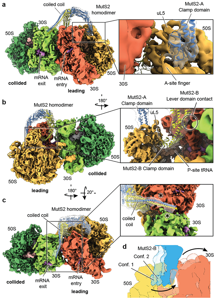
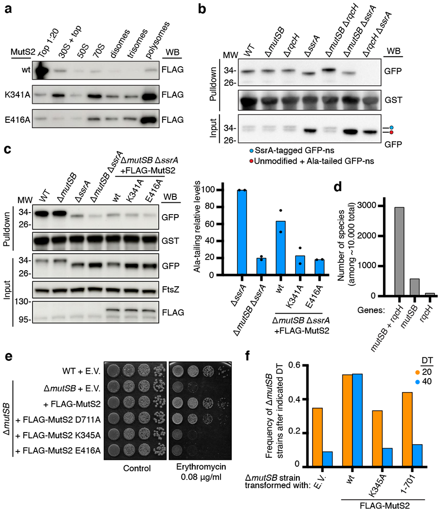
Ribosome stalling during translation is detrimental to cellular fitness, but how this is sensed and elicits recycling of ribosomal subunits and quality control of associated mRNA and incomplete nascent chains is poorly understood1,2. Here we uncover Bacillus subtilis MutS2, a member of the conserved MutS family of ATPases that function in DNA mismatch repair3, as an unexpected ribosome-binding protein with an essential function in translational quality control. Cryo-electron microscopy analysis of affinity-purified native complexes shows that MutS2 functions in sensing collisions between stalled and translating ribosomes and suggests how ribosome collisions can serve as platforms to deploy downstream processes: MutS2 has an RNA endonuclease small MutS-related (SMR) domain, as well as an ATPase/clamp domain that is properly positioned to promote ribosomal subunit dissociation, which is a requirement both for ribosome recycling and for initiation of ribosome-associated protein quality control (RQC). Accordingly, MutS2 promotes nascent chain modification with alanine-tail degrons-an early step in RQC-in an ATPase domain-dependent manner. The relevance of these observations is underscored by evidence of strong co-occurrence of MutS2 and RQC genes across bacterial phyla. Overall, the findings demonstrate a deeply conserved role for ribosome collisions in mounting a complex response to the interruption of translation within open reading frames.


Conflict of interest statement

Competing interests

The authors declare no competing interests.

Figures

Extended Data Fig. 1.
Extended Data Fig. 1.. Cryo-EM data processing scheme
a, Two datasets were acquired and processed separately using Relion. To reject false-positive particles, auto-picked particles were subjected to a 3D classification. A subset of 50S depicting particles from dataset 1 was subjected to 3D auto-refinement. b, To enrich for collided disomes, 70S depicting particles were subsequently subjected to multiple rounds of 3D classification using a spherical mask with a diameter of 435 Å (dataset 1) or focused on the 30S subunit of the collided disome (dataset 2). After the first round of classification particles were merged. c, Particles of collided disomes were re-extracted centered on the leading and the colliding ribosome. 3D auto-refinements were focused on the 30S subunit or the full 70S of the respective ribosome. d, MutS2 conformations were sorted by two rounds of 3D classification using a mask encompassing the MutS2-homodimer. To increase local resolution for the MustS2-A clamp domain, the two separate conformations were merged and subjected to 3D auto-refinements based on either the leading or the colliding ribosome. e, To enrich for particles that depict collided trisomes, particles from collided disomes centered on the collided ribosome were subjected to a 3D classification focused on the third ribosome. Retained particles were re-extracted centered on the third ribosome (Collided 2) and subjected to 3D auto-refinement.
Extended Data Fig. 2.
Extended Data Fig. 2.. Structural similarity between the predicted MutS2 structure and the X-ray structure of a bacterial MutS protein
a, Overall structures of B. subtilis MutS2 (this study) and Neisseria gonorrhoeae MutS (PDB 5X9W). Segments of MutS2 that were not resolved by cryo-EM, including the KOW and SMR domains, are shown in grey for one monomer as predicted by AlphaFold. b-c, Superposition of the two models with respect to the ATPase domain (b), the individual MutS clamp and lever domains (c,d) and all conserved segments of the individual monomer structures (e,f). Dashed boxes indicate the zoomed area for panels b-f.
Extended Data Fig. 3.
Extended Data Fig. 3.. Global and local resolution estimation
For each 3D auto-refinement run, densities have been colored according to local resolution. Mask-corrected Fourier shell correlation (FSC) has been calculated between two independently refined half-sets of the data (‘gold standard’; FSC threshold at 0.143). Shown particle sets are “Collided Disomes” (a, b, c, d,), “+MutS2” (g, h), MutS2 conformation 1 and 2 (respectively e, f) and 50S subunits obstructed with a nascent chain-linked P-site tRNA (i). Particles are either centered on the leading (a, b, e, f, g) or on the colliding ribosome (c, d, h) and refined using either a 30S (a, c) or a 70S mask of the respective ribosome (b, d, e, f, g, h).
Extended Data Fig. 4.
Extended Data Fig. 4.. Molecular details of contacts stabilizing the disome interface
a, The 30S subunit heads interact via complementary charged patches on uS9 of the leading ribosome and uS10 of the collided ribosome (‘head contact’). Surface representations for uS9 and uS10 are shown and colored according to electrostatic potential (blue: positive, red: negative, white: neutral). The interacting patches are indicated. b, On the opposing side of the inter-ribosomal mRNA trajectory, helix 25 of the leading ribosome 30S subunit rRNA is accommodated in a groove on the 30S subunit of the collided ribosome formed by uS2 and uS8 (‘body contact’). In particular, helix 25 of the leading ribosome directly interacts with a surface exposed α-helix (Leu43 - Glu63) and the partially unordered C-terminus of uS2 of the collided ribosome. c, In close proximity, two additional contacts between the two collided ribosomes (‘platform contacts’) complete the network of interactions clustered around the mRNA entry and exit sites. First, the L1-stalk adopting an extreme out-conformation on the leading ribosome directly contacts the 30S subunit rRNA of the collided ribosome. Second, uS11 of the leading ribosome contacts uS4 of the collided ribosome, mainly via hydrophobic interactions (Val14 and Ile18 of S11; Val157 and Gly23 of S4) and aromatic stacking (His40 of S11; Phe160 of S4). d, A more peripheral interaction is mediated by ribosomal protein bL9 of the leading ribosome (‘bL9 contact’). e, The binding site of bL9 on the B. subtilis 50S subunit has been significantly remodeled compared to the E. coli ribosome (PDB 6BY1). While the interaction area between bL28 and the N-terminal half of bL9 is reduced in B. subtilis, this is compensated by an expansion of the 50S subunit rRNA (helix 15), which together with the L1-stalk forms an extended binding groove for bL9.
Extended Data Fig. 5.
Extended Data Fig. 5.. Structural and compositional remodeling of the mRNA exit site on the leading ribosome
a, Upper panel: Trajectory of unstrained mRNA exiting the mRNA channel in a defined direction to interact with the anti-Shine-Dalgarno (SD) rRNA sequence of the 16S rRNA 3’end, thereby forming an RNA duplex reminiscent of the SD helix during translation initiation (PDB 3J9W, EMDB 6306). Middle panel: The unstrained mRNA exiting the collided ribosome interacts with the anti-SD rRNA to form an RNA duplex. Bottom panel: In the collided disome, the mRNA under strain follows a vastly different trajectory, which is accompanied by remarkable structural remodeling of the mRNA exit site of the leading ribosome. In particular, the rRNA anti-SD sequence of the leading ribosome can no longer interact with the mRNA and becomes disordered, which renders the binding site for the bS21 C-terminus on the leading ribosome accessible and at the same time reduces the interaction surface for uS2 on the 30S subunit. Superposition: Superposition of mRNAs exiting the ribosomes and the respective SD-helices in the translating and collided ribosomes, as well as bS21 in the leading ribosome. b, Comparison of uS2 density in the leading and collided ribosomes at the same density threshold level. In all panels, local resolution filtered densities based on 3D auto-refinements focused on either the 30S or the 70S of the respective ribosome are shown (See Extended Data Fig. 1).
Extended Data Fig. 6.
Extended Data Fig. 6.. The C-terminal half of bL9 sterically excludes binding of EF-G on the collided ribosome
a, Binding site of the bL9 on the 30S subunit of the collided ribosome. b, Atomic model and simulated density of EF-G (PDB 7N2V) mapped onto the 30S subunit of the collided ribosome by fitting the 30S-EF-G complex as a rigid body. Overlapping segments of bL9 and EF-G are shown in transparent grey. c, As in ‘b’, but not showing the EF-G atomic model and with overlapping segments of bL9 and EF-G colored in purple.
Extended Data Fig. 7.
Extended Data Fig. 7.. Conformational plasticity of the MutS2-B clamp region
a, Two views on the local resolution-filtered density of the MutS2 dimer after 3D auto-refinement of all MutS2-containing particles. Highly fragmented density for the MutS2-B clamp and lever domains (left panel) indicated conformational heterogeneity. b, c, Computational particle sorting focused on MutS2-B produced two structurally distinct subpopulations slightly differing in the positioning of the MutS-domains III and IV, in which the clamp region either binds to ribosomal protein L5 (b) or the nascent chain-associated P-site tRNA of the leading ribosome (c). d, Overlay of the two conformations from ‘b’ and ‘c’. In all panels, local resolution-filtered cryo-EM densities are shown as obtained after 3D autorefinement centered on the leading ribosome using either all MutS2-containing particles (ribosome densities in all panels, MutS2 density in panel A) or using only particles representing one of the two different MutS2 conformations (MutS2 density in panels b, c).
Extended Data Fig. 8.
Extended Data Fig. 8.. Conformational plasticity of the A-site finger during the translational elongation cycle
a-c, Atomic models for rRNA, tRNAs and ribosomal protein uL5. a, The leading ribosome of the B. subtilis collided disome. b, The E. coli ribosome in accommodation state IV-A (PDB 6WDB). c, The pre-translocation state VI-B (PDB 6WDG). d, Superposition of the structures shown in (a-c) according to ribosomal protein uL5, demonstrating remodeling of the MutS2-A binding site during the translation elongation cycle.
Extended Data Fig. 9.
Extended Data Fig. 9.. Structure of the 50S ribosomal subunit obstructed with a nascent chain-linked P-site tRNA
a, Local resolution-filtered cryo-EM density of the 50S ribosomal subunit obstructed with the nascent chain-linked P-site tRNA. Due to conformational flexibility, cryo-EM density for peripheral segments of the P-site tRNA is fragmented. b, Density was sliced open to allow for an unobstructed view on the P-site tRNA and the associated incomplete nascent chain. c, Model of a nascent chain-linked P-site tRNA (PDB 7AQC) superposed to the cryo-EM density. Zoom on the CCA-tail of the P-site-tRNA with linked nascent chain.
Extended Data Fig. 10.
Extended Data Fig. 10.. Evidence from genomic analyses link mutSB and rqcH
a, mutSB and rqcH strongly co-occur. Distribution of rqcH and mutSB quantitated separately for the indicated bacterial phyla. Both absolute numbers and frequencies are presented. For the latter, the higher the frequency the darker the background red color. b, mutSB localizes in the vicinity of rqcH in diverse bacteria. Genes are represented by arrows reflecting the direction of transcription, with mutSB and rqcH indicated in blue and red, respectively. In the instances where mutSB and rqcH are separated by genes represented as grey arrows, those genes are highly diverse, have no obvious relationship to translational quality control, and are generally unrelated between different species.
Extended Data Fig. 11.
Extended Data Fig. 11.. Model for MutS2 function in sensing ribosome collisions and eliciting downstream responses
The model depicts, from top to bottom: ribosomes translating an mRNA with a stalling site within the open reading frame (“Translation”); the leading ribosome becoming stalled (“Internal stalling”); a trailing ribosome colliding with the stalled ribosome (“Ribosomal collision”); MutS2 sensing the collision and promoting both separation of the ribosomal subunits (“Ribosome splitting”) and endonucleolytic cleavage (“mRNA cleavage”). Left side: Ribosomal splitting generates a 50S subunit still obstructed with a nascent chain-tRNA conjugate, which is sensed by RqcH and RqcP, resulting in elongation of the nascent chain with a C-terminal Ala tail (“Ala tailing”). Nascent-chain release is accompanied by 50S recycling (“Ribosome recycling”, dotted line). Right side: mRNA cleavage can result in mRNA decay (dotted line) or in trailing ribosomes becoming stalled at the mRNA 3’end, which are sensed by SsrA/tmRNA. The SsrA reaction leads to ribosome recycling (“Ribosome recycling”, dotted line) and to nascent-chain modification with a C-terminal SsrA tag (“SsrA tagging”). Both Ala-tails and the SsrA tag act as degrons, recognized by ClpXP and other proteases (“Proteolysis”). See the main text for additional details. Objects: the mRNA is shown in red, with a stalling site within the open reading frame represented by ‘!’ within a triangle and the mRNA stop codon shown as a ‘Stop’ traffic sign; the direction of translation is indicated by arrows; the stalled ribosome is shown in orange (light, 50S subunit; dark, 30S subunit); trailing and collided ribosomes are shown in green (dark, 50S subunit; light, 30S subunit). Quality control factors are indicated by their names.
Fig. 1.
Fig. 1.. MutS2 binds to ribosomes and protects cells from translational stress
a, mutSB protects cells against erythromycin and interacts genetically with the translational quality control pathways, RQC and SsrA. b, MutS2 binds to 70S ribosomes. Coomassie staining of FLAG-MutS2 and RqcH-FLAG co-IP’ed proteins. E.V., empty vector (n>3). c, Proteins co-IP’ed with FLAG-MutS2 identified by label-free LC-MS/MS. Individual proteins are represented as dots, with the Rel stringent response factor and RNA degradosome factors in green, 30S proteins in blue, and 50S proteins in red. d, Erythromycin-stimulated association of MutS2 with micrococcal nuclease (MNase)-resistant disome fractions. 3xFLAG-MutS2 expressing cells were exposed (right) or not (left) to erythromycin and lysates were treated with MNase before fractionation in sucrose gradient and analysis by anti-FLAG blot (top) or Coomassie staining (bottom). For the top fraction, only 1/20th of the volume was loaded. ‘30S + top’ indicates contamination of 30S with top fractions (n=2). e, Erythromycin-stimulated association of MutS2 with heavy polysome fractions (n=2). As in ‘d’, but without MNase treatment. For gel source data, see Supplementary Fig. 1.
Fig. 2.
Fig. 2.. Cryo-EM structure of the bacterial collided disome
a, Structure of the collided bacterial disome at ~ 3.5 Å resolution. The canonical position of uS2 on the stalled ribosome is indicated. b, The disome interface is formed by multiple contacts between leading and collided ribosomes. See Extended Data Fig. 4 for details. c, In the collided disome, ribosomes adopt distinct rotational states associated with specific tRNA occupancy. In all panels, local resolution filtered cryo-EM densities are shown. Density for 30S subunits and mRNA was obtained by focused refinement on the 30S subunits. Density for the 50S subunits and tRNAs was obtained by focused refinement on the full 70S particles (Extended Data Figs. 1 and 3).
Fig. 3.
Fig. 3.. Structural basis for ribosome collision sensing by MutS2
a, Frontal view on the structure of the MutS2-disome complex. The MutS2 homodimer density is shown with transparency and the atomic model superposed. Right: zoom onto the MutS2-A clamp domain binding to the A-site finger and the central protuberance of the leading ribosome. b, View rotated by 180°. The MutS2 homodimer encompasses the central protuberance from the A-site and E-site facing sides. Right: zoom on MutS2-B clamp domain insertion into the gap between ribosomal subunits and the L1-stalk. The additional contact between the MutS2-B lever domain and the collided ribosome 30S subunit is indicated. c, Top view onto the MutS2-disome complex showing how the MutS2 coiled-coil aligns with a composite binding site formed by the two 30S subunits. The interaction sites between the coiled-coil and helices 39 of the 30S subunit rRNAs are indicated (asterisks). d, Model for ribosomal subunit splitting mediated by conformational changes of MutS2. The two observed MutS2 conformations (transparent blue) and one additional extrapolated conformation (solid blue) are shown. Local resolution-filtered ribosome densities are shown as obtained after ribosome-wise particle refinement of all MutS2-containing particles. MutS2 density is shown as obtained after particle refinement centered on the stalled ribosome, for either all MutS2-containing particles (panel ‘a’, zoom) or only particles representing MutS2 conformation 2 (all other panels) (see Extended Data Figs. 1 and 3).
Fig. 4.
Fig. 4.. MutS2 promotes ribosome splitting upstream of RQC
a, ATPase domain mutation-stimulated association of MutS2 with heavy polysome fractions. As in Fig. 1d, but with no erythromycin (n= 2). b, A novel assay for Ala-tailing reveals a role for MutS2 upstream of RqcH. Lysates of strains episomally expressing a ribosome stalling reporter (GFP-ns) were utilized for GST-Pirh2 pulldown and analyzed by anti-GFP blot (n= 2). c, MutS2 wt, but not ATPase domain mutants, rescues the Ala-tailing defect of ΔssrA ΔmutSB cells. Left, as in ‘b’, except that a genome-integrated GFP-ns reporter was used. Right, effect of mutSB deletion on the ssrA background. The ratio of modified to input GFP-ns is shown. Data are presented as mean (n= 2). d, mutSB and rqcH strongly co-occur among bacterial genomes. Bar graphs indicate the number of species having both mutSB and rqcH or only one of the genes, among completely sequenced genomes. e, MutS2 protects cells against erythromycin in an ATPase domain-dependent manner. Strains expressing FLAG-MutS2 (wt and mutants) or empty vector (E.V.), analyzed as in Fig. 1a. f, MutS2 ATPase and SMR domains are both required for cellular fitness. Mutant strains were mixed with the WT strain at 1:1 OD600 ratio, and subjected to competitive growth in two separate experiments for either 20 (orange bars) or ~40 (blue bars) doubling times (DT), before determining the frequency of each strain in the final population by colony PCR. For gel source data, see Supplementary Fig. 1.

Comment in

References

    1. Joazeiro CAP Ribosomal Stalling During Translation: Providing Substrates for Ribosome-Associated Protein Quality Control. Annu. Rev. Cell Dev. Biol 33, 343–368 (2017). - PubMed
    1. Meydan S & Guydosh NR A cellular handbook for collided ribosomes: surveillance pathways and collision types. Curr. Genet 67, 19–26 (2021). - PMC - PubMed
    1. Modrich P Mechanisms in E. coli and Human Mismatch Repair (Nobel Lecture). Angew. Chem. Int. Ed. Engl 55, 8490–501 (2016). - PMC - PubMed
    1. Moore SD & Sauer RT The tmRNA system for translational surveillance and ribosome rescue. Annu. Rev. Biochem 76, 101–24 (2007). - PubMed
    1. Joazeiro CAP Mechanisms and functions of ribosome-associated protein quality control. Nat. Rev. Mol. Cell Biol 20, 368–383 (2019). - PMC - PubMed

Publication types