. 2022 Feb 21:13:811682.
doi: 10.3389/fphar.2022.811682. eCollection 2022.

Honokiol Ameliorates Post-Myocardial Infarction Heart Failure Through Ucp3-Mediated Reactive Oxygen Species Inhibition

Affiliations

Honokiol Ameliorates Post-Myocardial Infarction Heart Failure Through Ucp3-Mediated Reactive Oxygen Species Inhibition

Jianyu Liu et al. Front Pharmacol. .

Erratum in

Abstract

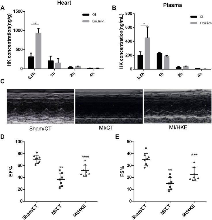
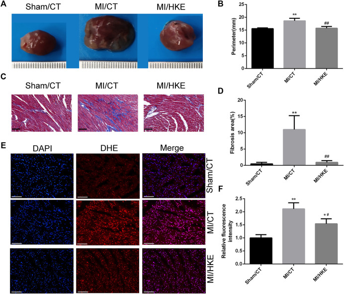
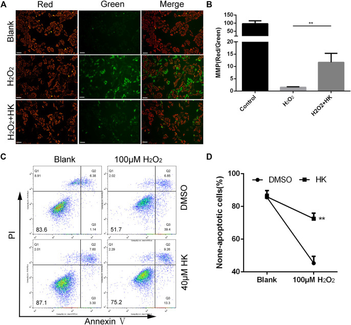
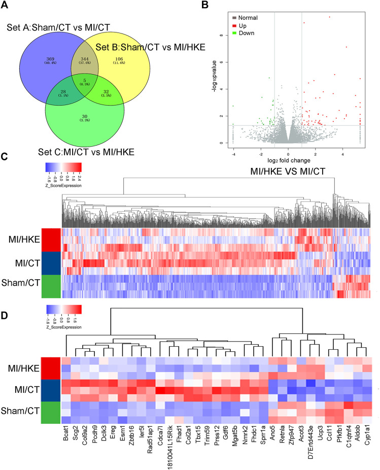
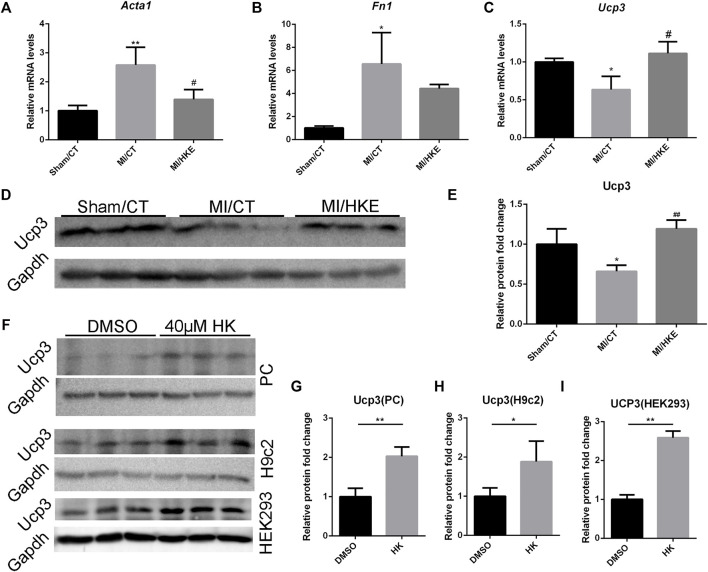
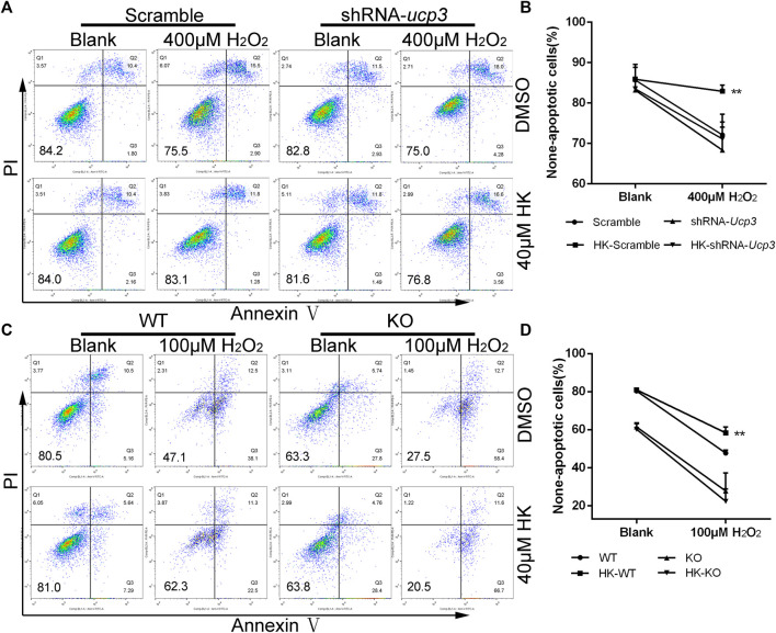
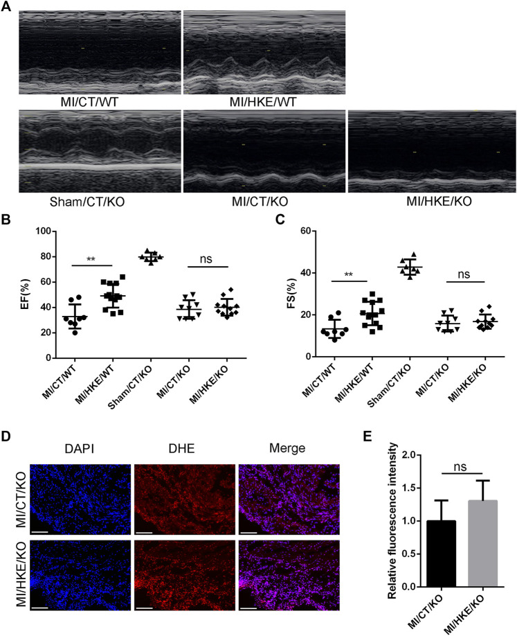
Post-myocardial infarction heart failure (post-MI HF) is one of the leading global causes of death, and current prevention and treatment methods still cannot avoid the increasing incidence. Honokiol (HK) has previously been reported to improve myocardial ischemia/reperfusion injury and reverse myocardial hypertrophy by activating Sirt1 and Sirt3. We suspect that HK may also have a therapeutic effect on post-MI HF. In this study, we aimed to investigate the efficacy and mechanism of HK in the treatment of post-MI HF. We found that HK inhibited myocardial reactive oxygen species (ROS) production, reduced myocardial fibrosis, and improved cardiac function in mice after MI. HK also reduced the abnormality of mitochondrial membrane potential (MMP) and apoptosis of cardiomyocytes caused by peroxide in neonatal cardiomyocytes. RNAseq results revealed that HK restored the transcriptome changes to a certain extent and significantly enhanced the expression of mitochondrial inner membrane uncoupling protein isoform 3 (Ucp3), a protein that inhibits the production of mitochondrial ROS, protects cardiomyocytes, and relieves heart failure after myocardial infarction (MI). In cardiomyocytes with impaired Ucp3 expression, HK cannot protect against the damage caused by peroxide. More importantly, in Ucp3 knockout mice, HK did not change the increase in the ROS level and cardiac function damage after MI. Taken together, our results suggest that HK can increase the expression of the cardioprotective protein Ucp3 and maintain MMP, thereby inhibiting the production of ROS after MI and ameliorating heart failure.

Keywords: UCP3; heart failure; honokiol; myocardial infarction; reactive oxygen species.


Conflict of interest statement

The authors declare that the research was conducted in the absence of any commercial or financial relationships that could be construed as a potential conflict of interest.

Figures

FIGURE 1
FIGURE 1
HKE improves cardiac function in post-MI HF. HKE or soybean oil dissolved HK were injected IP (20 mg/kg), and the concentration of HK in heart (A) and plasma (B) were monitored for 0.5, 1, 2, and 4 h after injection (n = 4 per group). Four weeks after MI, EF and FS were determined by echocardiography (C–E) (n = 7 or 8 per group). Values are shown as means ± SD, *p < 0.05, **p < 0.01 vs. Sham/CT group, #p < 0.05, ##p < 0.01 vs. MI/CT group.
FIGURE 2
FIGURE 2
HKE inhibits myocardial remodeling, reduces the ROS, and alleviates myocardial fibrosis in vitro. Typical heart appearance in groups Sham/CT, MI/CT, and MI/HKE (A). The perimeter of the largest part cross section of the heart was measured to evaluate myocardial remodeling (B). Masson’s Trichrome staining was conducted to evaluate myocardial fibrosis, the fibrotic area was indicated as a blue region, the red frame encircles the fibrosis away from the infarct area in the MI group and similar locations in other groups, scale bar: 50 μm (C). The fibrosis of red frame area was measured using ImageJ software (D) (n = 3 per group). Heart slides were stained with DHE to analyze the production of ROS in situ, scale bar: 100 μm (E). The fluorescence intensity was measured using ImageJ software (F) (n = 3 per group). Values are shown as means ± SD, *p < 0.05, **p < 0.01 vs. Sham/CT group or control, # p < 0.05, ## p < 0.01 vs. MI/CT.
FIGURE 3
FIGURE 3
HK normalizes MMP and protects against the apoptosis caused by peroxide in vitro. For MMP detection, primary cardiomyocytes from neonatal mice hearts were cultured in a six-well dish for 12 h in the presence or absence of 40 μM HK, the medium was then replaced with normal medium containing 50 μM hydrogen peroxide for 12 h to induce oxidative damage. Cardiomyocytes were stained with JC-1 to monitoring the MMP in vitro, scale bar: 100 μm (A). The fluorescence intensity was measured using ImageJ software (B) (n = 3 per group). For apoptosis detection, cardiomyocytes were cultured in six well dish for 12 h in the presence or absence of 40 μM HK, the medium was then replaced with normal medium containing 100 μM hydrogen peroxide for 12 h to induce oxidative damage. Annexin V-FITC/PI Apoptosis Detection Kit were used for apoptosis assay by FACS analysis, data show quantification of non-apoptotic cells (C,D) (n = 3 per group). A total of 8,000 cells were collected for each sample. Values are shown as means ± SD. *p < 0.05, **p < 0.01 vs. H2O2 treatment group.
FIGURE 4
FIGURE 4
RNAseq and differential gene expression analysis. Total RNA was isolated from heart tissue for RNAseq (n = 3 per group). The different expressed gene numbers between each group were shown by Venn diagram (A). The volcano plot shows the different expressed genes between MI/CT and MI/HKE (B). Cluster analysis of all differential genes (C). Cluster analysis of 33 differential genes in the intersection of Set A and Set C (D).
FIGURE 5
FIGURE 5
HKE restores fibrosis-related genes and Ucp3. The mRNA expression of Acta1, Fn1, and Ucp3 in myocardial tissue was detected using real-time PCR (n = 3 per group). 2-△△Ct method was used to analyze relative gene expression levels (A–C). The Ucp3 abundance in myocardial tissue were detected by Western blot. Gapdh was included as a loading control (n = 3 per group) (D,E). After treatment with 40 μM HK or equal volume of DMSO for 24 h, The Ucp3 abundance in primary cardiomyocytes (PC), H9c2 and HEK293 were detected by Western blot. Gapdh was included as a loading control (n = 3 per group) (F–I). Values are shown as means ± SD, *p < 0.05, **p < 0.01 vs. Sham/CT or DMSO, # p < 0.05, ## p < 0.01 vs. MI/CT group.
FIGURE 6
FIGURE 6
Ucp3 mediates the protection of HK against oxidative damage in cardiomyocyte H9c2 and neonatal mice cardiomyocyte. For apoptosis detection, Wild-type H9c2 and H9c2 cells with disturbed Ucp3 expression or primary cardiomyocytes from WT and Ucp3-KO mice were cultured in six well dish for 12 h in the presence or absence of 40μM HK, respectively. The H9c2 medium was then replaced with normal medium containing 400 mM hydrogen peroxide (A,B) and primary cardiomyocytes replaced with normal medium containing 100 mM hydrogen peroxide (C,D) for 12 h to induce oxidative damage. Annexin V-FITC/PI Apoptosis Detection Kit was used for apoptosis assay by FACS analysis, data show quantification of none-apoptotic cells (n = 3 per group). Values are shown as means ± SD. *p < 0.05, **p < 0.01 vs. 100/400 μM H2O2 treatment WT/Scramble group.
FIGURE 7
FIGURE 7
Ucp3 mediates the protection of HKE against post-MI HF. Construction of MI model with Ucp3 knockout and WT mice,4 weeks after MI, EF and FS were determined by echocardiography (A–C). Heart slides were stained with DHE to analyze the production of ROS in situ, scale bar: 100 μm (D). The fluorescence intensity was measured using ImageJ software (E) (n = 3 per group). Values are shown as means ± SD, *p < 0.05, **p < 0.01, ns: no significance.

Similar articles

Cited by

References

    1. Abraham W. T., Fisher W. G., Smith A. L., Delurgio D. B., Leon A. R., Loh E., et al. (2002). Cardiac Resynchronization in Chronic Heart Failure. N. Engl. J. Med. 346 (24), 1845–1853. 10.1056/NEJMoa013168 - DOI - PubMed
    1. Akhmedov A. T., Rybin V., Marín-García J. (2015). Mitochondrial Oxidative Metabolism and Uncoupling Proteins in the Failing Heart. Heart Fail. Rev. 20 (2), 227–249. 10.1007/s10741-014-9457-4 - DOI - PubMed
    1. Atanasov A. G., Wang J. N., Gu S. P., Bu J., Kramer M. P., Baumgartner L., et al. (2013). Honokiol: a Non-adipogenic PPARγ Agonist from Nature. Biochim. Biophys. Acta 1830 (10), 4813–4819. 10.1016/j.bbagen.2013.06.021 - DOI - PMC - PubMed
    1. Banik K., Ranaware A. M., Deshpande V., Nalawade S. P., Padmavathi G., Bordoloi D., et al. (2019). Honokiol for Cancer Therapeutics: A Traditional Medicine that Can Modulate Multiple Oncogenic Targets. Pharmacol. Res. 144, 192–209. 10.1016/j.phrs.2019.04.004 - DOI - PubMed
    1. Bardy G. H., Lee K. L., Mark D. B., Poole J. E., Packer D. L., Boineau R., et al. (2005). Amiodarone or an Implantable Cardioverter-Defibrillator for Congestive Heart Failure. N. Engl. J. Med. 352 (3), 225–237. 10.1056/NEJMoa043399 - DOI - PubMed