. 2022 Feb 21:13:828269.
doi: 10.3389/fphar.2022.828269. eCollection 2022.

The Bioflavonoids Rutin and Rutin Succinate Neutralize the Toxins of B. jararaca Venom and Inhibit its Lethality

Affiliations

The Bioflavonoids Rutin and Rutin Succinate Neutralize the Toxins of B. jararaca Venom and Inhibit its Lethality

Ana Teresa Azevedo Sachetto et al. Front Pharmacol. .

Abstract

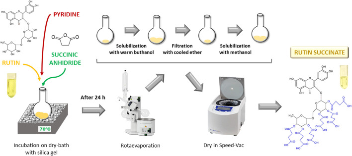
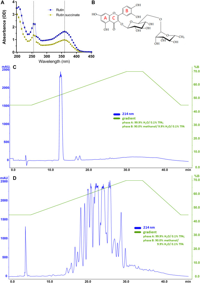
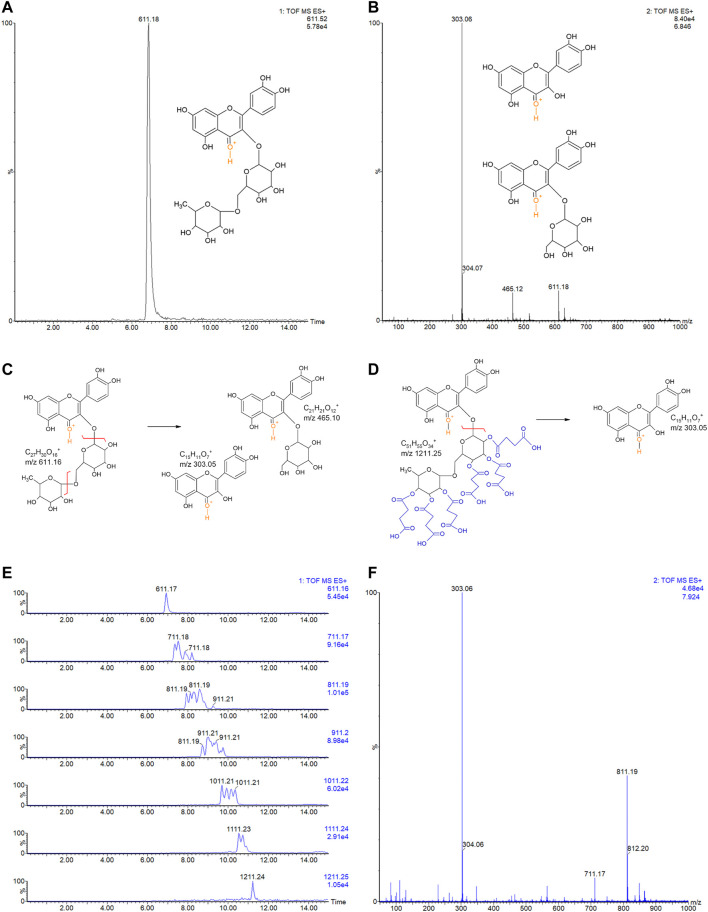
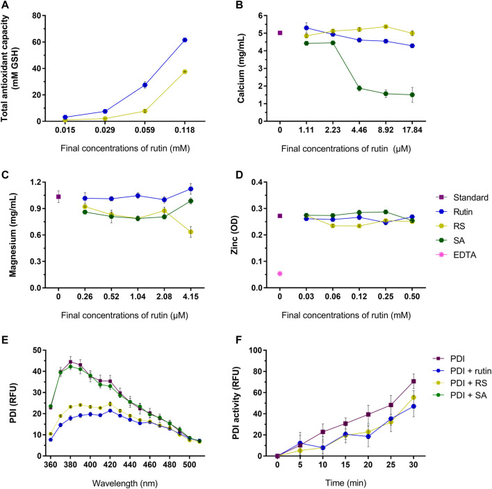
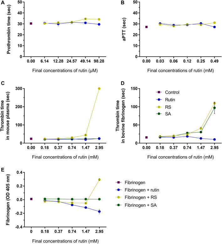
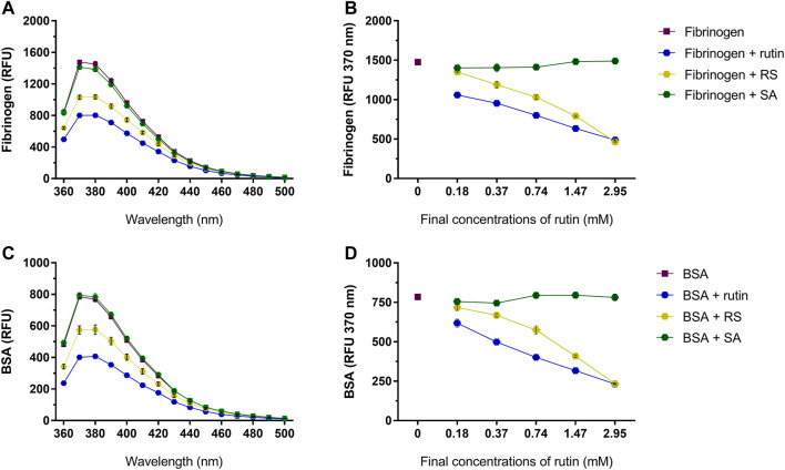
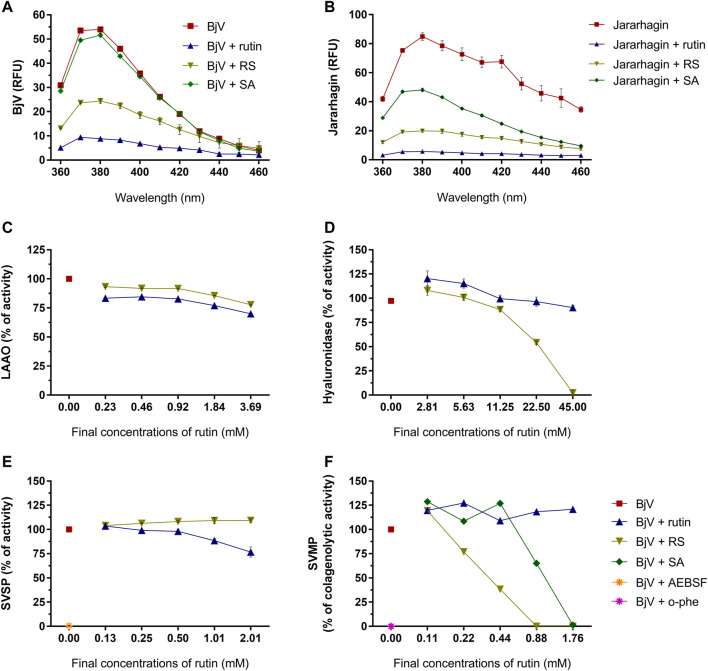
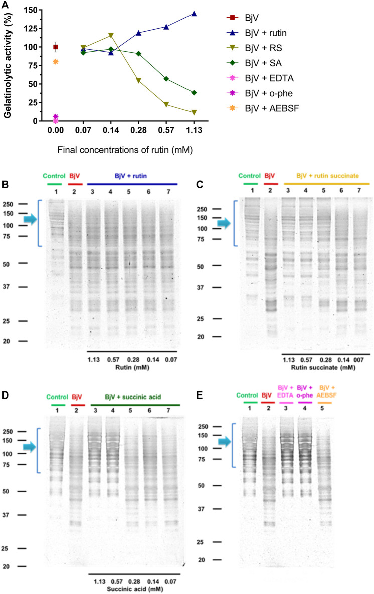
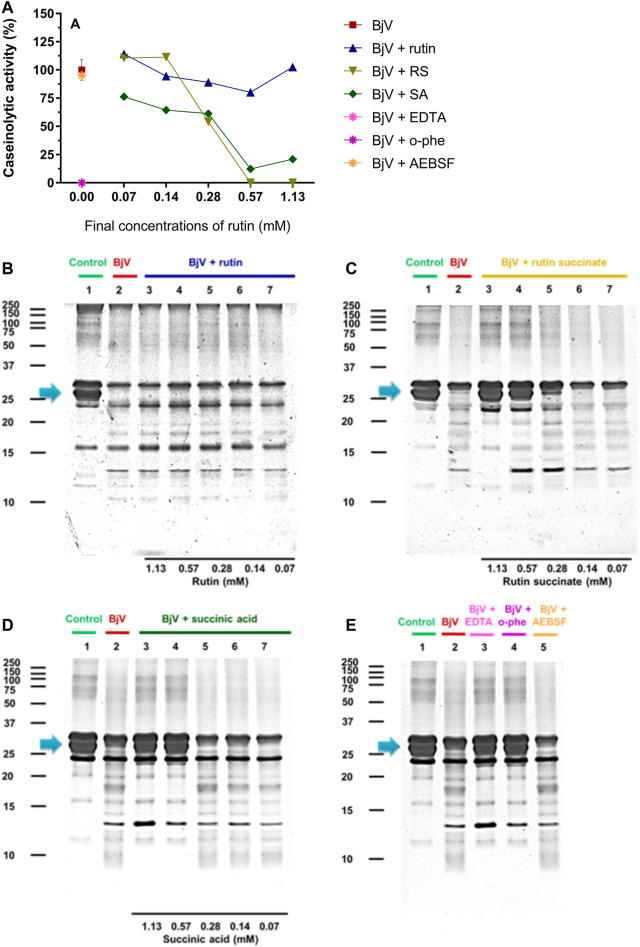
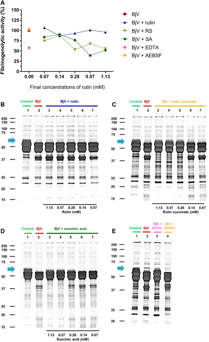
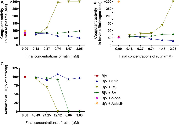
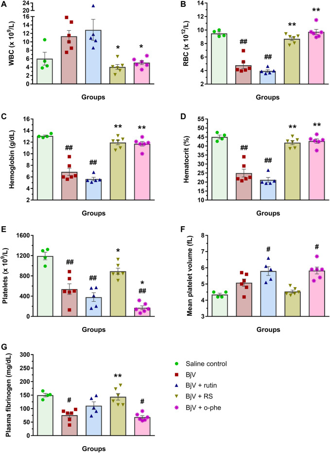
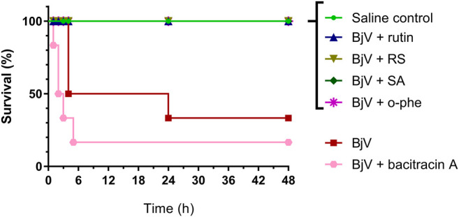
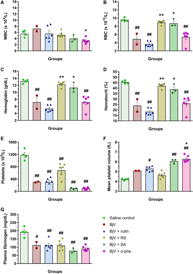
The venom of the Brazilian pit viper Bothrops jararaca (BjV) is a complex mixture of molecules, and snake venom metalloproteinases (SVMP) and serine proteinases (SVSP) are the most abundant protein families found therein. Toxins present in BjV trigger most of the deleterious disturbances in hemostasis observed in snakebites, i.e., thrombocytopenia, hypofibrinogenemia and bleedings. The treatment of patients bitten by snakes still poses challenges and the bioflavonoid rutin has already been shown to improve hemostasis in an experimental model of snakebite envenomation. However, rutin is poorly soluble in water; in this study, it was succinylated to generate its water-soluble form, rutin succinate (RS), which was analyzed comparatively regarding the chemical structure and characteristic features of rutin. Biological activities of rutin and RS were compared on hemostatic parameters, and against toxic activities of crude BjV in vitro. In vivo, C57BL/6 mice were injected i.p. with either BjV alone or pre-incubated with rutin, RS or 1,10-phenanthroline (o-phe, an SVMP inhibitor), and the survival rates and hemostatic parameters were analyzed 48 h after envenomation. RS showed the characteristic activities described for rutin - i.e., antioxidant and inhibitor of protein disulfide isomerase - but also prolonged the clotting time of fibrinogen and plasma in vitro. Differently from rutin, RS inhibited typical proteolytic activities of SVMP, as well as the coagulant activity of BjV. Importantly, both rutin and RS completely abrogated the lethal activity of BjV, in the same degree as o-phe. BjV induced hemorrhages, falls in RBC counts, thrombocytopenia and hypofibrinogenemia in mice. Rutin and RS also improved the recovery of platelet counts and fibrinogen levels, and the development of hemorrhages was totally blocked in mice injected with BjV incubated with RS. In conclusion, RS has anticoagulant properties and is a novel SVMP inhibitor. Rutin and RS showed different mechanisms of action on hemostasis. Only RS inhibited directly BjV biological activities, even though both flavonoids neutralized B. jararaca toxicity in vivo. Our results showed clearly that rutin and RS show a great potential to be used as therapeutic compounds for snakebite envenomation.

Keywords: antivenom; coagulation; hemostasis; rutin; snake venom.


Conflict of interest statement

The authors declare that the research was conducted in the absence of any commercial or financial relationships that could be construed as a potential conflict of interest.

Figures

FIGURE 1
FIGURE 1
Schematic representation of rutin succinate synthesis.
FIGURE 2
FIGURE 2
(A) The absorbance spectra of rutin and RS. (B) Molecular structure of rutin with indications of aromatic rings A, B and C; Chromatographic profiles obtained for (C) rutin and (D) RS analyzed by HPLC using a C18 column (250 × 4.6 mm). Chromatographic conditions: ACE C18 column (250 × 4.6 mm); mobile phase A: 99.9% H2O/0.1% TFA and B: 90.0% methanol/9.9% H2O/0.1% TFA); flow rate of 1.0 ml/min; λ = 214 nm; and injection volume of 20 μl.
FIGURE 3
FIGURE 3
(A) LC-MSE chromatogram of rutin (10 ng, RT 6.84). (B) CID-MS/MS spectrum of rutin: main fragment ions observed at m/z 303.06+ and 465.12+. Remaining precursor ion at 611.18+ due to the DIA analysis. Proposed fragmentation routes of (C) rutin and (D) RS by ESI-CID-MS/MS. (E) LC-MSE chromatograms of RS (500 ng) with multiple succinylations (1–6), and (F) representative CID-MS/MS spectrum of RS with 2 and 3 succinylations, at RT 7.924. Remaining precursor ions (at m/z 711.17+ and m/z 811.19+) due to the DIA analysis.
FIGURE 4
FIGURE 4
(A) Total antioxidant capacity of rutin and RS at different concentrations, analyzed by the CUPRAC method. Data were expressed as GSH equivalents (mM). Quenching of (B) calcium, (C) magnesium and (D) zinc by the pre-incubation with rutin, RS, SA or Na2EDTA at different concentrations. Data were expressed as concentration of free calcium or magnesium (mg/ml) or as optical density units (OD) for zinc. (E) Fluorescence spectra of PDI or PDI pre-incubated with rutin, RS or AS. Data were expressed as relative fluorescence units (RFU). (F) PDI reductase activity, evaluated by change in the fluorescence of di-eosine-GSSG probe (150 nM); PDI was tested alone or PDI pre-incubated with rutin, RS and SA (60 µM). Data were expressed as relative fluorescence units (RFU). Assays for (A–D) were carried out in duplicate and for (E,F) in triplicate.
FIGURE 5
FIGURE 5
Activity of rutin and RS at different concentrations on (A) prothrombin time in mouse plasma; (B) aPTT in mouse plasma. Activity of rutin, RS and SA at different concentrations on (C) thrombin time in mouse plasma; (D) thrombin time in bovine fibrinogen; (E) formation of complexes with fibrinogen. Data are representative of assays in triplicate.
FIGURE 6
FIGURE 6
Activity of rutin, RS and SA at different concentrations on the fluorescence spectra of (A,B) bovine fibrinogen and (C,D) BSA. Data are expressed as RFU, and assays were carried out in triplicate.
FIGURE 7
FIGURE 7
Activity of rutin, RS and SA on the fluorescence spectra of (A) BjV and (B) jararhagin. Data were expressed as RFU. Activity of rutin, RS, SA, AEBSF (SVSP inhibitor) and o-phe (SVMP inhibitor) on enzymatic activities of BjV protein families in vitro: (C) LAAO, (D) hyaluronidase, (E) SVSP and (F) SVMP. SA and o-phe were tested only for SVMP activity, and AEBSF was tested only for SVSP activity. Data were expressed as percentage of the maximum enzymatic activity; assays were carried out in duplicates/triplicates.
FIGURE 8
FIGURE 8
(A–D) Activity of rutin, RS and SA at different concentration on BjV-induced gelatinolytic activity in vitro. (A) data were expressed as percentage of gelatinolytic activity induced by BjV alone; (B–E) gelatin degradation by BjV evaluated by means of SDS-PAGE gels (10%). Gelatin incubated without BjV (lanes 1) or with BjV solutions at 1.0 mg/ml (lanes 2); [(B), BjV + rutin], lanes 3–7, rutin concentrations ranging from 1.13 to 0.07 mM; [(C), BjV + RS], lanes 3–7, RS concentrations ranging from 1.13 to 0.07 mM; [(D), BjV + SA], lanes 3–7, SA concentrations ranging from 1.13 to 0.07 mM; (E) BjV + Na2EDTA (lane 3), BjV + o-phe (lane 4) and BjV + AEBSF (lane 5). Arrows refer to interest bands of 75 kDa–250 kDa, relative to non-degraded gelatin.
FIGURE 9
FIGURE 9
(A–D) Activity of rutin, RS and SA at different concentration on BjV-induced caseinolytic activity in vitro. (A) data were expressed as percentage of caseinolytic activity induced by BjV alone; (B–E) casein degradation by BjV evaluated by means of SDS-PAGE gels (10%). Casein incubated without BjV (lanes 1) or with BjV solutions at 1.0 mg/ml (lanes 2); [(B), BjV + rutin], lanes 3–7, rutin concentrations ranging from 1.13 to 0.07 mM; [(C), BjV + RS], lanes 3–7, RS concentrations ranging from 1.13 to 0.07 mM; [(D), BjV + SA], lanes 3–7, SA concentrations ranging from 1.13 to 0.07 mM; (E) BjV + Na2EDTA (lane 3), BjV + o-phe (lane 4) and BjV + AEBSF (lane 5). Arrows refer to interest bands of 25 kDa–30 kDa (approximately), relative to non-degraded α-casein.
FIGURE 10
FIGURE 10
(A–D) Activity of rutin, RS and SA at different concentration on BjV-induced fibrinogenolytic activity in vitro. (A) Data were expressed as percentage of fibrinogenolytic activity induced by BjV alone; (B–E) fibrinogen degradation by BjV was evaluated by means of SDS-PAGE gels (10%). Fibrinogen incubated without BjV (lanes 1) or with BjV solutions at 1.0 mg/ml (lanes 2); [(B), BjV + rutin], lanes 3–7, rutin concentrations ranging from 1.13 to 0.07 mM; [(C), BjV + RS], lanes 3–7, RS concentrations ranging from 1.13 to 0.07 mM; [(D), BjV + SA], lanes 3–7, SA concentrations ranging from 1.13 to 0.07 mM; (E) BjV + Na2EDTA (lane 3), BjV + o-phe (lane 4) and BjV + AEBSF (lane 5). Arrows refers to interest bands of 60 kDa (approximately), relative to non-degraded fibrinogen α-chain.
FIGURE 11
FIGURE 11
Activity of rutin, RS or AS at different concentrations on BjV activities in vitro: (A) clotting of mouse plasma or (B) bovine fibrinogen. Data were expressed as clotting activity in seconds after the addition of BjV (0.25 mg/ml). (C) Prothrombin activation. Data were expressed as percentage of activity, and assays were carried out in triplicates.
FIGURE 12
FIGURE 12
(A) WBC counts, (B) RBC counts, (C) hemoglobin, (D) hematocrit, (E) platelet counts, (F) MPV and (G) plasma fibrinogen in mice 48 h after the injection of saline, BjV, BjV + rutin, BjV + RS or BjV + o-phe (BjV dose: 2×LD50). One-way ANOVA was used, followed by Bonferroni post-hoc test; #p < 0.05 and ##p < 0.001 when compared to saline control group; *p < 0.05 and **p < 0.001 when compared to BjV group. Data are expressed as mean ± SEM. (n = 4–6/group).
FIGURE 13
FIGURE 13
Survival curves of mice during 48 h after the injection of saline, BjV, BjV + rutin, BjV + RS, BjV + SA, BjV + o-phe and BjV + bacitracin A (BjV dose: 3×LD50). Log-rank test was used, and survival curves showed significant difference (p = 0.005). Data were expressed as percentage of survival (n = 3–6/group).
FIGURE 14
FIGURE 14
(A) WBC counts, (B) RBC counts, (C) hemoglobin, (D) hematocrit, (E) platelet counts, (F) MPV and (G) plasma fibrinogen of mice 48 h after the injection of saline, BjV, BjV + rutin, BjV + RS, BjV + SA and BjV + o-phe (BjV dose: 3LD50). One-way ANOVA was used, followed by Bonferroni post-hoc test; #p < 0.05 and ##p < 0.001 when compared to saline control group; *p < 0.05 and **p < 0.001 when compared to BjV group. Data are expressed as mean ± SEM. (n = 3–6/group).

Similar articles

Cited by

References

    1. Abreu T. F., Sumitomo B. N., Nishiyama M. Y., Oliveira U. C., Souza G. H., Kitano E. S., et al. (2017). Peptidomics of Acanthoscurria Gomesiana Spider Venom Reveals New Toxins with Potential Antimicrobial Activity. J. Proteomics 151, 232–242. 10.1016/j.jprot.2016.07.012 - DOI - PubMed
    1. Ado K., Takeda N., Kikuchi M., Taniguchi Y. (2006). The Pressure Effect on the Structure and Functions of Protein Disulfide Isomerase. Biochim. Biophys. Acta 1764, 586–592. 10.1016/j.bbapap.2006.01.015 - DOI - PubMed
    1. Afanas'ev I. B., Dorozhko A. I., Brodskii A. V., Kostyuk V. A., Potapovitch A. I. (1989). Chelating and Free Radical Scavenging Mechanisms of Inhibitory Action of Rutin and Quercetin in Lipid Peroxidation. Biochem. Pharmacol. 38, 1763–1769. 10.1016/0006-2952(89)90410-3 - DOI - PubMed
    1. Alluis B., Pérol N., El hajji H., Dangles O. (2000). Water-soluble Flavonol (=3-Hydroxy-2-Phenyl-4h-1-Benzopyran-4-One) Derivatives: Chemical Synthesis, Colouring, and Antioxidant Properties. Hca 83, 428–443. 10.1002/(sici)1522-2675(20000216)83:2<428:aid-hlca428>3.0.co;2-j - DOI
    1. Antunes T. C., Yamashita K. M., Barbaro K. C., Saiki M., Santoro M. L. (2010). Comparative Analysis of Newborn and Adult Bothrops jararaca Snake Venoms. Toxicon 56, 1443–1458. 10.1016/j.toxicon.2010.08.011 - DOI - PubMed

LinkOut - more resources