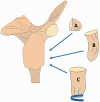
A review of bone grafting techniques for glenoid reconstruction
- PMID: 35265177
- PMCID: PMC8899324
- DOI: 10.1177/17585732211008474
A review of bone grafting techniques for glenoid reconstruction
Abstract
Background: Traumatic anterior shoulder dislocations can cause bony defects of the anterior glenoid rim and are often associated with recurrent shoulder instability. For large glenoid defects of 20-30% without a mobile bony fragment, glenoid reconstruction with bone grafts is often recommended. This review describes two broad categories of glenoid reconstruction procedures found in literature: coracoid transfers involving the Bristow and Latarjet procedures, and free bone grafting techniques.
Methods: An electronic search of MEDLINE and PubMed was conducted to find original articles that described glenoid reconstruction techniques or modifications to existing techniques.
Results: Coracoid transfers involve the Bristow and Latarjet procedures. Modifications to these procedures such as arthroscopic execution, method of graft attachment and orientation have been described. Free bone grafts have been obtained from the iliac crest, distal tibia, acromion, distal clavicle and femoral condyle.
Conclusion: Both coracoid transfers and free bone grafting procedures are options for reconstructing large bony defects of the anterior glenoid rim and have had similar clinical outcomes. Free bone grafts may offer greater flexibility in graft shaping and choice of graft size depending on the bone stock chosen. Novel developments tend towards minimising invasiveness using arthroscopic approaches and examining alternative non-rigid graft fixation techniques.
Keywords: Shoulder; bone graft; glenoid defect; glenoid reconstruction; recurrent instability.
© 2021 The British Elbow & Shoulder Society.
Conflict of interest statement
Declaration of Conflicting Interests: The author(s) declared the following potential conflicts of interest with respect to the research, authorship, and/or publication of this article: GACM: Journal of Shoulder and Elbow Surgery: Editorial or governing board. Shoulder and Elbow: Editorial or governing board. Smith & Nephew: Paid consultant and research support. Techniques in Shoulder and Elbow Surgery: Editorial or governing board. All other authors: Nil.
Figures


References
-
- Zacchilli MA, Owens BD. Epidemiology of shoulder dislocations presenting to emergency departments in the United States. J Bone Joint Surg Am 2010; 92: 542–549. - PubMed
-
- Provencher MT, Bhatia S, Ghodadra NS, et al. Recurrent shoulder instability: current concepts for evaluation and management of glenoid bone loss. J Bone Joint Surg Am 2010; 92(Suppl 2): 133–151. - PubMed
-
- Griffith JF, Antonio GE, Yung PS, et al. Prevalence, pattern, and spectrum of glenoid bone loss in anterior shoulder dislocation: CT analysis of 218 patients. AJR Am J Roentgenol 2008; 190: 1247–1254. - PubMed
-
- Sugaya H, Moriishi J, Dohi M, et al. Glenoid rim morphology in recurrent anterior glenohumeral instability. J Bone Joint Surg Am 2003; 85: 878–884. - PubMed
-
- Yiannakopoulos CK, Mataragas E, Antonogiannakis E. A comparison of the spectrum of intra-articular lesions in acute and chronic anterior shoulder instability. Arthroscopy 2007; 23: 985–990. - PubMed
Publication types
LinkOut - more resources
Full Text Sources
