. 2022 Feb 27:2022:7837837.
doi: 10.1155/2022/7837837. eCollection 2022.

Differential Lung Protective Capacity of Exosomes Derived from Human Adipose Tissue, Bone Marrow, and Umbilical Cord Mesenchymal Stem Cells in Sepsis-Induced Acute Lung Injury

Affiliations

Differential Lung Protective Capacity of Exosomes Derived from Human Adipose Tissue, Bone Marrow, and Umbilical Cord Mesenchymal Stem Cells in Sepsis-Induced Acute Lung Injury

Huimin Deng et al. Oxid Med Cell Longev. .

Abstract

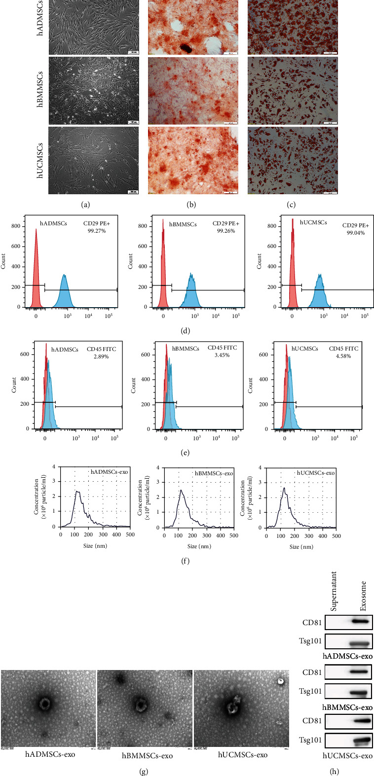
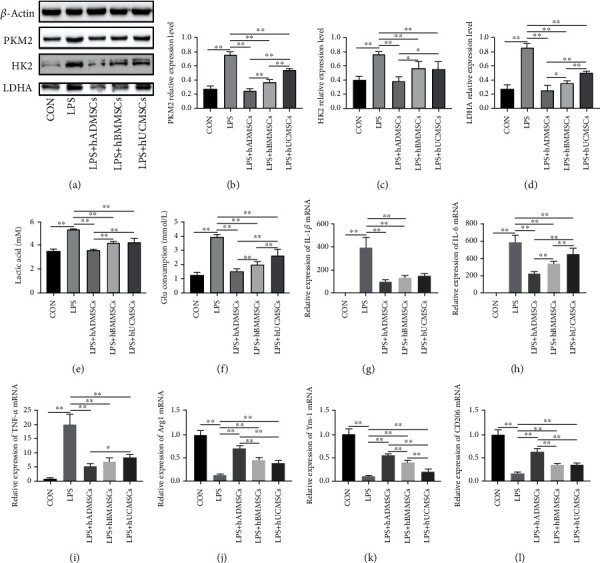
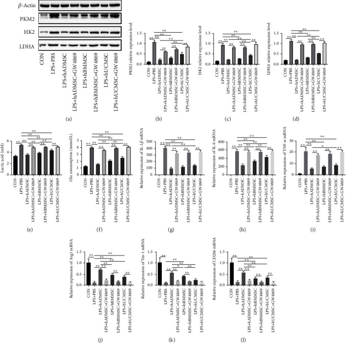
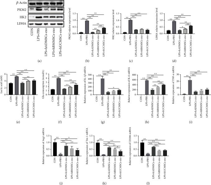
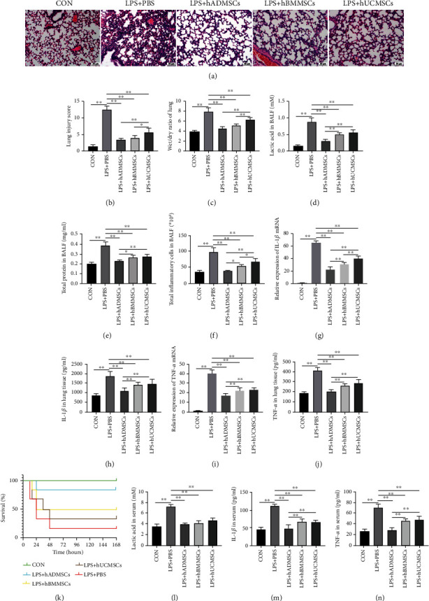
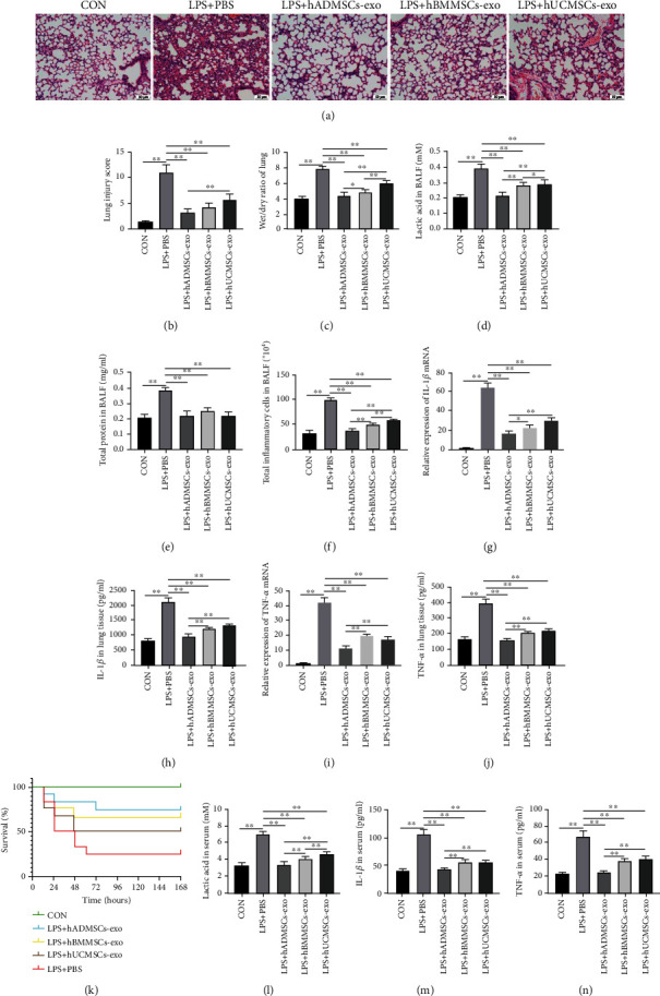
Exosomes derived from human mesenchymal stem cells (hMSCs) have the capacity to regulate various biological events associated with sepsis-induced acute respiratory distress syndrome (ARDS), including cellular immunometabolism, the production of proinflammatory cytokines, allowing them to exert therapeutic effects. However, little is known about which type of hMSC-derived exosomes (hMSC-exo) is more effective and suitable for the treatment of sepsis-induced ARDS. The purpose of this study is to compare the efficacy of hMSC-derived exosomes from human adipose tissue (hADMSC-exo), human bone marrow (hBMMSC-exo), and human umbilical cord (hUCMSC-exo) in the treatment of sepsis-induced ARDS. We cocultured lipopolysaccharide- (LPS-) stimulated RAW264.7 macrophage cells with the three kinds of hMSCs and found that all hMSCs reduced the glycolysis level and the content of lactic acid in macrophages. Accordingly, the expression of proinflammatory cytokines also decreased. Notably, the protective effects of hMSCs from adipose tissue were more obvious than those of bone marrow and umbilical cord hMSCs. However, this protective effect was eliminated when an exosome inhibitor, GW4869, was added. Subsequently, we extracted and cocultured hMSC-derived exosomes with LPS-stimulated RAW264.7 cells and found that all three kinds of exosomes exerted a similar protective effect as their parental cells, with exosomes from adipose hMSCs showing the strongest protective effect. Finally, an experimental sepsis model in mice was established, and we found that all three types of hMSCs have obvious lung-protective effects, in reducing lung injury scores, lactic acid, and proinflammatory cytokine levels in the lung tissues and decreasing the total protein content and inflammatory cell count in the bronchoalveolar lavage fluid (BALF), and also can attenuate the systemic inflammatory response and improve the survival rate of mice. Intravenous injection of three types of hMSC-exo, in particular those derived from adipose hADMSCs, also showed lung-protective effects in mice. These findings revealed that exosomes derived from different sources of hMSCs can effectively downregulate sepsis-induced glycolysis and inflammation in macrophages, ameliorate the lung pathological damage, and improve the survival rate of mice with sepsis. It is worth noting that the protective effect of hADMSC-exo is better than that of hBMMSC-exo and hUCMSC-exo.


Conflict of interest statement

The authors declare that they have no competing interests.

Figures

Figure 1
Figure 1
Characterization of human mesenchymal stem cells (hMSCs) and hMSC-derived exosomes (hMSC-exo). (a) Morphological observation of hMSCs derived from adipose tissue (hADMSCs), bone marrow (hBMMSCs), or umbilical cord (hUCMSCs). hMSCs had a long spindle shape and were arranged in an organized fashion. Scale bar, 100 μm. (b, c) The multidifferentiation potential of hMSCs in vitro. Alizarin red S staining was used to evaluate (b) osteogenic differentiation, while oil red O staining was used to evaluate (c) adipogenic differentiation capacity. Scale bar, 100 μm. (d, e) Flow cytometric analysis of hBMSC surface markers. Note that all hMSCs were (d) positive for CD29 and (e) negative for CD45. The red shape represents the target antibody, and the blue shape represents the isotype control antibody. (f) Nanoparticle tracking assay showed the size distribution of exosomes. (g) hADMSC-exo, hBMMSC-exo, and hUCMSC-exo under the transmission electron microscope. Scale bar, 200 nm. (h) Western blot showing that exosomes derived from the three types of hMSCs were TSG101- and CD81-positive.
Figure 2
Figure 2
Human mesenchymal stem cells (hMSCs) inhibited lipopolysaccharide- (LPS-) induced glycolysis and proinflammatory cytokine production in macrophages. (a) Western blot experiment showed that hMSCs derived from adipose tissue (hADMSCs), bone marrow (hBMMSCs), and umbilical cord (hUCMSCs) inhibited key enzymes involved in glycolysis, including (b) PKM2, (c) HK2, and (d) LDHA. Moreover, the end products of glycolysis decreased, such as (e) lactic acid, as did (f) the consumption of glucose. hADMSCs, hBMMSCs, and hUCMSCs significantly reduced the mRNA expression of (g) interleukin- (IL-) 1β, (h) IL-6, and (i) tumor necrosis factor- (TNF-) α and increased the mRNA levels of (j) Arg1, (k) Ym-1, and (l) CD206 in LPS-treated RAW264.7 cells. All mRNA levels were normalized to the level of β-actin mRNA. Data are expressed as mean ± standard deviation (n = 6 in each group). p values were calculated using one-way ANOVA. p < 0.05; ∗∗p < 0.01.
Figure 3
Figure 3
Human mesenchymal stem cells (hMSCs) suppressed glycolysis and production of proinflammatory cytokines in lipopolysaccharide- (LPS-) treated macrophages by secreting exosomes. (a) Western blot experiments showed that exosomes from hMSCs derived from adipose tissue (AD), bone marrow (hBMMSCs), and umbilical cord (hUCMSCs) inhibited key enzymes involved in glycolysis, including (b) PKM2, (c) HK2, and (d) LDHA. Moreover, the end products of glycolysis decreased, such as (e) lactic acid, as did (f) the consumption of glucose. The exosome inhibitor GW4869 eliminated the inhibitory effect. hADMSCs, hBMMSCs, and hUCMSCs significantly reduced the mRNA expression of (g) IL-1β, (h) IL-6, and (i) TNF-α and increased the mRNA levels of (j) Arg1, (k) Ym-1, and (l) CD206 in LPS-treated RAW264.7 cells. These effects were also reversed by the exosome inhibitor GW4869. All mRNA levels were normalized to the level of β-actin mRNA. Data are expressed as mean ± standard deviation (n = 6 in each group). p values were calculated by one-way ANOVA. p < 0.05; ∗∗p < 0.01.
Figure 4
Figure 4
Human mesenchymal stem cell- (hMSC-) derived exosomes suppressed glycolysis and production of proinflammatory cytokines in lipopolysaccharide- (LPS-) treated macrophages. (a) Western blot experiments showed that exosomes from hMSCs derived from adipose tissue (hADMSCs), bone marrow (hBMMSCs), and umbilical cord (hUCMSCs) inhibited key involved in the glycolysis, including (b) PKM2, (c) HK2, and (d) LDHA. Moreover, the end products of glycolysis, such as (e) lactic acid, as well as (f) the consumption of glucose, decreased. hADMSC-derived exosomes, hBMMSC-derived exosomes, and hUCMSC-derived exosomes significantly reduced the mRNA expression of (g) IL-1β, (h) IL-6, and (i) TNF-α and increased the mRNA levels of (j) Arg1, (k) Ym-1, and (l) CD206 in LPS-treated RAW264.7 cells. All mRNA levels were normalized to the level of β-actin mRNA. The data are expressed as the mean ± standard deviation. n = 6 in each group. p values were calculated by one-way ANOVA. p < 0.05; ∗∗p < 0.01.
Figure 5
Figure 5
Human mesenchymal stem cells (hMSCs) alleviated sepsis-induced ALI and systemic inflammation and improved survival in mice. (a) Hematoxylin and eosin staining of lung tissue sections from the different experimental groups. (b) Lung injury score analysis. (c) Wet-to-dry ratio of lung tissues. (d) Lactic acid content in bronchoalveolar lavage fluid (BALF). (e) Protein concentration in BALF. (f) Inflammatory cell counts in BALF. mRNA expression of (g) interleukin- (IL-) 1β and (i) tumor necrosis factor- (TNF-) α in lung tissue. Levels of (h) IL-1β and (j) TNF-α in lung tissue measured by ELISA. (k) Survival of mice (n = 12 mice in each group); p < 0.001 among the curves as determined using the log-rank (Mantel-Cox) test. Levels of (l) lactic acid, (m) IL-1β, and (n) TNF-α in serum, as measured by ELISA. Data in (b–j) and (l–n) are expressed as mean ± standard deviation (n = 6 in each group); p values were calculated using one-way ANOVA. p < 0.05; ∗∗p < 0.01.
Figure 6
Figure 6
Human mesenchymal stem cell- (hMSC-) derived exosomes attenuated sepsis-induced acute lung injury and systemic inflammation and improved survival in mice. (a) Hematoxylin and eosin staining of lung tissue sections. (b) Lung injury score analysis. (c) Wet-to-dry ratio of lung tissues. (d) Lactic acid content in bronchoalveolar lavage fluid (BALF). (e) Protein concentration in BALF. (f) Inflammatory cell counts in BALF. mRNA expression of (g) interleukin- (IL-) 1β and (i) tumor necrosis factor- (TNF-) α in lung tissue. Levels of (h) IL-1β and (j) TNF-α in lung tissue, as measured by ELISA. (k) Survival rate of mice (n = 12 mice in each group); p = 0.0033 among the curves as determined using the log-rank (Mantel-Cox) test. Levels of (l) lactic acid, (m) IL-1β, and (n) TNF-α in mouse serum, as measured by ELISA. All data in (b–j) and (l–n) are expressed as mean ± standard deviation (n = 6 in each group); p values were calculated using one-way ANOVA. p < 0.05; ∗∗p < 0.01.

Similar articles

Cited by

References

    1. Singer M., Deutschman C. S., Seymour C. W., et al. The third international consensus definitions for sepsis and septic shock (Sepsis-3) JAMA . 2016;315(8):801–810. doi: 10.1001/jama.2016.0287. - DOI - PMC - PubMed
    1. Cecconi M., Evans L., Levy M., Rhodes A. Sepsis and septic shock. Lancet . 2018;392(10141):75–87. doi: 10.1016/s0140-6736(18)30696-2. - DOI - PubMed
    1. Park I., Kim M., Choe K., et al. Neutrophils disturb pulmonary microcirculation in sepsis-induced acute lung injury. The European Respiratory Journal . 2019;53(3):p. 1800786. doi: 10.1183/13993003.00786-2018. - DOI - PMC - PubMed
    1. Máca J., Jor O., Holub M., et al. Past and present ARDS mortality rates: a systematic review. Respiratory Care . 2017;62(1):113–122. doi: 10.4187/respcare.04716. - DOI - PubMed
    1. Fan E., Brodie D., Slutsky A. S. Acute respiratory distress Syndrome. JAMA . 2018;319(7):698–710. doi: 10.1001/jama.2017.21907. - DOI - PubMed