

Frequency of caries in triangular-shaped radiolucencies on periapical radiographs of maxillary deciduous second molars
- PMID: 35265287
- PMCID: PMC8804553
Frequency of caries in triangular-shaped radiolucencies on periapical radiographs of maxillary deciduous second molars
Abstract
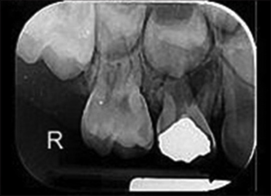
Background: Dentists might face various artifacts (such as triangular-shaped radiolucencies [TSRs]) during the assessment of radiographs and should be able to differentiate them from caries to avoid unnecessary treatments.
Materials and methods: In this cross-sectional study, 109 maxillary second primary molars were evaluated in cooperative children aged 4-9 years, who had distal caries in their maxillary first primary molars. First, TSRs were recorded on periapical radiographs of each maxillary second primary molar's proximal surface. Then, after excavating distal caries in the adjacent teeth "D," a pedodontist examined the mesial surfaces of teeth "E." Chi-square test was used to compare the distribution of caries in different variables, and the kappa coefficient was applied to evaluate clinical and radiographic agreements. A P < 0.05 was considered statistically significant.
Results: Forty-four cases were found to be carious both clinically and radiographically, and 54 cases were noncarious by both methods, while for 11 cases, the diagnosis was controversial. No statistically significant difference was found between radiographic and clinical caries detection methods in children whose periapical radiographs contained TSRs, and most of the subjects had similar diagnoses. Value of caries detection sensitivity, specificity, positive predictive value, and negative predictive value in TSRs was 88%, 92%, 90%, and 90%, respectively.
Conclusion: Considering high radiographic sensitivity for caries detection in TSRs, clinicians should be more cautious about them being carious or not, and both radiographic and clinical examinations are necessary. Further, to avoid misinterpretation in radiographs, additional education is necessary for young dentists.
Keywords: Artifact; deciduous tooth; dental decay; dental radiography.
Copyright: © 2021 Dental Research Journal.
Conflict of interest statement
The authors of this manuscript declare that they have no conflicts of interest, real or perceived, financial or nonfinancial in this article.
Figures
Similar articles
-
Frequency of non-carious triangular-shaped radiolucencies on bitewing radiographs.Dentomaxillofac Radiol. 2008 Jan;37(1):23-7. doi: 10.1259/dmfr/79243767. Dentomaxillofac Radiol. 2008. PMID: 18195251
-
Development of caries in permanent first molars adjacent to primary second molars with interproximal caries: four-year prospective radiographic study.Pediatr Dent. 2004 Jul-Aug;26(4):362-8. Pediatr Dent. 2004. PMID: 15344633
-
Efficacy of sealing the mesial surfaces of first permanent molars with respect to the status of the distal surfaces of the second primary molars in children at high caries-risk.Eur Arch Paediatr Dent. 2014 Apr;15(2):65-73. doi: 10.1007/s40368-013-0066-z. Epub 2013 Jul 9. Eur Arch Paediatr Dent. 2014. PMID: 23835900
-
Panoramic radiography in dental diagnostics.Swed Dent J Suppl. 1996;119:1-26. Swed Dent J Suppl. 1996. PMID: 8971997 Review.
-
A systematic review of clinical diagnostic criteria of early childhood caries.J Public Health Dent. 1999 Summer;59(3):171-91. doi: 10.1111/j.1752-7325.1999.tb03267.x. J Public Health Dent. 1999. PMID: 10649590
References
-
- Stuart C, White P, Michael J. Oral Radiology: Principles and Interpretation. 7th ed. India: Elsevier; 2014.
-
- Brocklebank L. Dental Radiology, Understanding the X-Ray Image. England: Oxford Medical Publications; 1997.
-
- Kuhnisch J, Pasler F, Bucher K, Hickel R, Heinrich-Weltzein R. Frequency of non-carious triangular-shaped radiolucencies on bite-wing radiographs. Dentomaxillofac Radiol. 2008;37:7–23. - PubMed
-
- Khayam E, Daneshkazemi A, Hozhabri H, Moeini M, Namiranian N, Ratki SK, et al. Evaluation of the relative frequency of non-carious triangular-shaped radiolucencies in the first and second permanent molars bite-wing radiography. Indian J Dent. 2013;4:4–141.
LinkOut - more resources
Full Text Sources