

Using Marker-Controlled Watershed Transform to Detect Baker's Cyst in Magnetic Resonance Imaging Images: A Pilot Study
- PMID: 35265470
- PMCID: PMC8804590
- DOI: 10.4103/jmss.JMSS_49_20
Using Marker-Controlled Watershed Transform to Detect Baker's Cyst in Magnetic Resonance Imaging Images: A Pilot Study
Abstract
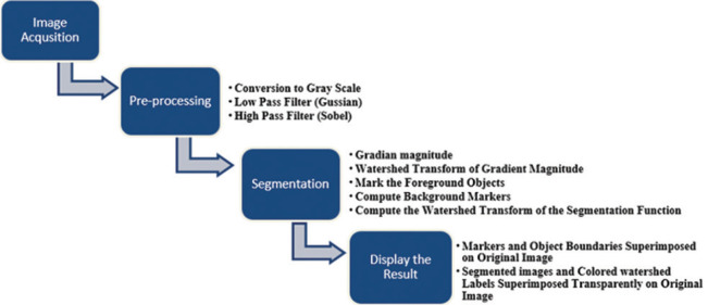
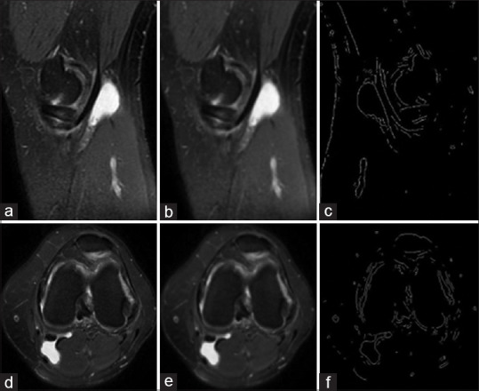
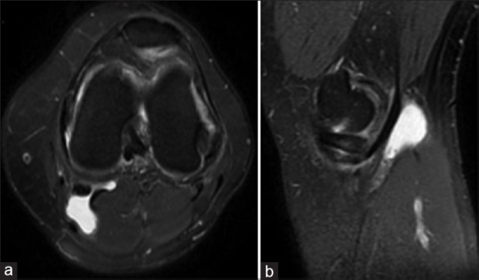
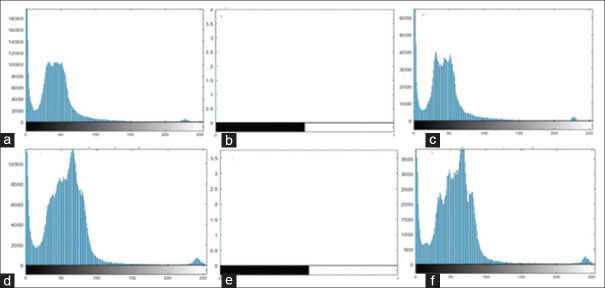
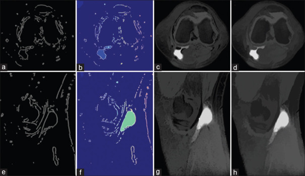
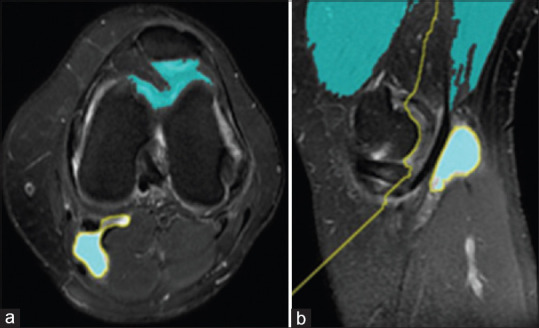
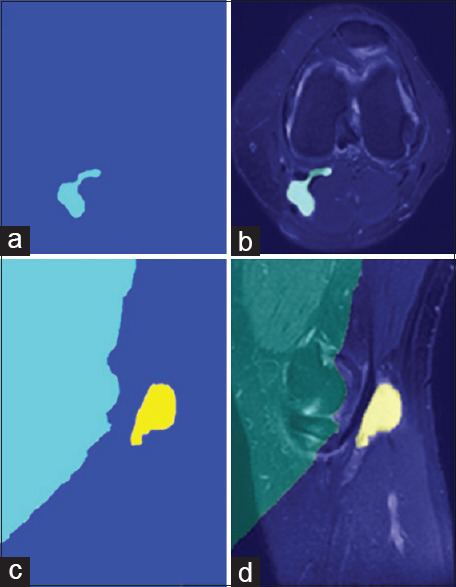
Nowadays, magnetic resonance imaging (MRI) has a high ability to distinguish between soft tissues because of high spatial resolution. Image processing is extensively used to extract clinical data from imaging modalities. In the medical image processing field, the knee's cyst (especially Baker) segmentation is one of the novel research areas. There are different methods for image segmentation. In this paper, the mathematical operation of the watershed algorithm is utilized by MATLAB software based on marker-controlled watershed segmentation for the detection of Baker's cyst in the knee's joint MRI sagittal and axial T2-weighted images. The performance of this algorithm was investigated, and the results showed that in a short time Baker's cyst can be clearly extracted from original images in axial and sagittal planes. The marker-controlled watershed segmentation was able to detect Baker's cyst reliable and can save time and current cost, especially in the absence of specialists it can help us for the easier diagnosis of MRI pathologies.
Keywords: Baker's cyst; image processing; magnetic resonance imaging; marker-controlled watershed transform.
Copyright: © 2021 Journal of Medical Signals & Sensors.
Conflict of interest statement
There are no conflicts of interest.
Figures

References
-
- Drake R, Vogl AW, Mitchell AW. 41st ed. United State: Elsevier Health Sciences; 2017. Gray's Anatomy for Students; pp. 1386–92.
-
- Grey M, Alinani J. 2nd ed. United State McGraw-Hill; 2003. CT and MRI Pathology: A Pocket Atlas. Part VII; p. 388.
-
- Janzen DL, Peterfy C, Forbes JR, Tirman P, Genant H. Cystic lesions around the knee joint: MR imaging findings. AJR Am J Roentgenol. 1994;163:155–61. - PubMed
-
- Thomas S, Pullagura M, Robinson E, Cohen A, Banaszkiewicz P. sports traumatology, arthroscopy. Value Magnetic Resonance Imaging Curr Manage ACL Meniscal Injur. 2007;15:533–6. - PubMed
-
- Lefevre N, Naouri JF, Herman S, Gerometta A, Klouche S, Bohu Y. A current review of the meniscus imaging: Proposition of a useful tool for its radiologic analysis. 2016;47:???.https://doi.org/100.1155/2016/8329296. - PMC - PubMed
LinkOut - more resources
Full Text Sources