

Fetal intracranial hemorrhage: prenatal sonographic diagnosis criteria and postnatal outcomes
- PMID: 35266380
- PMCID: PMC9743346
- DOI: 10.4274/jtgga.galenos.2021.2021-0042
Fetal intracranial hemorrhage: prenatal sonographic diagnosis criteria and postnatal outcomes
Abstract
Objective: The aim of this study was to improve knowledge of prenatally diagnosed fetal intracranial hemorrhage (ICH), defining the ultrasound (US) examination results, the contribution of fetal magnetic resonance imagination (MRI) to the diagnosis, and the pregnancy outcomes, from a series of fetal ICH cases.
Material and methods: This retrospective, observational study included eleven fetuses diagnosed with ICH from April 2016 to August 2020. The data regarding the medical records, prenatal US and MRI findings, treatment, and prognosis of fetal ICH cases were collected from the hospital database and analyzed.
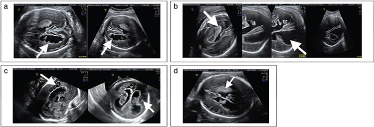
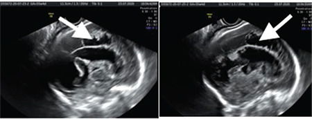
Results: Fetal ICHs were grade 3 in six cases, and grade 4 in the remaining five cases. The mean gestational age at diagnosis was 30.2 weeks. Nine (81.8%) of the cases were diagnosed in the third trimester and two (18.2%) in the second trimester. Fetal cranial MRI was performed in 7/11 (63.6%) following ultrasonographic diagnosis. MRI confirmed fetal ICH diagnosis and previous US findings regarding location and grade in all cases. Five patients (45.5%) diagnosed with grade 3 (n=1) and grade 4 (n=4) ICH underwent pregnancy termination. Of the remaining six cases, one (9.1%) diagnosed with grade 3 fetal ICH resulted in an intrauterine fetal demise. Four cases classified as grade 3 fetal ICH and one case with grade 4 fetal ICH were born alive at term.
Conclusion: The clinical manifestations of fetal ICH are diverse and have a wide spectrum of severity and prognostic implications. Fetal ICH cases were mainly detected in the third trimester, with a minority detected in the second trimester. These cases can be safely diagnosed and graded by US examination, but the underlying etiology frequently cannot be determined. Fetal cranial MRI may aid in diagnosis confirmation if this is unclear from US in order to provide appropriate counseling to the parents.
Keywords: Fetal intracranial hemorrhage; prenatal diagnosis; ultrasound.
Conflict of interest statement
Figures
References
-
- Sanapo L, Whitehead MT, Bulas DI, Ahmadzia HK, Pesacreta L, Chang T, et al. Fetal intracranial hemorrhage: role of fetal MRI. Prenat Diagn. 2017;37:827–36. - PubMed
-
- Monteagudo A; Society for Maternal-Fetal Medicine (SMFM) Intracranial Hemorrhage. Am J Obstet Gynecol. 2020;223:B34–7. - PubMed
-
- Adiego B, Martínez-Ten P, Bermejo C, Estévez M, Recio Rodriguez M, Illescas T. Fetal intracranial hemorrhage. Prenatal diagnosis and postnatal outcomes. J Matern Fetal Neonatal Med. 2019;32:21–30. - PubMed
-
- Qi W, Luo JY, Li ZL, Zhang QJ, Liu ZD, Liao QP, et al. Clinical analysis of eight cases of fetal intracranial hemorrhage in pregnancy. J Matern Fetal Neonatal Med. 2021;16:2609–15. 1-7. - PubMed
-
- Carletti A, Colleoni GG, Perolo A, Simonazzi G, Ghi T, Rizzo N, et al. Prenatal diagnosis of cerebral lesions acquired in utero and with a late appearance. Prenat Diagn. 2009;29:389–95. - PubMed
LinkOut - more resources
Full Text Sources
Miscellaneous