. 2022 Apr 19;66(4):e0210921.
doi: 10.1128/aac.02109-21. Epub 2022 Mar 10.

An Optimized Dihydrodibenzothiazepine Lead Compound (SBI-0797750) as a Potent and Selective Inhibitor of Plasmodium falciparum and P. vivax Glucose 6-Phosphate Dehydrogenase 6-Phosphogluconolactonase

Affiliations

An Optimized Dihydrodibenzothiazepine Lead Compound (SBI-0797750) as a Potent and Selective Inhibitor of Plasmodium falciparum and P. vivax Glucose 6-Phosphate Dehydrogenase 6-Phosphogluconolactonase

Isabell Berneburg et al. Antimicrob Agents Chemother. .

Abstract

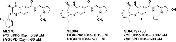
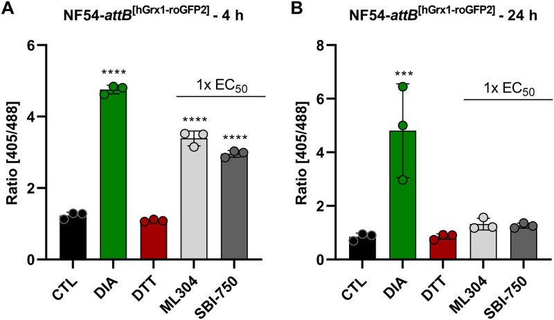
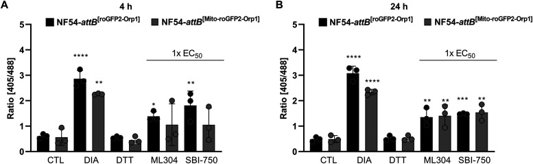
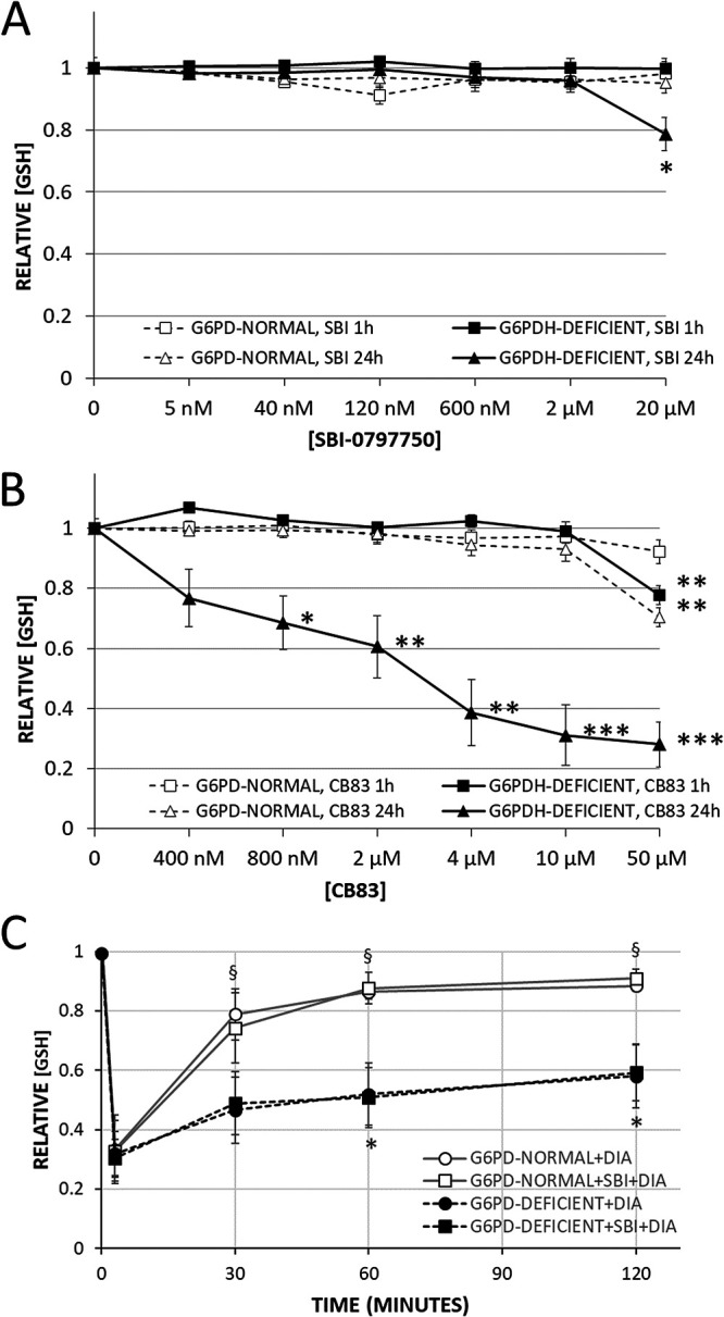
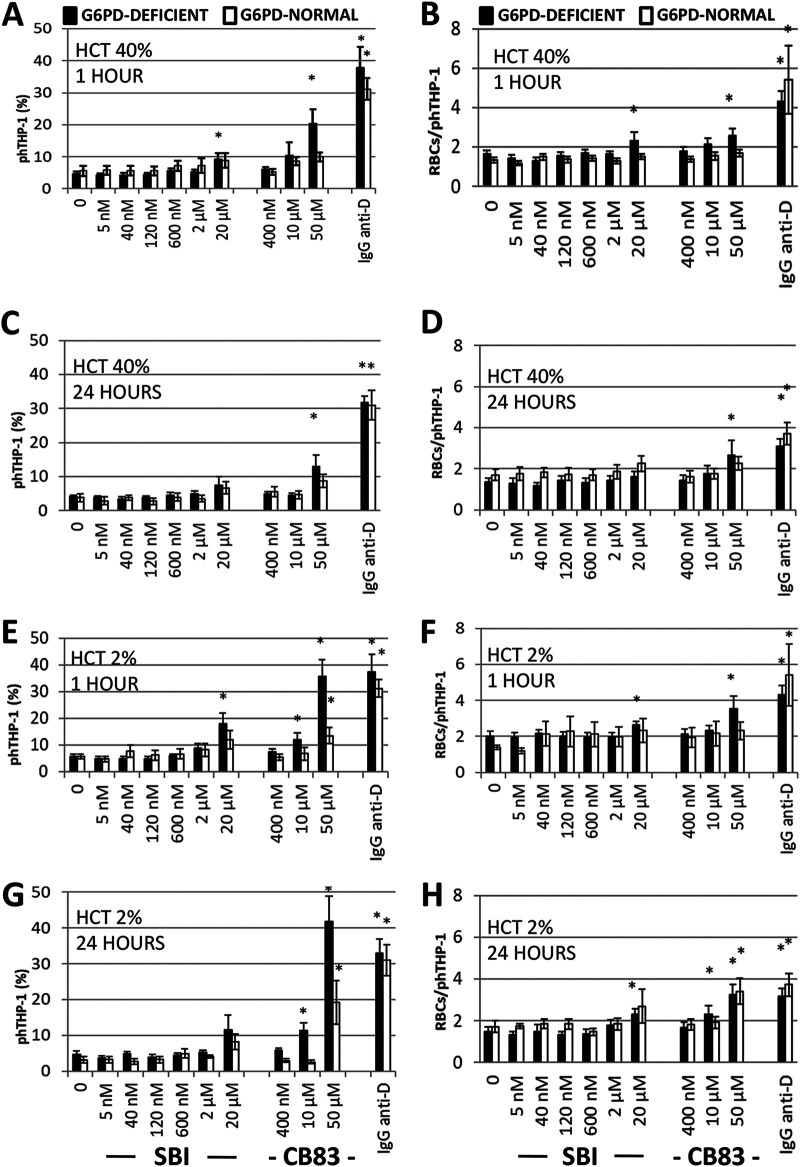
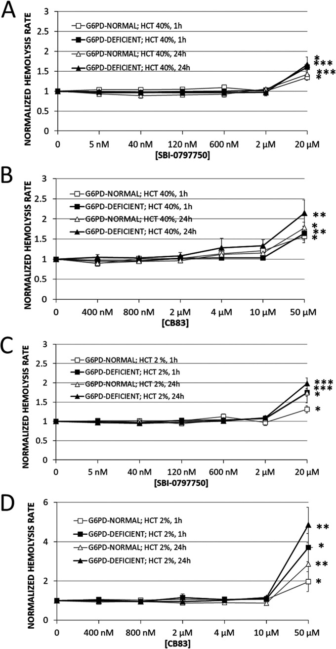
In Plasmodium, the first two and rate-limiting enzymes of the pentose phosphate pathway, glucose 6-phosphate dehydrogenase (G6PD) and the 6-phosphogluconolactonase, are bifunctionally fused to a unique enzyme named GluPho, differing structurally and mechanistically from the respective human orthologs. Consistent with the enzyme's essentiality for malaria parasite proliferation and propagation, human G6PD deficiency has immense impact on protection against severe malaria, making PfGluPho an attractive antimalarial drug target. Herein we report on the optimized lead compound N-(((2R,4S)-1-cyclobutyl-4-hydroxypyrrolidin-2-yl)methyl)-6-fluoro-4-methyl-11-oxo-10,11-dihydrodibenzo[b,f][1,4]thiazepine-8-carboxamide (SBI-0797750), a potent and fully selective PfGluPho inhibitor with robust nanomolar activity against recombinant PfGluPho, PvG6PD, and P. falciparum blood-stage parasites. Mode-of-action studies have confirmed that SBI-0797750 disturbs the cytosolic glutathione-dependent redox potential, as well as the cytosolic and mitochondrial H2O2 homeostasis of P. falciparum blood stages, at low nanomolar concentrations. Moreover, SBI-0797750 does not harm red blood cell (RBC) integrity and phagocytosis and thus does not promote anemia. SBI-0797750 is therefore a very promising antimalarial lead compound.

Keywords: G6PDH; Plasmodium; Plasmodium falciparum; Plasmodium vivax; inhibitors; malaria.


Conflict of interest statement

The authors declare no conflict of interest.

Figures

FIG 1
FIG 1
Previously described PfGluPho inhibitors ML276 and ML304 and optimized lead compound SBI-0797750.
FIG 2
FIG 2
Mechanism of inhibition of SBI-0797750 against PfGluPho. (A and B) Various compound concentrations were titrated against G6P. SBI-0797750 acts as a competitive inhibitor against PfGluPho, since the Km value for G6P increases with increasing compound concentrations, while Vmax stays constant. (C and D) Titration of NADP+ against different compound concentrations. The Km value for NADP+ increases, while Vmax decreases, indicating a mixed-type inhibition. PfGluPho activity is in relative fluorescence units per second (modified from reference 20).
FIG 3
FIG 3
Reversibility of the PfGluPho inhibition by SBI-0797750. Reversibility of PfGluPho inhibition by SBI-0797750 (SBI-750) was determined by incubating the enzyme with a high compound concentration of 1.65 μM (predilution), followed by dilution to 3.3 nM (postdilution). The diluted sample had the same activity as a control containing the same final compound concentration (CTL, 3.3 nM), while the undiluted sample was completely inhibited. Treatment without SBI-750 was defined as 100% activity (CTL, 0 nM). Values are means and SD from three independent determinations, each including two measurements.
FIG 4
FIG 4
Mid- and long-term effects of ML304 and its derivative SBI-0797750 on the redox ratio of P. falciparum NF54-attB[hGrx1-roGFP2] parasites. In 4-h and 24-h experiments, P. falciparum NF54-attB[hGrx1-roGFP2] parasites were incubated with 1× EC50 ML304 and SBI-0797750 (SBI-750). Via CLSM, a significant increase of the 405/488-nm fluorescence ratio of the redox sensor could be observed in the 4-h-incubation experiment (A). In the 24-h-incubation experiment, neither ML304 nor SBI-0797750 significantly changed the redox ratio (B). Nontreated parasites served as controls. All experiments included fully oxidized (1 mM DIA) and fully reduced (10 mM DTT) parasites. CLSM data were obtained from 10 to 20 trophozoites for each experiment and each incubation time. Values are means and SD from three independent experiments. A one-way ANOVA with 95% confidence intervals with the Dunnett’s multiple-comparison test was applied for statistical analysis of significance (***, P < 0.001; ****, P < 0.0001).
FIG 5
FIG 5
Mid- and long-term effects of ML304 and its derivative SBI-0797750 on the redox ratio of P. falciparum NF54-attB[roGFP2-Orp1] and NF54-attB[Mito-roGFP2-Orp1]-transfected parasites. Four and 24 h of incubation of NF54-attB[roGFP2-Orp1] transfectants with 1× EC50 SBI-0797750 (SBI-750) and ML304 significantly increased the fluorescence ratio of the cytosolic sensor, as determined using CLSM. In NF54-attB[Mito-roGFP2-Orp1] transfectants, ML304 and SBI-0797750 increased the redox ratio after 4 h (A), both of which reached significance after 24 h of incubation (B). Nontreated parasites served as controls. All experiments included fully oxidized (1 mM DIA) and fully reduced (10 mM DTT) parasites. CLSM data comprised 10 to 20 trophozoites analyzed per experiment for each incubation. Values are means and SD (error bars) for three independent experiments. A one-way ANOVA with 95% confidence intervals with the Dunnett’s multiple-comparison test was applied for statistical analysis of significance (*, P < 0.05; **, P < 0.01; ***, P < 0.001; ****, P < 0.0001).
FIG 6
FIG 6
Effect of SBI-0797750 and CB83 on steady-state concentration and recovery of GSH after oxidative challenge in G6PD-normal and G6PD-deficient RBCs. G6PD-normal and -deficient RBCs were incubated with SBI-0797750 (A) or CB83 (B) at indicated concentrations or kept under the same conditions without inhibitor treatment (0 nM). GSH concentrations in RBCs were measured after 1 and 24 h of incubation as indicated. GSH concentrations were normalized by referring them to the hemolysis of respective untreated RBCs (0 nM). Mean GSH concentrations and SE in untreated RBCs (0 nM) were 2.86 ± 0.13 mM and 2.65 ± 0.13 mM for G6PD-normal RBCs (n = 5) and 1.89 ± 0.13 mM and 1.24 ± 0.23 mM for G6PD-deficient ones (n = 8) after 1 and 24 h of incubation, respectively. Normalized GSH concentrations in RBCs (relative [GSH]) of G6PD-normal (n = 5) and G6PD-deficient (n = 8) donors are shown as means and SE. Significant differences relative to untreated RBCs are indicated: *, P < 0.05; **, P < 0.01; ***, P < 0.001. (C) GSH was measured in RBCs after preincubation with or without 2 μM SBI-0797750, and G6PD-normal and -deficient RBCs were subsequently challenged with 0.5 and 1.0 mM DIA (final concentrations), respectively, at time zero. RBC suspensions were incubated for 3 min at 4°C and afterward at 37°C. GSH values of RBCs measured at 3, 30, 60, and 120 min after DIA supplementation were referred to the corresponding starting GSH value (t = 0) of the same RBC suspension measured immediately before DIA supplementation (relative [GSH]). Mean GSH concentrations and SE of G6PD-normal and -deficient RBCs at time zero were 2.71 ± 0.15 mM and 1.66 ± 0.15 mM, respectively, without SBI-0797750 and 2.82 ± 0.17 mM and 1.68 ± 0.14 mM, respectively, with SBI-0797750. Relative GSH concentrations in RBCs of G6PD-normal (n = 5) and G6PD-deficient (n = 8) donors are shown as means and SE. Significant differences relative to respective values at 3 min (*, P < 0.05) and differences between G6PD-normal and -deficient RBCs (§, P < 0.05) are indicated.
FIG 7
FIG 7
Effect of SBI-0797750 and CB83 on the phagocytosis of G6PD-normal and G6PD-deficient RBCs by human phagocytes. G6PD-normal and -deficient RBCs were incubated at a hematocrit of either 40% (A to D) or 2% (E to H) with SBI-0797750 (SBI) or CB83 at the indicated concentrations for 1 h and 24 h or kept under the same conditions without inhibitor treatment (0 nm). RBCs were fluorescence stained with carboxyfluorescein diacetate succinimidyl ester (CFDA-SE). Erythrophagocytosis by THP-1 phagocytes expressed as a proportion of phagocytosis-positive phagocytes (phTHP-1 [%]) and the number of RBCs phagocytosed per phagocyte (RBCs/phTHP-1) were assessed via FACS. IgG anti-D-opsonized RBCs were included as a positive phagocytosis control. For details, see Materials and Methods. Columns represent means and SE of RBC phagocytosis from G6PD-normal (n = 5) and G6PD-deficient (n = 8) donors. Significant differences compared to untreated RBCs are indicated: *, P < 0.05.
FIG 8
FIG 8
Effect of SBI-0797750 and CB83 on hemolysis in G6PD-normal and -deficient RBCs. G6PD-normal and -deficient RBCs were incubated at a hematocrit of either 40% (A and B) or 2% (C and D) with SBI-0797750 (SBI) (A and C) or CB83 (B and D) at indicated concentrations or kept under the same conditions without inhibitor treatment (0 nM). The hemolysis rate was assessed in terms of hemoglobin release from RBCs and was measured in the supernatant after 1 h and 24 h of incubation. Supernatant hemoglobin was referred to total hemoglobin in the RBC suspension to obtain the hemolysis rate. These rates were normalized by referring any measured value to the hemolysis rate assessed in respective untreated RBCs (0 nM). Mean hemolysis rates and SE in untreated RBCs (0 nM) incubated at a hematocrit of 40% were 0.9% ± 0.09% and 1.47% ± 0.16% for G6PD-normal (n = 5) and 1.17% ± 0.23% and 1.56% ± 0.36% for G6PD-deficient RBCs (n = 8) after 1 h and 24 h of incubation, respectively (A and B). Mean hemolysis rates and SE in untreated RBCs (0 nM) incubated at a hematocrit of 2% were 1.95% ± 0.28% and 2.94% ± 0.48% for G6PD-normal (n = 5) and 2.40% ± 0.26% and 3.56% ± 0.59% for G6PD-deficient RBCs (n = 8) after 1 h and 24 h of incubation, respectively (C and D). Normalized hemolysis rates of RBCs are shown as means and SE of G6PD-normal (n = 5) and G6PD-deficient (n = 8) donors. Significant differences to untreated RBCs are indicated: *, P < 0.05; **, P < 0.01; ***, P ≤ 0.001.

References

    1. WHO. 2020. World Malaria Report 2020. https://www.who.int/publications/i/item/9789240015791.
    1. Cowman AF, Healer J, Marapana D, Marsh K. 2016. Malaria: biology and disease. Cell 167:610–624. 10.1016/j.cell.2016.07.055. - DOI - PubMed
    1. Naing C, Whittaker MA, Nyunt Wai V, Mak JW. 2014. Is Plasmodium vivax malaria a severe malaria?: a systematic review and meta-analysis. PLoS Negl Trop Dis 8:e3071. 10.1371/journal.pntd.0003071. - DOI - PMC - PubMed
    1. WHO. 2017. World malaria report 2017. http://apps.who.int/iris/bitstream/handle/10665/259492/9789241565523-eng....
    1. Mbengue A, Bhattacharjee S, Pandharkar T, Liu H, Estiu G, Stahelin RV, Rizk SS, Njimoh DL, Ryan Y, Chotivanich K, Nguon C, Ghorbal M, Lopez-Rubio J-J, Pfrender M, Emrich S, Mohandas N, Dondorp AM, Wiest O, Haldar K. 2015. A molecular mechanism of artemisinin resistance in Plasmodium falciparum malaria. Nature 520:683–687. 10.1038/nature14412. - DOI - PMC - PubMed

Publication types

MeSH terms