

Impact of rapid maxillary expansion on palatal morphology at different dentition stages
- PMID: 35267098
- PMCID: PMC9276570
- DOI: 10.1007/s00784-022-04434-9
Impact of rapid maxillary expansion on palatal morphology at different dentition stages
Abstract
Objective: Rapid maxillary expansion (RME) is an established and frequently used procedure to overcome maxillary constriction. In-depth studies about morphological changes of the alveolar process and its immediate surroundings are missing. Therefore, the aim of the present study was to examine the treatment effects of a dentally anchored, rapid maxillary expander at different dentition stages upon palatal width, height and shape.
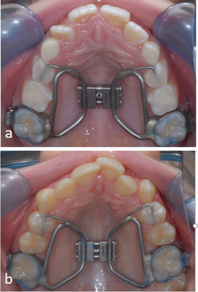
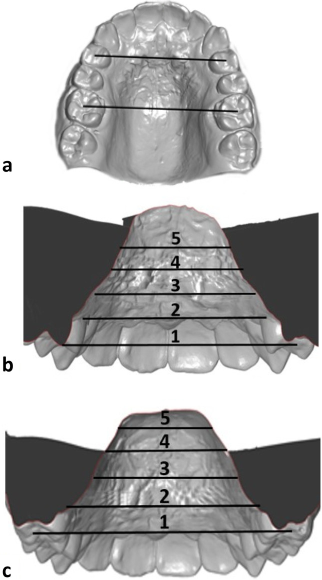
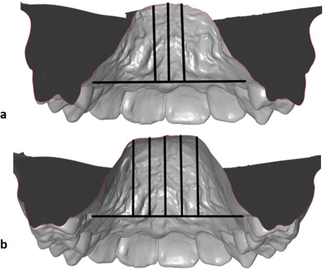
Material and methods: The dental casts of 114 patients-taken immediately before and after RME-were three-dimensionally analysed. Depending on the dentition stage, the patients were divided into two groups (each n = 57, group 1, early mixed dentition; group 2, late mixed or permanent dentition).
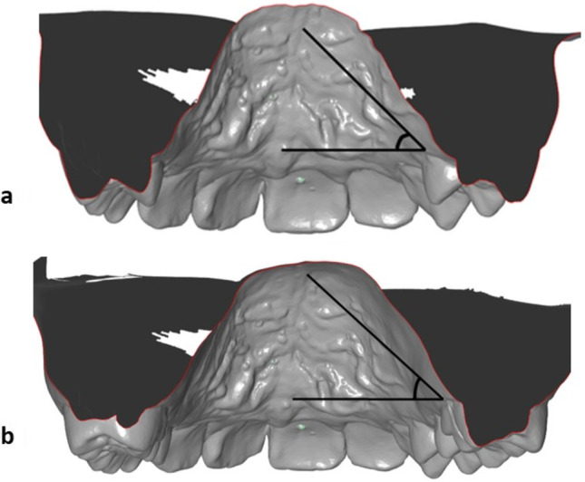
Results: The width increases were highly significant, both in the overall and in the individual groups (p < 0.001). While the width increase was greater in the posterior area than anteriorly in the early group, the widening in the late group happened significantly greater anteriorly than posteriorly. Palatal height increased anteriorly and posteriorly in both groups to a significant extent (p < 0.001). The height increase was more pronounced in the anterior region than in the posterior region in the late group. The palatine index according to Kim revealed a change in palatal morphology both anteriorly and posteriorly in the early group but only anteriorly in the late group.
Conclusions: Maxillary expansion occurs more parallel in early treatment compared to V-shaped opening in the later treatment approach.
Clinical relevance: RME is more advantageous in an early dentition.
Keywords: Median palatine suture; Palatal morphology; Rapid maxillary expansion (RME); Transverse palatine suture.
© 2022. The Author(s).
Conflict of interest statement
The authors declare no competing interests.
Figures
References
-
- Angell EC. Treatment of irregularities of the permanent adult teeth. Dent Cosmos. 1860;1:540–545.
-
- Korbmacher H, Huck L, Merkle T, Kahl-Nieke B. Clinical profile of rapid maxillary expansion – outcome of a national inquiry. J Orofac Orthop. 2005;66:455–468. - PubMed
-
- Grabowski R, Hinz R, Stahl de Castrillon F, editors. Das kieferorthopädische Risikokind: Gebissentwicklung und Funktionsstörungen - KFO-Prävention und Frühbehandlung. Herne: Zahnärztlicher Fach-Verlag; 2009.
-
- Biederman W. Rapid correction of class III malocclusion by midpalatal expansion. Am J Orthod. 1973;63:47–55. - PubMed
-
- Haas AJ. Rapid expansion of the maxillary dental arch and nasal cavity by opening the midpalatal suture. Angle Orthod. 1961;31:73–90.
MeSH terms
LinkOut - more resources
Full Text Sources
