. 2022 Feb 27;14(5):1233.
doi: 10.3390/cancers14051233.

Role of the Skin Microenvironment in Melanomagenesis: Epidermal Keratinocytes and Dermal Fibroblasts Promote BRAF Oncogene-Induced Senescence Escape in Melanocytes

Affiliations

Role of the Skin Microenvironment in Melanomagenesis: Epidermal Keratinocytes and Dermal Fibroblasts Promote BRAF Oncogene-Induced Senescence Escape in Melanocytes

Shreyans Sadangi et al. Cancers (Basel). .

Abstract

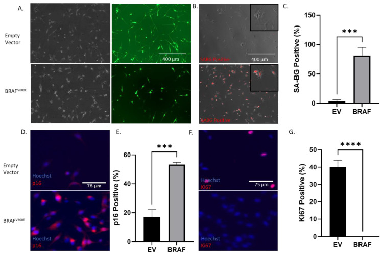
BRAFV600E is the most common mutation driver in melanoma. This mutation is known to cause a brief burst of proliferation followed by growth arrest and senescence, which prevent an uncontrolled cell proliferation. This phenomenon is known as oncogene-induced senescence (OIS) and OIS escape is thought to lead to melanomagenesis. Much attention has been focused on the melanocyte-intrinsic mechanisms that contribute to senescence escape. Additional genetic events such as the loss of tumor suppressor PTEN and/or epigenetic changes that contribute to senescence escape have been described. However, the role of the skin microenvironment-specifically, the role of epidermal keratinocytes-on melanomagenesis is not fully understood. In this study, we employ a microfluidic platform to study the interaction between melanocytes expressing the BRAFV600E mutation as well as keratinocytes and dermal fibroblasts. We demonstrate that keratinocytes suppress senescence-related genes and promote the proliferation of transformed melanocytes. We also show that a keratinocyte-conditioned medium can alter the secretion of both pro- and anti-tumorigenic factors by transformed melanocytes. In addition, we show that melanocytes and keratinocytes from donors of white European and black African ancestry display different crosstalks; i.e., white keratinocytes appear to promote a more pro-tumorigenic phenotype compared with black keratinocytes. These data suggest that keratinocytes exert their influence on melanomagenesis both by suppressing senescence-related genes in melanocytes and by affecting the balance of the melanocyte-secreted factors that favor tumorigenesis.

Keywords: melanoma; microfluidics; senescence; tumor microenvironment.


Conflict of interest statement

D.J.B. holds equity in Bellbrook Labs LLC, Tasso Inc., Salus Discovery LLC, Flambeau Diagnostics LLC, Stacks to the Future LLC, Lynx Biosciences Inc. and Onexio Biosystems.

Figures

Figure 1
Figure 1
BRAFV600E induces growth arrest in melanocytes. (A) Upper panel shows Caucasian melanocytes transduced with the control lentivirus carrying an empty vector coding for green fluorescence protein (GFP). Lower Panel shows Caucasian melanocytes transduced with the BRAFV600E mutant lentivirus (60% transduction efficiency). (B) Senescence-associated beta-galactosidase (SA-β-gal) staining of Caucasian melanocytes after transduction with a BRAFV600E lentivirus. Upper panel shows transduction with the empty vector whereas the lower image shows BRAFV600E-transduced melanocytes after the addition of the beta-gal substrate, DCM beta-gal. Insets show magnified images. (C) Bar graph shows the percentage of SA-β-galactosidase (SA-β-gal)-positive cells shown in (B). (D,E) Immunofluorescence (IF) images showing p16 staining, a cell cycle inhibitor commonly upregulated during senescence. BRAFV600E-transduced melanocytes (lower image) show a higher expression of p16 than control melanocytes (upper image). (F,G) IF images showing the cell proliferation marker Ki67. BRAFV600E-transduced melanocytes (lower image) show no expression of Ki67, suggesting the growth arrest of cells. Experiments were repeated at least 3 independent times. Data were analyzed using a Student’s t-test with a Welch correction. Graphs show mean ± standard deviation. *** denotes p < 0.005; **** denotes p < 0.0005.
Figure 2
Figure 2
Effect of keratinocytes on melanocyte oncogene-induced senescence (OIS). (A) Schematic of the melanocyte and keratinocyte co-culture. Melanocytes were seeded in the central chamber and keratinocytes in the lateral chamber. The microdevice design allowed us to confine each cell type into the desired chamber whilst allowing cell–cell crosstalk. Cells were analyzed for SA-β-gal activity 7 days after co-culture. (B) BRAFV600E-transduced Caucasian melanocytes were seeded on the central chamber of the microfluidic device and cultured with autologous keratinocytes (+) or culture media only (-) in the lateral chambers. Images show the SA-β-gal red signal overlaid with a brightfield. We observed that the presence of keratinocytes lowered the number of oncogene-induced senescence (OIS)-positive BRAFV600E melanocytes. (C) BRAFV600E-transduced Caucasian melanocytes were seeded on a 96-well plate and cultured with autologous Caucasian keratinocyte-conditioned media or fresh keratinocyte media. Left and right panels show melanocytes in phase contrast in media and keratinocyte-conditioned media, respectively, suggesting a keratinocyte-induced OIS decrease. Melanocytes were 12 days post-transduction and 7 days after co-culture. ** denotes p < 0.005; *** denotes p < 0.0005. Experiments were repeated at least 3 independent times. Data were analyzed using a Student’s t-test with a Welch correction. Graphs show mean ± standard deviation.
Figure 3
Figure 3
Transcriptomic changes due to the presence of Caucasian ancestry keratinocytes. (A) Schematic showing the co-culture of Caucasian ancestry melanocytes and keratinocytes. A collagen mixture was added to the second and fourth chambers and allowed to polymerize (approximately 15 min). Transformed melanocytes (5 days post-transduction) were seeded in the central chamber and keratinocytes were seeded in the extreme lateral chambers. Cells were co-cultured for 7 days following which RNA was extracted and followed with a PCR analysis. (B) Bar graphs showing a fold change of selected genes in the absence (−) and presence (+) of keratinocytes. * denotes < 0.05; ** denotes < 0.005; *** denotes < 0.0005. Data were analyzed using a Student’s t-test with a Welch correction. Graphs show mean ± standard deviation.
Figure 4
Figure 4
VEGF-A secretion is modulated by BRAFV600E expression and keratinocytes. (A) Experimental design. Transformed and untransformed Caucasian melanocytes were exposed to a keratinocyte-conditioned medium from autologous and allogenic keratinocytes for 3 days and the spent media were harvested for a growth factor analysis. (B) Effect of oncogene transformation on VEGF-A secretion. BRAFV600E expression enables the secretion of VEGF-A by melanocytes in isolation. (C) Effect of keratinocytes on VEGF-A secretion by normal melanocytes. Caucasian keratinocytes enable the secretion of VEGF-A in normal melanocytes in autologous and allogeneic melanocytes. (D) Effect of keratinocytes on VEGF-A secretion by transformed melanocytes. African-American keratinocytes suppress VEGF-A secretion by transformed African-American melanocytes. + in (C) and (D). * denotes p < 0.05; ** denotes p < 0.005; *** denotes p < 0.0005; **** denotes p < 0.00005. Experiments were repeated at least 3 independent times. Data were analyzed using a Student’s t-test with a Welch correction. Graphs show mean ± standard deviation.
Figure 5
Figure 5
LIF secretion is modulated by BRAFV600E expression and keratinocytes. (A) Experimental design. Transformed Caucasian melanocytes were exposed to autologous and allogeneic keratinocytes for 3 days and the spent keratinocyte-conditioned medium was harvested for a growth factor analysis. (B) Effect of oncogene transformation on LIF. BRAFV600E expression enables the secretion of VEGF-A by melanocytes in isolation. (C) Effect of keratinocytes on LIF secretion by normal melanocytes. As opposed to VEGF-A, keratinocytes had a minor effect on LIF secretion by normal melanocytes. (D) Effect of keratinocytes on LIF secretion by BRAF-transformed melanocytes. Similar to the empty vector, keratinocytes showed a minor effect on LIF secretion by BRAF-transduced melanocytes. * denotes p < 0.05; ** denotes p < 0.005; **** denotes p < 0.00005. Experiments were repeated at least 3 independent times. Data were analyzed using a Student’s t-test with a Welch correction. Graphs show mean ± standard deviation. NS denotes non-significant difference.

References

    1. American Cancer Society Key Statistics for Melanoma Skin Cancer. 2021. [(accessed on 5 September 2021)]. Available online: https://www.cancer.org/cancer/melanoma-skin-cancer/about/key-statistics.....
    1. Vultur A., Herlyn M. SnapShot: Melanoma. Cancer Cell. 2013;23:706. doi: 10.1016/j.ccr.2013.05.001. - DOI - PubMed
    1. Davies H., Bignell G.R., Cox C., Stephens P., Edkins S., Clegg S., Teague J., Woffendin H., Garnett M.J., Bottomley W., et al. Mutations of the BRAF gene in human cancer. Nature. 2002;417:949–954. doi: 10.1038/nature00766. - DOI - PubMed
    1. Pollock P., Harper U.L., Hansen K.S., Yudt L.M., Stark M., Robbins C.M., Moses T.Y., Hostetter G., Wagner U., Kakareka J., et al. High frequency of BRAF mutations in nevi. Nat. Genet. 2003;33:19–20. doi: 10.1038/ng1054. - DOI - PubMed
    1. Michaloglou C., Vredeveld L.C.W., Soengas M.S., Denoyelle C., Kuilman T., Van Der Horst C.M.A.M., Majoor D.M., Shay J.W., Mooi W.J., Peeper D.S. BRAFE600-associated senescence-like cell cycle arrest of human naevi. Nature. 2005;436:720–724. doi: 10.1038/nature03890. - DOI - PubMed