. 2022 Feb 22;14(5):857.
doi: 10.3390/polym14050857.

3D Printed Scaffold Based on Type I Collagen/PLGA_TGF-β1 Nanoparticles Mimicking the Growth Factor Footprint of Human Bone Tissue

Affiliations

3D Printed Scaffold Based on Type I Collagen/PLGA_TGF-β1 Nanoparticles Mimicking the Growth Factor Footprint of Human Bone Tissue

Federica Banche-Niclot et al. Polymers (Basel). .

Abstract

In bone regenerative strategies, the controlled release of growth factors is one of the main aspects for successful tissue regeneration. Recent trends in the drug delivery field increased the interest in the development of biodegradable systems able to protect and transport active agents. In the present study, we designed degradable poly(lactic-co-glycolic)acid (PLGA) nanocarriers suitable for the release of Transforming Growth Factor-beta 1 (TGF-β1), a key molecule in the management of bone cells behaviour. Spherical TGF-β1-containing PLGA (PLGA_TGF-β1) nanoparticles (ca.250 nm) exhibiting high encapsulation efficiency (ca.64%) were successfully synthesized. The TGF-β1 nanocarriers were subsequently combined with type I collagen for the fabrication of nanostructured 3D printed scaffolds able to mimic the TGF-β1 presence in the human bone extracellular matrix (ECM). The homogeneous hybrid formulation underwent a comprehensive rheological characterisation in view of 3D printing. The 3D printed collagen-based scaffolds (10 mm × 10 mm × 1 mm) successfully mimicked the TGF-β1 presence in human bone ECM as assessed by immunohistochemical TGF-β1 staining, covering ca.3.4% of the whole scaffold area. Moreover, the collagenous matrix was able to reduce the initial burst release observed in the first 24 h from about 38% for the PLGA_TGF-β1 alone to 14.5%, proving that the nanocarriers incorporation into collagen allows achieving sustained release kinetics.

Keywords: 3D printed scaffolds; TGF-β1; bone; drug delivery; polymeric nanoparticles; tissue regeneration; type I collagen.


Conflict of interest statement

The authors declare no conflict of interest.

Figures

Figure 1
Figure 1
Schematic representation of PLGA_TGF-β1 synthesis process.
Figure 2
Figure 2
(A) Schematic representation of Coll/PLGA_TGF-β1_GEN scaffold realization process and (B) mechanism of genipin chemical crosslinking of collagen.
Figure 3
Figure 3
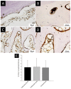
TGF-β1 staining in bone tissue and semi-quantitative analysis (AD): TGF-β1 staining in cortical (A,B) and trabecular (C,D) bone tissue. ((A,C) 10× magnification; (B,D) 20× magnification); black arrows highlight the protein accumulation in bone ECM. (E) Histogram representing area percentage of TGF-β1 staining.
Figure 4
Figure 4
PLGA_TGF-β1 nanoparticles characterisation: FESEM images at different magnifications: 25,000× (A) and 200,000× (B) and DLS size distribution graph and numerical results (C) (abbreviations: d.Ave: average diameter, PDI: polydispersity index). (D) TGF-β1 release profile from PLGA nanoparticles (red line) and composite scaffold (blue line) under physiological conditions (PBS, 37 °C, pH 7.4).
Figure 5
Figure 5
Rheological assessment of Coll/PLGA_TGF-β1 system: flow ramp at 10 °C (A) and sol–gel transition at 37 °C (B) of the Coll/PLGA_TGF-β1 system. Amplitude sweep tests at 37 °C (C,D) and temperature ramps (E,F) performed on Coll/PLGA_TGF-β1 before (C,E) and after (D,F) the chemical crosslinking with genipin.
Figure 6
Figure 6
Coll/PLGA_TGF-β1 scaffolds characterisation: Optical images (A,B), ATR-FTIR spectra (C) and FESEM images (CF) of 3D printed Coll/PLGA_TGF-β1 scaffolds. 3D mesh-like structure obtained by the optimised extrusion printing of Coll/PLGA TGF-β1 suspension before (A) and after (B) the genipin crosslinking treatment. (C) ATR-FTIR spectra of the different materials: collagen (black line), PLGA_ TGF-β1 nanoparticles (red line), Coll/PLGA_TGF-β1 (blue line, * collagen and • PLGA_ TGF-β1 peaks). FESEM images of Coll/PLGA_TGF-β1 scaffold at different magnification: 25,000× (D), 100,000× (E) and 250,000× (F). (E) Orange arrows indicate collagen fibrils properly reconstructed after incubation at 37 °C and white arrows highlight the successful embedding of PLGA_TGF-β1 nanoparticles into the collagen matrix.
Figure 7
Figure 7
Histological staining in 3D-printed scaffolds (AD): (A) Haematoxylin and Eosin staining; (BD) Sirius Red staining underlining collagen fibres observed by brightfield microscope (B,C) and fluorescence microscope (D) ((A,B) 10× magnification; (C,D) 40× magnification). TGF-β1 staining in 3D-printed scaffold and semi-quantitative analysis (EH): TGF-β1 staining representing PLGA_ TGF-β1 NPs inclusion in the scaffold structure at different magnification: 10× (E); 20× (F); 40× (G); (E) black arrows highlight the protein accumulation inside PLGA_TGF-β1 NPs throughout the collagen-based scaffold. (H) Histogram representing area percentage of TGF-β1 staining in 3D-printed scaffold measured by semi-quantitative analysis.

Similar articles

Cited by

References

    1. Moreira C.A., Dempster D.W., Baron R., Feingold K.R., Anawalt B., Boyce A., Chrousos G., de Herder W.W., Dhatariya K., Dungan K., et al. Endotext [Internet] MDText.com, Inc.; South Dartmouth, MA, USA: 2000. Anatomy and ultrastructure of bone-histogenesis, growth and remodeling. - PubMed
    1. Florencio-Silva R., Sasso G.R.D.S., Sasso-Cerri E., Simões M.J., Cerri P.S. Biology of bone tissue: Structure, function, and factors that influence bone cells. BioMed Res. Int. 2015;2015:421746. doi: 10.1155/2015/421746. - DOI - PMC - PubMed
    1. Bismar H., Kloppinger T., Kloppinger K., Schuster E.M., Balbach S., Diel I., Ziegler R., Pfeilschifter J. Transforming growth factor (TGF-) levels in the conditioned media of human bone cells: Relationship to donor age, bone volume, and concentration of TGF-in human bone matrix in vivo. Bone. 1999;24:565–569. doi: 10.1016/S8756-3282(99)00082-4. - DOI - PubMed
    1. Macfarlane E.G., Haupt J., Dietz H.C., Shore E.M. TGF-β family signaling in connective tissue and skeletal diseases. Cold Spring Harb. Perspect. Biol. 2017;9:a022269. doi: 10.1101/cshperspect.a022269. - DOI - PMC - PubMed
    1. Licini C., Vitale-Brovarone C., Mattioli-Belmonte M. Collagen and non-collagenous proteins molecular crosstalk in the pathophysiology of osteoporosis. Cytokine Growth Factor Rev. 2019;49:59–69. doi: 10.1016/j.cytogfr.2019.09.001. - DOI - PubMed

LinkOut - more resources