. 2022 Feb 25;14(5):919.
doi: 10.3390/polym14050919.

Evaluation of the Performance of a ZnO-Nanoparticle-Coated Hydrocolloid Patch in Wound Healing

Affiliations

Evaluation of the Performance of a ZnO-Nanoparticle-Coated Hydrocolloid Patch in Wound Healing

Van Anh Thi Le et al. Polymers (Basel). .

Abstract

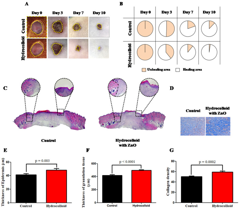
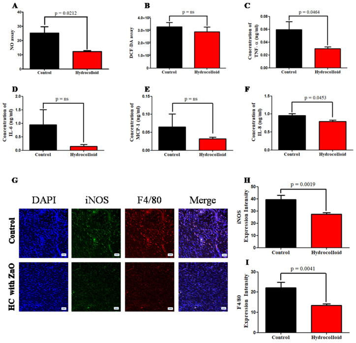
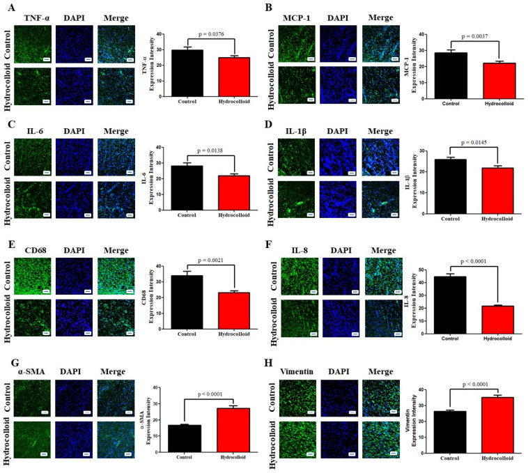
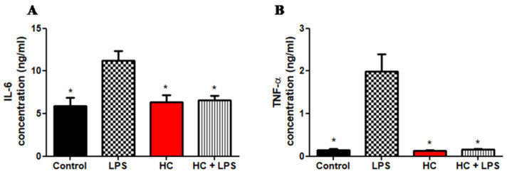
Hydrocolloid dressings are an important method for accelerating wound healing. A combination of a hydrocolloid and nanoparticles (NPs), such as gold (Au), improves the wound healing rate, but Au-NPs are expensive and unable to block ultraviolet (UV) light. Herein, we combined zinc oxide nanoparticles (ZnO-NPs) with hydrocolloids for a less expensive and more effective UV-blocking treatment of wounds. Using Sprague-Dawley rat models, we showed that, during 10-day treatment, a hydrocolloid patch covered with ZnO-NPs (ZnO-NPs-HC) macroscopically and microscopically stimulated the wound healing rate and improved wound healing in the inflammation phase as shown by reducing of pro-inflammatory cytokines (CD68, IL-8, TNF-α, MCP-1, IL-6, IL-1β, and M1) up to 50%. The results from the in vitro models (RAW264.7 cells) also supported these in vivo results: ZnO-NPs-HCs improved wound healing in the inflammation phase by expressing a similar level of pro-inflammatory mediators (TNF-α and IL-6) as the negative control group. ZnO-NPs-HCs also encouraged the proliferation phase of the healing process, which was displayed by increasing expression of fibroblast biomarkers (α-SMA, TGF-β3, vimentin, collagen, and M2) up to 60%. This study provides a comprehensive analysis of wound healing by measuring the biomarkers in each phase and suggests a cheaper method for wound dressing.

Keywords: ZnO-NPs; hydrocolloid; inflammation; proliferation; wound healing.


Conflict of interest statement

The authors declare no conflict of interest.

Figures

Figure 1
Figure 1
A hydrocolloid patch covered with ZnO-NPs accelerates wound healing on both macroscopic and microscopic scales. (A) Representative images (1× magnification) of the wound of the hydrocolloid-with-ZnO-NPs patch and the control group throughout the healing process. (B) Wound healing closure rates of these two groups. The rates presented as a percentage of the initial wound area on day 0. (C) 4× magnification images of H&E staining on day 10 after treatment with a hydrocolloid patch covered with ZnO-NPs and without treatment. (D) 10× magnification images of MT staining on day 10. The blue color indicates the distribution of the collagen. (E) Quantified epidermis thickness gap on day 10. (F) Quantified granulation tissue gap on day 10. (G) Quantified collagen density on day 10.
Figure 2
Figure 2
A hydrocolloid patch covered with ZnO nanoparticles accelerates the inflammatory phase progression and decreases the inflammatory responses of wound healing. (A) Nitrate assay. (B) DCF-DA assay. The concentration gap of pro-inflammation responses on day 10 was confirmed by ELISA between two groups. (C) Tumor necrosis factor-alpha, TNF-α. (D) Interleukin 6, IL-6. (E) Monocyte chemoattractant protein-1, MCP-1. (F) Interleukin 8, IL-8. (G) Fluorescent micrographs showing cytokine staining of macrophage 1 on day 10 of the wound healing process. The quantitative density gap of antibodies of macrophage 1 of the two groups on day 10. (H) Inducible nitric oxide synthase, iNOS. (I) F4/80. Scale bars are 50 µm.
Figure 3
Figure 3
A hydrocolloid patch covered with ZnO nanoparticles bolsters inflammation and proliferative phases, which is confirmed by immunofluorescence staining. Fluorescent micrographs showing different cytokine staining of tissues growing on the posterior wound after 10 days and the quantitative density gap of the inflammation response of the two groups on day 10. (A) Tumor necrosis factor-alpha, TNF-α. (B) Interleukin 8, IL-8. (C) Interleukin 6, IL-6. (D) Interleukin 1 beta, IL-1β. (E) Cluster of Differentiation 68, CD68. (F) Monocyte chemoattractant protein-1, MCP-1. The quantitative density gap of proliferative response of the two groups on day 10. (G) Alpha smooth muscle actin, α-SMA. (H) Vimentin. Scale bars are 50 µm.
Figure 4
Figure 4
A hydrocolloid patch covered with ZnO nanoparticles accelerates the pro-inflammatory property of the RAW 246.7 cell culture. The concentration gap of pro-inflammation responses between four groups (control, LPS, HC, and HC + LPS) was confirmed by ELISA. (A) Interleukin 6, IL-6. (B) Tumor necrosis factor-alpha, TNF-α. The asterisks indicates p < 0.05.
Figure 5
Figure 5
A hydrocolloid patch covered with ZnO nanoparticles accelerates proliferative phases and increases the proliferative responses of wound healing. (A) Representative images of Western blotting. Quantitative densitometry analysis of proliferative response expression. (B) Alpha smooth muscle actin, α-SMA. (C) Vimentin. (D) Transforming growing factor-beta 3, TGF-β3. (E) Collagen III. (F) Collagen I. (G) Fluorescent micrographs showing cytokine staining of macrophage 2 on day 10 of the wound healing process. The quantitative density gap of macrophage 1 antigens of the two groups on day 10. (H) Arginase 1, Arg-1. (I) CD163. Scale bars are 50 µm.

References

    1. Li M., Hou Q., Zhong L., Zhao Y., Fu X. Macrophage Related Chronic Inflammation in Non-Healing Wounds. Front. Immunol. 2021;12:2289. doi: 10.3389/fimmu.2021.681710. - DOI - PMC - PubMed
    1. Guo S., DiPietro L.A. Critical review in oral biology & medicine: Factors affecting wound healing. J. Dent. Res. 2010;89:219–229. doi: 10.1177/0022034509359125. - DOI - PMC - PubMed
    1. Eming S.A., Martin P., Tomic-Canic M. Wound repair and regeneration: Mechanisms, signaling, and translation. Sci. Transl. Med. 2014;6:265sr6. doi: 10.1126/scitranslmed.3009337. - DOI - PMC - PubMed
    1. Xiao T., Yan Z., Xiao S., Xia Y. Proinflammatory cytokines regulate epidermal stem cells in wound epithelialization. Stem Cell Res. Ther. 2020;11:232. doi: 10.1186/s13287-020-01755-y. - DOI - PMC - PubMed
    1. Wilkinson H.N., Hardman M.J. Wound healing: Cellular mechanisms and pathological outcomes: Cellular Mechanisms of Wound Repair. Open Biol. 2020;10:200223. doi: 10.1098/rsob.200223. - DOI - PMC - PubMed

LinkOut - more resources