

Defining Wound Healing Progression in Cetacean Skin: Characteristics of Full-Thickness Wound Healing in Fraser's Dolphins (Lagenodelphis hosei)
- PMID: 35268108
- PMCID: PMC8908859
- DOI: 10.3390/ani12050537
Defining Wound Healing Progression in Cetacean Skin: Characteristics of Full-Thickness Wound Healing in Fraser's Dolphins (Lagenodelphis hosei)
Abstract
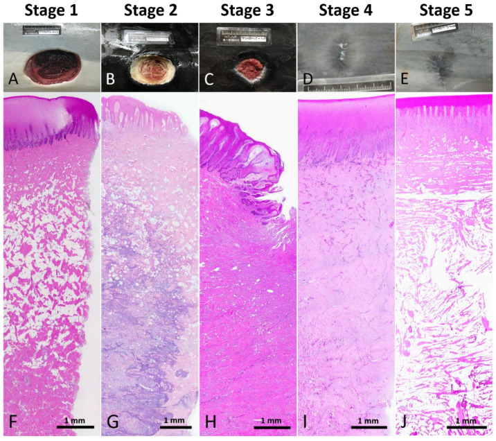
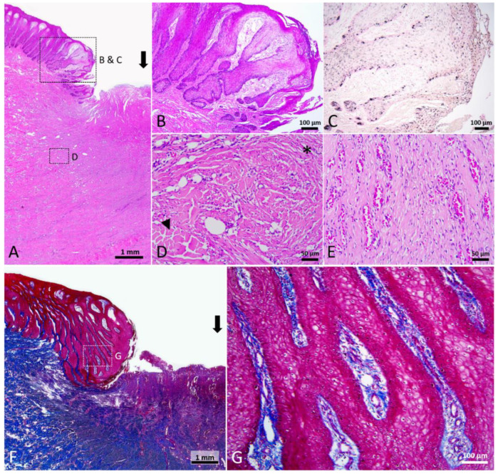
Cetaceans are tight-skinned mammals that exhibit an extraordinary capacity to heal deep soft tissue injuries. However, essential information of large full-thickness wound healing in cetaceans is still lacking. Here, the stages of full-thickness wound healing were characterized in Fraser's dolphins (Lagenodelphis hosei). The skin samples were collected from normal skin and full-thickness cookiecutter shark (Isistius brasiliensis)-bite wounds of stranded carcasses. We defined five stages of wound healing according to macroscopic and histopathological examinations. Wounds in Stage 1 and 2 were characterized by intercellular and intracellular edema in the epidermal cells near the wound edge, mixed inflammatory cell infiltration, and degradation of collagen fibers. In Stage 3 wounds, melanocytes, melanin granules, rete and dermal ridges were noticed in the neo-epidermis, and the adipose tissue in adjacent blubber was replaced by cells and fibers. Wounds in Stage 4 and 5 were characterized by gradual restoration of the normal skin architecture including rete and dermal ridges, collagen bundles, and adipose tissue. These phenomena were quite different from previous studies in terrestrial tight-skinned mammals, and therefore, further in-depth research into the mechanisms of dolphin wound healing would be needed to gain new insights into veterinary and human regenerative medicine.
Keywords: adipose tissue; dolphins; melanocytes; regenerative medicine; rete ridges; wound healing.
Conflict of interest statement
The authors declare no conflict of interest.
Figures


Similar articles
-
Histopathological Study on Collagen in Full-Thickness Wound Healing in Fraser's Dolphins (Lagenodelphis hosei).Animals (Basel). 2023 May 18;13(10):1681. doi: 10.3390/ani13101681. Animals (Basel). 2023. PMID: 37238111 Free PMC article.
-
Successful Repigmentation of Full-Thickness Wound Healing in Fraser's Dolphins (Lagenodelphis hosei).Animals (Basel). 2022 Jun 8;12(12):1482. doi: 10.3390/ani12121482. Animals (Basel). 2022. PMID: 35739819 Free PMC article.
-
Histological characterization of γδ T cells in cutaneous wound healing in Fraser's dolphins (Lagenodelphis hosei).Dev Comp Immunol. 2025 Feb;163:105326. doi: 10.1016/j.dci.2025.105326. Epub 2025 Jan 22. Dev Comp Immunol. 2025. PMID: 39855438
-
Scar-free healing: from embryonic mechanisms to adult therapeutic intervention.Philos Trans R Soc Lond B Biol Sci. 2004 May 29;359(1445):839-50. doi: 10.1098/rstb.2004.1475. Philos Trans R Soc Lond B Biol Sci. 2004. PMID: 15293811 Free PMC article. Review.
-
Rete ridges: Morphogenesis, function, regulation, and reconstruction.Acta Biomater. 2023 Jan 1;155:19-34. doi: 10.1016/j.actbio.2022.11.031. Epub 2022 Nov 24. Acta Biomater. 2023. PMID: 36427683 Review.
Cited by
-
Codamozza-Fluker: The Compelling Case of a Flukeless Fin Whale Traveling Throughout the Mediterranean Sea and the Need for Basin-Wide Conservation Efforts.Ecol Evol. 2025 May 21;15(5):e71313. doi: 10.1002/ece3.71313. eCollection 2025 May. Ecol Evol. 2025. PMID: 40406589 Free PMC article.
-
Histopathological Study on Collagen in Full-Thickness Wound Healing in Fraser's Dolphins (Lagenodelphis hosei).Animals (Basel). 2023 May 18;13(10):1681. doi: 10.3390/ani13101681. Animals (Basel). 2023. PMID: 37238111 Free PMC article.
-
Successful Repigmentation of Full-Thickness Wound Healing in Fraser's Dolphins (Lagenodelphis hosei).Animals (Basel). 2022 Jun 8;12(12):1482. doi: 10.3390/ani12121482. Animals (Basel). 2022. PMID: 35739819 Free PMC article.
-
Cetacean epidermal specialization: A review.Anat Histol Embryol. 2022 Sep;51(5):563-575. doi: 10.1111/ahe.12829. Epub 2022 Jun 27. Anat Histol Embryol. 2022. PMID: 35758554 Free PMC article. Review.
-
Proteome profiling reveals opportunities to investigate biomarkers of oxidative stress and immune responses in blubber biopsies from free-ranging baleen whales.Conserv Physiol. 2024 Aug 19;12(1):coae059. doi: 10.1093/conphys/coae059. eCollection 2024. Conserv Physiol. 2024. PMID: 39161698 Free PMC article.
References
-
- Häkkinen L., Larjava H., Koivisto L. Granulation tissue formation and remodeling. Endod. Top. 2011;24:94–129. doi: 10.1111/etp.12008. - DOI
Grants and funding
LinkOut - more resources
Full Text Sources
