Review
. 2022 Feb 24;12(5):573.
doi: 10.3390/ani12050573.

Relevance of Aquaporins for Gamete Function and Cryopreservation

Affiliations
Review

Relevance of Aquaporins for Gamete Function and Cryopreservation

Ariadna Delgado-Bermúdez et al. Animals (Basel). .

Abstract

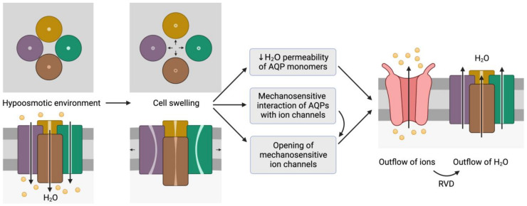
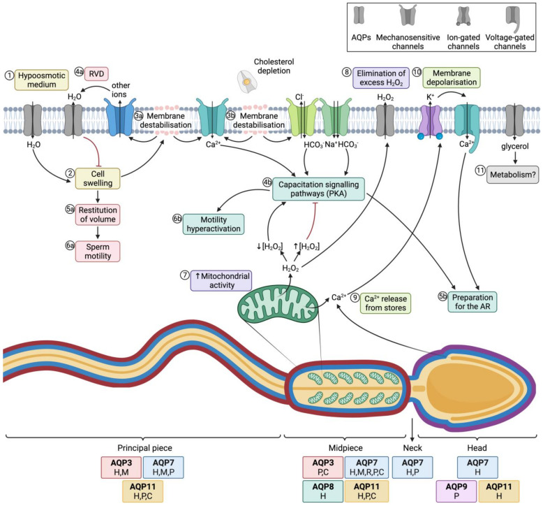
The interaction between cells and the extracellular medium is of great importance, and drastic changes in extracellular solute concentrations drive water movement across the plasma membrane. Aquaporins (AQPs) are a family of transmembrane channels that allow the transport of water and small solutes across cell membranes. Different members of this family have been identified in gametes. In sperm, they are relevant to osmoadaptation after entering the female reproductive tract, which is crucial for sperm motility activation and capacitation and, thus, for their fertilizing ability. In addition, they are relevant during the cryopreservation process, since some members of this family are also permeable to glycerol, one of the most frequently used cryoprotective agents in livestock. Regarding oocytes, AQPs are very important in their maturation but also during cryopreservation. Further research to define the exact sets of AQPs that are present in oocytes from different species is needed, since the available literature envisages certain AQPs and their roles but does not provide complete information on the whole set of AQPs. This is of considerable importance because, in sperm, specific AQPs are known to compensate the role of non-functional members.

Keywords: cryopreservation; mammals; oocyte; physiology; sperm; water channels.


Conflict of interest statement

The authors declare no conflict of interest. The funders had no role in the design of the study; in the collection, analyses, or interpretation of data; in the writing of the manuscript; or in the decision to publish the results.

Figures

Figure 2
Figure 2
Representation of the mechanosensitive mechanism of aquaporins (AQPs). When cells are in a hypoosmotic environment, water enters them in response to the concentration gradient of solutes. Due to the entry of water, cells may undergo swelling, which can cause the distortion of AQP structures and thus compromise water permeability. In addition, AQPs may have mechanosensitive interactions with ion channels that can trigger their opening to trigger the outflow of ions. In fact, some ion channels are mechanosensitive and open in response to cell swelling, regardless of AQP signaling. The outflow of ions generates a driving force that elicits the outflow of water; this process is also known as regulatory volume decrease (RVD). This schematic representation is inspired by the model proposed by Hill et al. [20] and its representation by Ozu et al. [16].
Figure 4
Figure 4
Aquaporins (AQPs) and sperm function after ejaculation. (1) After entering the female tract, sperm encounter a hypoosmotic medium, which causes water influx and thus (2) cell swelling. (3a) The increase of cell volume causes membrane destabilization (which affects the sperm plasma membrane, but also alters mitochondrial and acrosomal membranes). (4a) As a consequence, mechanosensory ion channels are open and outflowing ion currents activate regulatory volume decrease (RVD) events, such as (4a) water outflow. (5a) Cell volume is then restituted, which allows (6a) normal sperm motility. (3b) In the female reproductive tract, membrane destabilization occurs in conjunction with cholesterol depletion. The resulting membrane reorganization elicits the entrance of calcium and bicarbonate through different ion channels, which activates capacitation signaling pathways (4b) that involve the activation of protein kinase A (PKA). (5b) Downstream events prepare the spermatozoon for the acrosome reaction (AR) and (6b) drive hyperactivated motility. (7) In this context, mitochondrial activity is elevated and reactive oxygen species (ROS), such as H2O2, are produced. This molecule at low concentrations is essential to elicit capacitation, but at high concentrations it acts as an inhibitor of this process. (8) Aquaporins (AQPs) play an essential role, allowing the outflow of the excess amounts of H2O2 towards the extracellular medium. (9) Another downstream event of the capacitation signaling pathway consists of the release of Ca2+ from intracellular stores, which triggers the opening of calcium-activated K+ channels that causes, in turn, plasma membrane hyperpolarization (10); membrane hyperpolarization subsequently opens voltage-gated Ca2+ channels. This increase in intracellular Ca2+ is crucial for acrosome reaction [64,65] (11) Glycerol transport through GLPs can be used by metabolic pathways. The sperm regions where AQPs have been identified in different mammalian species are highlighted in the diagram. Boxes indicate the species in which each AQP has been identified (C, cattle; P, pig; H, human; M, mouse; R, rat).
Figure 1
Figure 1
Structural characteristics of the family of aquaporins (AQPs). (A) Aquaporins present six transmembrane α-helices (TM1-6) that are connected through loops (A–E). Loops B and E are half-transmembrane helices oriented towards the center of the pore and present an NPA (asparagine, proline, alanine) motif that is highly conserved. In loop E, there is also a highly conserved arginine (R). Some residues from loop B have been suggested to be involved in AQPs’ mechanosensitivity (light residues). Transmembrane helices TM1, TM2, TM4 and TM5 interact with the TM of the adjacent monomers from an AQP tetramer (dark dot lines). (B) Each monomer folds in an hour-glass conformation, and the region near the R from loop E is formed by aromatic residues. This region is known as the aromatic/arginine (ar/R) region but also as a selectivity filter since it forms the narrowest point of the AQP pore. After folding, the two NPA motifs are in the same region, which is also highly conserved. (C) The quaternary structure of AQPs consists of the formation of tetramers, where each monomer has its own functional pore. This figure is based on PDB structure 6QZJ from AQP7.
Figure 3
Figure 3
Classification of aquaporins (AQPs) based on their homology and specific permeability to different molecules. The three main groups into which AQPs are classified are: orthodox AQPs (blue), aquaglyceroporins (GLPs; yellow) and superaquaporins (superAQPs; red). The structural characteristics of these three groups are graphically represented. In addition to the classical classification, some AQPs present permeability to H2O2 and they are considered peroxiporins (green). Similarly, some members of this family of proteins are considered ammoniaporins (pink) since they are permeable to ammonia and/or to NH3. Finally, certain members present permeability to other molecules, such as Cl (dark red), metalloids (grey) or lactate (orange). NPA motif (asparagine, proline, alanine); NPC motif (asparagine, proline, cysteine). Image based on [21,22,23].
Figure 5
Figure 5
Blocking of aquaporins (AQPs) through mercury causes cell swelling. From a structural point of view, the plasma membrane is disrupted due to stretching and mitochondrial and acrosomal membranes swell. Axoneme structure is also altered, which underlies sperm motility impairment. Mitochondria show collapsed cristae, which decreases energy production. In addition, mitochondrial membrane disruption causes a decrease in mitochondrial membrane potential, release of Ca2+ and higher levels of reactive oxygen species (ROS). Since AQPs are permeable to H2O2, their blockade avoids detoxification and ROS accumulate intracellularly. As previously stated, small amounts of ROS are needed for capacitation, but high concentrations of these molecules have an inhibitory effect on capacitation signaling pathways, which can be assessed through the levels of tyrosine phosphorylation. On the other hand, plasma membrane disruption increases its permeability, which reduces its hyperpolarization, an essential event to trigger the acrosome reaction. Finally, disruption of the acrosomal membrane triggers premature exocytosis and thus sperm cannot undergo a physiological acrosome reaction.
Figure 6
Figure 6
Potential mechanism of translational regulation of aquaporins (AQPs) in mammalian oocytes. Mechanosensitive transmembrane proteins, in response to osmotic stress, trigger signaling pathways that involve protein kinase C (PKC) and phosphoinositide 3-kinase (PI3K). These kinases activate Aurora A, which phosphorylates cytoplasmic polyadenylation element-binding protein (CPEB) from translationally inactive (dormant) mRNAs. These mRNAs then become translationally active, and the resulting AQP7 protein is translocated to the plasma membrane. It has not yet been elucidated whether this mechanism is also involved in the translational regulation of the other AQPs identified in mammalian oocytes (P, pig; Ho, horse; C, cattle; D, dog; H, human; M, mouse; R, rat).

Similar articles

Cited by

References

    1. Watson H. Biological membranes. Essays Biochem. 2015;59:43–70. doi: 10.1042/bse0590043. - DOI - PMC - PubMed
    1. Denker B.M., Smith B.L., Kuhajda F.P., Agre P. Identification, purification, and partial characterization of a novel Mr 28,000 integral membrane protein from erythrocytes and renal tubules. J. Biol. Chem. 1988;263:15634–15642. doi: 10.1016/S0021-9258(19)37635-5. - DOI - PubMed
    1. Smith B.L., Agre P. Erythrocyte Mr 28,000 transmembrane protein exists as a multisubunit oligomer similar to channel proteins. J. Biol. Chem. 1991;266:6407–6415. doi: 10.1016/S0021-9258(18)38133-X. - DOI - PubMed
    1. Preston G.M., Carroll T.P., Guggino W.B., Agre P. Appearance of water channels in Xenopus oocytes expressing red cell CHIP28 protein. Science. 1992;256:385–387. doi: 10.1126/science.256.5055.385. - DOI - PubMed
    1. Törnroth-Horsefield S., Hedfalk K., Fischer G., Lindkvist-Petersson K., Neutze R. Structural insights into eukaryotic aquaporin regulation. FEBS Lett. 2010;584:2580–2588. doi: 10.1016/j.febslet.2010.04.037. - DOI - PubMed

LinkOut - more resources