

The Arrangement of the Peripheral Olfactory System of Pleuragramma antarcticum: A Well-Exploited Small Sensor, an Aided Water Flow, and a Prominent Effort in Primary Signal Elaboration
- PMID: 35268231
- PMCID: PMC8909514
- DOI: 10.3390/ani12050663
The Arrangement of the Peripheral Olfactory System of Pleuragramma antarcticum: A Well-Exploited Small Sensor, an Aided Water Flow, and a Prominent Effort in Primary Signal Elaboration
Abstract
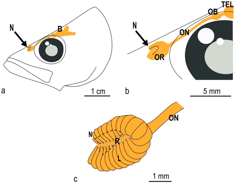
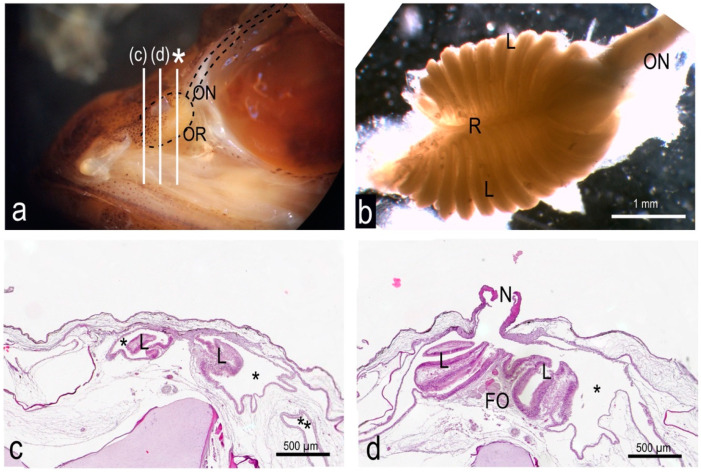
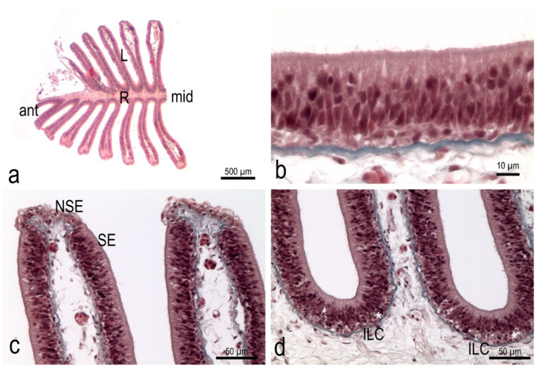
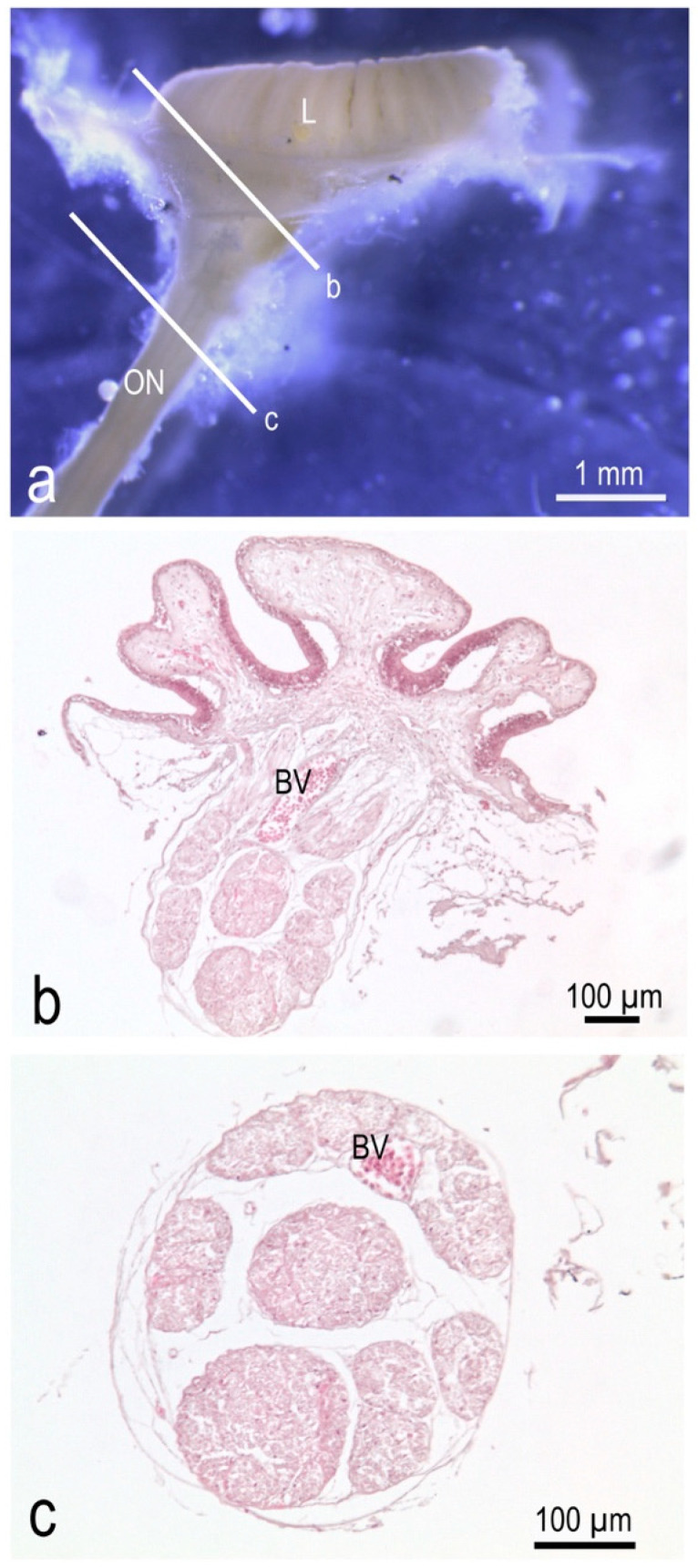
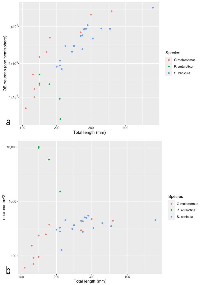
The olfactory system is constituted in a consistent way across vertebrates. Nasal structures allow water/air to enter an olfactory cavity, conveying the odorants to a sensory surface. There, the olfactory neurons form, with their axons, a sensory nerve projecting to the telencephalic zone-named the olfactory bulb. This organization comes with many different arrangements, whose meaning is still a matter of debate. A morphological description of the olfactory system of many teleost species is present in the literature; nevertheless, morphological investigations rarely provide a quantitative approach that would help to provide a deeper understanding of the structures where sensory and elaborating events happen. In this study, the peripheral olfactory system of the Antarctic silverfish, which is a keystone species in coastal Antarctica ecosystems, has also been described, employing some quantitative methods. The olfactory chamber of this species is connected to accessory nasal sacs, which probably aid water movements in the chamber; thus, the head of the Antarctic silverfish is specialized to assure that the olfactory organ keeps in contact with a large volume of water-even when the fish is not actively swimming. Each olfactory organ, shaped like an asymmetric rosette, has, in adult fish, a sensory surface area of about 25 mm2, while each olfactory bulb contains about 100,000 neurons. The sensory surface area and the number of neurons in the primary olfactory brain region show that this fish invests energy in the detection and elaboration of olfactory signals and allow comparisons among different species. The mouse, for example-which is considered a macrosmatic vertebrate-has a sensory surface area of the same order of magnitude as that of the Antarctic silverfish, but ten times more neurons in the olfactory bulb. Catsharks, on the other hand, have a sensory surface area that is two orders of magnitude higher than that of the Antarctic silverfish, while the number of neurons has the same order of magnitude. The Antarctic silverfish is therefore likely to rely considerably on olfaction.
Keywords: Antarctic silverfish; fish olfaction; isotropic fractionator; olfactory bulb; olfactory nerve; olfactory rosette.
Conflict of interest statement
The authors declare no conflict of interest.
Figures


Similar articles
-
Olfaction in fish.Prog Neurobiol. 1975;5(4):271-335. doi: 10.1016/0301-0082(75)90014-3. Prog Neurobiol. 1975. PMID: 830087 Review.
-
Divergence of brain and retinal anatomy and histology in pelagic antarctic notothenioid fishes of the sister taxa Dissostichus and Pleuragramma.J Morphol. 2011 Apr;272(4):419-41. doi: 10.1002/jmor.10926. Epub 2011 Jan 18. J Morphol. 2011. PMID: 21246598
-
Quantification of neurons in the olfactory bulb of the catsharks Scyliorhinus canicula (Linnaeus, 1758) and Galeus melastomus (Rafinesque, 1810).Zoology (Jena). 2020 Aug;141:125796. doi: 10.1016/j.zool.2020.125796. Epub 2020 Apr 26. Zoology (Jena). 2020. PMID: 32464514
-
Brain and sense organ anatomy and histology of the Falkland Islands mullet, Eleginops maclovinus (Eleginopidae), the sister group of the Antarctic notothenioid fishes (Perciformes: Notothenioidei).J Morphol. 2008 Jan;269(1):84-103. doi: 10.1002/jmor.10571. J Morphol. 2008. PMID: 17902153
-
Zonal organization of the mammalian main and accessory olfactory systems.Philos Trans R Soc Lond B Biol Sci. 2000 Dec 29;355(1404):1801-12. doi: 10.1098/rstb.2000.0736. Philos Trans R Soc Lond B Biol Sci. 2000. PMID: 11205342 Free PMC article. Review.
Cited by
-
Ultramicroscopic organization of the exterior olfactory organ in Anguilla vulgaris in relation to its spawning migration.Open Vet J. 2024 Jan;14(1):512-524. doi: 10.5455/OVJ.2024.v14.i1.46. Epub 2024 Jan 31. Open Vet J. 2024. PMID: 38633152 Free PMC article.
-
Anatomy and histology of the olfactory organ of the javelin goby Synechogobius hasta (Gobiiformes, Gobiidae).Appl Microsc. 2024 Dec 17;54(1):12. doi: 10.1186/s42649-024-00105-z. Appl Microsc. 2024. PMID: 39688788 Free PMC article.
References
-
- Burne R.H. The Anatomy of the Olfactory Organ of Teleostean Fishes. Proc. Zool. Soc. Lond. 1909;2:610–663.
-
- Døving K.B. Functional Properties of the Fish Olfactory System. Prog. Sens. Physiol. 1986;6:39–104.
-
- Helfman G.S., Collette B.B., Facey D.E., Bowen B.W. The Diversity of Fishes: Biology, Evolution, and Ecology. 2nd ed. Wiley-Blackwell, A John Wiley & Sons, Ltd.; Hoboken, NJ, USA: 2009.
-
- Kasumyan A.O. The Olfactory System in Fish: Structure, Function, and Role in Behavior. J. Ichthyol. 2004;44:S180–S223.
-
- Yamamoto M. Comparative Morphology of the Peripheral Olfactory Organ in Teleosts. In: Hara T.J., editor. Chemoreception in Fishes. Elsevier Scientific Pub. Co.; Amsterdam, The Netherlands: 1982. pp. 39–59.
Grants and funding
LinkOut - more resources
Full Text Sources
