. 2022 Mar 7;12(5):664.
doi: 10.3390/ani12050664.

The Characteristics and Distribution of α2D-, α2B- and α2C-Adrenoceptor Subtypes in Goats

Affiliations

The Characteristics and Distribution of α2D-, α2B- and α2C-Adrenoceptor Subtypes in Goats

Ming Xu et al. Animals (Basel). .

Abstract

α2-Adrenegic receptors (α2Rs) are important presynaptic modulators of central noradrenergic function (auto receptors) and postsynaptic mediators of many of the widespread effects of catecholamines and related drugs. Studies have shown that ruminants (such as goats and cattle) express special α2DR subtypes in addition to α2BR and α2CR. Real-time quantitative PCR and Western blotting were used to investigate the distribution and density of α2R in different nuclei of the goat central nervous system, selected regions of the spinal cord (L4-L6), and in various peripheral tissues. α2-AR subtype-specific antibodies were injected intrathecally and intracerebroventricularly into the tested goats to block the corresponding subtype of receptors. Pain threshold and physiological parameters were evaluated to explore the functional characteristics of α2BR, α2CR and α2DR in goats. Our results suggest that the expression of the mRNAs and proteins of all three α2R subtypes are widely but unevenly distributed in the goat CNS and peripheral tissues. Furthermore, α2DR plays a more important role in α2R-mediated analgesia in goats than α2BR and α2CR, whereas α2CR activation exerts a greater effect on body temperature than α2BR and α2DR.

Keywords: analgesia; goat; intracerebroventricular injection; intrathecal injection; α2 adrenoceptor subtypes.


Conflict of interest statement

The authors declare no conflict of interest. The funders had no role in the design of the study; in the collection, analyses, or interpretation of data; in the writing of the manuscript, or in the decision to publish the results.

Figures

Figure 1
Figure 1
Brain section: H0: the horizontal zero plane. The 0 plane represents the interaural line. A35, A25, A20, A15, A10 and A5 show transverse planes located 35, 30, 25, 20, 15, 10 and 5 mm rostral to the interaural line, and P5, P10, P15 and P20 show transverse planes located 5, 10, 15 and 20 mm caudal to the interaural line, respectively. S1-8, take the base edge of the olfactory bulb as the starting line, and make a sagittal plane, each 5mm. The locations of nuclei or areas in the brain blocks are presented at the top of the figure. The nuclei and areas identified include the caudate putamen nucleus (Cpu), nucleus accumbens (Acb), paraventricular thalamus (PVT), hypothalamic paraventricular nucleus (PVN), arcuate nucleus (Arc), amygdala (Amy), ventromedial hypothalamic nucleus (VMH), hippocampus (Hip), thalamic nucleus submedius (Sm), habenular nucleus (Hb), parafascicular nucleus (Pf), substantia nigra (SN), periaqueductal gray (PAG), parabrachial nucleus (PBN), locus coeruleus (LC)), nucleus raphe magnus (NRM), gigantocellular reticular nucleus (GRN) and nucleus of the solitary tract (NTS).
Figure 2
Figure 2
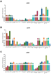
Real-time qPCR analyses of α2BR (A), α2CR (B) and α2DR (C) mRNA abundances in the Cpu, Acb, PVT, PVN, Arc, Amy, VMH, Hip, Sm, Hb, Pf, SN, PAG, PBN, LC, NRM, GRN, NTS, SCDH, Myo, liver, spleen, lung, kidney, aorta, SVC, IVC, arterioles, venules and Mφ of goats. The results were normalized to GAPDH as a housekeeping gene and are presented as the means ± SD (n = 3). Bars with different letters differ (p < 0.05).
Figure 3
Figure 3
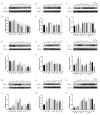
Western blot analyses of α2DR, α2BR and α2CR protein abundances in the Cpu, Acb, PVT, PVN, Arc, Amy, VMH, Hip, Sm, Hb, Pf, SN, PAG, PBN, LC, NRM, GRN, NTS, SCDH, Myo, liver, spleen, lung, kidney, aorta, SVC, IVC, arterioles, venules and Mφ of goats. (AC) Representative bands for α2BR and GAPDH. (GI) Representative bands for α2CR and GAPDH. (MO) Representative bands for α2DR and GAPDH. (DF,JL,PR) Densitometry analyses of protein bands. Data were normalized to GAPDH as the housekeeping protein and are presented as the means ± SD (n = 3). Bars with different letters differ (p < 0.05) Original Western blot figures are in Figure S5.
Figure 4
Figure 4
The effect of different α2R subtypes expressed in the spinal cord on the pain threshold of goats. Potassium iontophoresis (A) and HWT (B) thresholds of the tested goats represent the effect of the intrathecal injection of α2R subtype antibody on the potassium iontophoresis and HWT thresholds of goats, respectively. Data are presented as the means ± SD (n = 6 in each group). * p < 0.05 compared with the IgG group and ** p < 0.01 compared with the IgG group.
Figure 5
Figure 5
Monitoring of physiological indices in the goats from each group. (AG) Effects of different α2R subtypes expressed in the goat spinal cord on HR, RESP, SPO2, NIBP-SYS, NIBP-MEAN, NIBP-DIA and body temperature. (H) Decreased body temperature in the goats of each group. Data are presented as the means ± SD (n = 6 in each group).
Figure 6
Figure 6
The effects of different α2R subtypes expressed in the brain on the pain threshold of goats. (A,B) Effects of the intrathecal injection of α2R subtype-specific antibodies on potassium iontophoresis and the HWT threshold of goats, respectively. Data are presented as the means ± SD (n = 6 in each group). ** p < 0.01 compared with the IgG group. 3.6 Effects of α2Rs expressed in the brain on the physiological indices of goats.
Figure 7
Figure 7
Effects of different α2R subtypes expressed in the brain on the physiological indices of goats. (AG) Effects of different α2-AR subtypes expressed in the goat brain on HR, RESP, SPO2, NIBP-SYS, NIBP-MEAN, NIBP-DIA and body temperature. (H) The range of decrease in the body temperature of goats from each group. Data are presented as the means ± SD (n = 6 in each group). ** p < 0.01 compared with the IgG group.

Similar articles

Cited by

References

    1. Bantel C., Maze M., Stone L., Wilcox G. Encyclopedia of Pain. Springer; Berlin/Heidelberg, Germany: 2007. Alpha(α) 2-Adrenergic Agonists in Pain Treatment; pp. 58–61.
    1. Chan A.K., Cheung C.W., Chong Y.K. Alpha-2 agonists in acute pain management. Expert Opin. Pharmacother. 2010;11:2849–2868. doi: 10.1517/14656566.2010.511613. - DOI - PubMed
    1. McKelvey D., Hollingshead K.W. Veterinary Anesthesia and Analgesia. 3rd ed. Mosby; St Louis, MO, USA: 2003.
    1. Shah Z., Ding M.-X., Hu M.-L. A review on the current use of alpha2 agonists in small ruminants. Kafkas Univ. Vet. Fak. Derg. 2014;20:633–639.
    1. Carroll G.L., Hartsfield S.M. General anesthetic techniques in ruminants. Vet. Clin. N. Am. Food Anim. Pract. 1996;12:627–661. doi: 10.1016/S0749-0720(15)30391-1. - DOI - PubMed