Review
. 2022 Feb 22;11(5):1161.
doi: 10.3390/jcm11051161.

Baroreceptors in the Aortic Arch and Their Potential Role in Aortic Dissection and Aneurysms

Affiliations
Review

Baroreceptors in the Aortic Arch and Their Potential Role in Aortic Dissection and Aneurysms

Benedikt Reutersberg et al. J Clin Med. .

Abstract

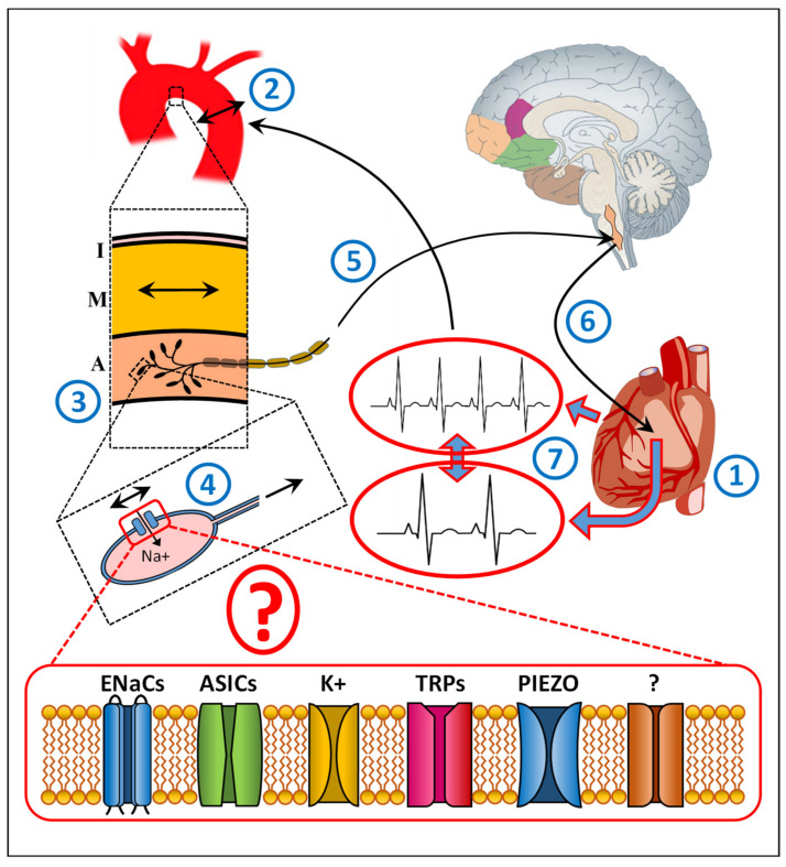
The arterial baroreflex is a key autonomic regulator of blood pressure whose dysfunction has been related to several cardiovascular diseases. Changes in blood pressure are sensed by specific mechanosensory proteins, called baroreceptors, particularly located in the outer layer of the carotid sinus and the inner curvature of the aortic arch. The signal is propagated along the afferent nerves to the central nervous system and serves as negative feedback of the heart rate. Despite extensive research, the precise molecular nature of baroreceptors remains elusive. Current knowledge assumes that baroreceptors are ion channels at the nerve endings within the outer layer of the arteries. However, the evidence is based mainly on animal experiments, and the specific types of mechanosensitive receptors responsible for the signal transduction are still unknown. Only a few studies have investigated mechanosensory transmission in the aortic arch. In addition, although aortic dissection, and particularly type A involving the aortic arch, is one of the most life-threatening cardiovascular disorders, there is no knowledge about the impact of aortic dissection on baroreceptor function. In this review, we aim not to highlight the regulation of the heart rate but what mechanical stimuli and what possible ion channels transfer the corresponding signal within the aortic arch, summarizing and updating the current knowledge about baroreceptors, specifically in the aortic arch, and the impact of aortic pathologies on their function.

Keywords: aortic dissection; baroreceptors; ion channels; mechanotransduction.


Conflict of interest statement

The authors declare no conflict of interest.

Figures

Figure 1
Figure 1
A schematic diagram illustrating the baroreceptor reflex of the aortic arch and the involvement of ion channels. The heart (1) regulates its rate according to the body’s needs. Changes in heart rate (1) alternate blood pressure. The corresponding changes exercise mechanical forces on the aortic wall (2). The changes in shear stress are sensed by specific mechanoreceptors, called baroreceptors, at the endings of sensory nerves innervating the outer wall (adventitia) of aortic arch (3). These mechanical deformations are affecting the mechanosensitive baroreceptors (4), assumed to be specific ion channels, which in turn provide an action potential in the corresponding nerve terminal, which is propagated along the afferent nerve fibers (5) toward the nodose ganglia in the brainstem. The incoming signal is part of the negative feedback system (6) that contributes to the regulation of heart rate (7) to restore heart rate to its normal level. I, Intima, the inner aortic wall layer containing endothelial cells; M, Media, the middle aortic layer containing particularly smooth muscle cells, regulating the vascular tone; A, Adventitia, the outer aortic layer containing the mechanosensory baroreceptors. ENaCs, Epithelial sodium channels, containing four subunits (α, β, γ, and δ) [7,8,9,10,11,12,13,14,15,16]; ASICs, Acid-sensing ion channels [12,13,14,15,16,17,18,19,20,21,22,23,24] belonging to the DEG/ENaC superfamily with at least seven ASIC isoforms; K+, Stretch-sensitive K+ channels, both calcium (Ca)-insensitive and Ca-sensitive [25,26]; TRP, transient receptor potential ion channels [6,27,28,29,30,31]: TRPA (ankyrin 1), TRPC (canonical 1–7), TRPM (melastatin 1–8), TRPN (NOMPC-like 1), TRPP (polycystin 2, 3, 5), TRPML (mucolipin 1–3), and TRPV (vanilloid 1–6): TRPV1, TRPC1, and TRPC3-7; PIEZO1, 2, mechanosensitive ion channels [32,33,34].

References

    1. Levy M.N. Neural control of cardiac function. Baillieres Clin. Neurol. 1997;6:227–244. - PubMed
    1. Talman W.T., Kelkar P. Neural control of the Heart Central and peripheral. Neurol. Clin. 1993;11:239–256. doi: 10.1016/S0733-8619(18)30151-8. - DOI - PubMed
    1. Okada Y., Galbreath M.M., Shibata S., Jarvis S.S., VanGundy T.B., Meier R.L., Vongpatanasin W., Levine B.D., Fu Q. Relationship between sympathetic baroreflex sensitivity and arterial stiffness in elderly men and women. Hypertension. 2012;59:98–104. doi: 10.1161/HYPERTENSIONAHA.111.176560. - DOI - PMC - PubMed
    1. Sun H., Li D.P., Chen S.R., Hittelman W.N., Pan H.L. Sensing of blood pressure increase by transient receptor potential vanilloid 1 receptors on baroreceptors. J. Pharmacol. Exp. Ther. 2009;331:851–859. doi: 10.1124/jpet.109.160473. - DOI - PMC - PubMed
    1. Hainsworth R. Cardiovascular control from cardiac and pulmonary vascular receptors. Exp. Physiol. 2014;99:312–319. doi: 10.1113/expphysiol.2013.072637. - DOI - PubMed

LinkOut - more resources