Review
. 2022 Feb 25;11(5):1251.
doi: 10.3390/jcm11051251.

Diagnosis of Congenital Uterine Abnormalities: Practical Considerations

Affiliations
Review

Diagnosis of Congenital Uterine Abnormalities: Practical Considerations

Kanna Jayaprakasan et al. J Clin Med. .

Abstract

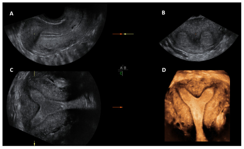
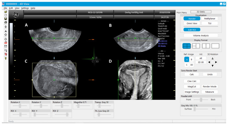
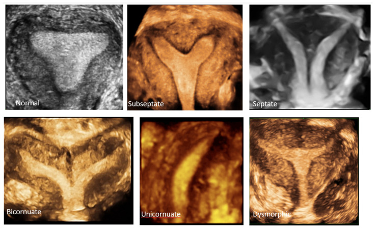
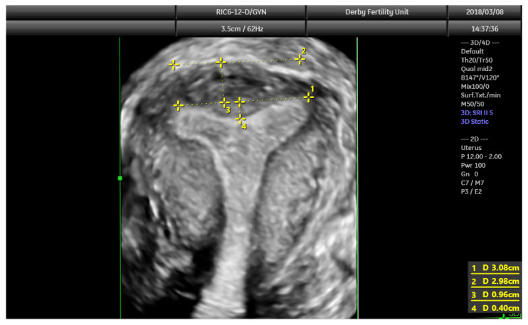
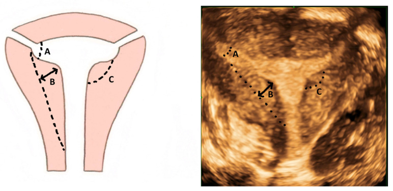
As most congenital uterine abnormalities are asymptomatic, the majority of them are detected incidentally. While most women with uterine anomalies have a normal reproductive outcome, some may experience adverse reproductive outcomes. Accurate diagnosis and correct classification help in the appropriate counselling of women about their potential reproductive prognosis and risks and for planning any intervention. Evaluation of the internal and external contours of the uterus is the key in making a diagnosis and correctly classifying a uterine anomaly. Considering this, the gold standard test has been the combined laparoscopy and hysteroscopy historically, albeit invasive. However, 3D ultrasound has now become the diagnostic modality of choice for uterine anomalies due to its high degree of diagnostic accuracy, less invasive nature and it being comparatively less expensive. While 2D ultrasound and HSG are adequate for screening for uterine anomalies, MRI and combined laparoscopy and hysteroscopy are reserved for diagnosing complex Mullerian anomalies. Imaging for renal anomalies is recommended if a uterine anomaly is diagnosed.

Keywords: 3D ultrasound; MRI; Mullerian duct; congenital uterine anomalies; diagnosis; hysteroscopy; laparoscopy.


Conflict of interest statement

The authors declare no conflict of interest.

Figures

Figure 1
Figure 1
Development of uterus and vagina (adapted from [3]).
Figure 2
Figure 2
ESHRE/ESGE classification of uterine anomalies (adapted from [6]).
Figure 3
Figure 3
Hysterosalpingogram showing acute angle in a septate uterus (A) and wider angle in a bicornuate uterus (B).
Figure 4
Figure 4
Longitudinal section of subseptate uterus in midsagittal plane (A); transverse plane of a subseptate uterus showing two endometrial echoes (B); transverse plane of a bicornuate uterus showing two endometrial echoes (C); transverse plane of uterus didelphys showing two uterine bodies (D).
Figure 5
Figure 5
Unicornuate uterus: 2D transverse view showing only one uterine angle (shoulder)—Right (A); a small left rudimentary uterine horn (B) and banana-shaped uterine cavity on a 3D coronal plane (C).
Figure 6
Figure 6
3D ultrasound scan of a subseptate uterus showing simultaneous display of longitudinal plane (A), transverse plane showing two endometrial echoes (B), coronal plane (C), unique for 3D ultrasound and Rendered view of coronal plane demonstrating subseptate uterus (D).
Figure 7
Figure 7
3D multiplanar view with rendering box using 4D view software: Green line of the rendering box placed on top and at the level of endometrial cavity in the longitudinal plane (A); transverse plane (B), Coronal plane (C) and rendered coronal view of uterus in the bottom right corner (D).
Figure 8
Figure 8
3D coronal plane of uterus: normal uterus, subseptate uterus, septate uterus, bicornuate, unicornuate uterus and dysmorphic (T-shaped) uterus.
Figure 9
Figure 9
3D coronal plane of uterus with assessments: interostial line (measurement 1); a parallel line along the serosal surface (measurement 2); uterine wall thickness (measurement 3) and septal indentation length. This uterus is not septate, but may be classified as arcuate uterus, which has no clinical relevance based on the recent ESHRE and ASRM guidelines.
Figure 10
Figure 10
Criteria for diagnosing T-shaped uterus according to CUME ([22]). A—T-angle ≤ 40°; B—lateral indentation depth ≥ 7 mm; C—lateral indentation angle ≤ 130°.

References

    1. Chan Y.Y., Jayaprakasan K., Zamora J., Thornton J.G., Raine-Fenning N., Coomarasamy A. The prevalence of congenital uterine anomalies in unselected and high-risk populations: A systematic review. Hum. Reprod. Update. 2011;17:761–771. doi: 10.1093/humupd/dmr028. - DOI - PMC - PubMed
    1. Saravelos S.H., Cocksedge K.A., Li T.C. Prevalence and diagnosis of congenital uterine anomalies in women with reproductive failure: A critical appraisal. Hum. Reprod. Update. 2008;14:415–429. doi: 10.1093/humupd/dmn018. - DOI - PubMed
    1. Lin P.C., Bhatnagar K.P., Nettleton G.S., Nakajima S.T. Female genital anomalies affecting reproduction. Fertil. Steril. 2002;78:899–915. doi: 10.1016/S0015-0282(02)03368-X. - DOI - PubMed
    1. Sadler T.W. Langman’s Medical Embryology. 12th ed. Lippincott Williams & Wilkins; Philadelphia, PA, USA: 2012. pp. 243–259.
    1. AFS. The American Fertility Society classifications of adnexal adhesions, distal tubal occlusion, tubal occlusion secondary to tubal ligation, tubal pregnancies, mullerian anomalies and intrauterine adhesions. Fertil. Steril. 1988;49:944–955. - PubMed