Review
. 2022 Feb 26;11(5):1285.
doi: 10.3390/jcm11051285.

Developing a Mechanistic Approach to Sudden Death Prevention in Mitral Valve Prolapse

Affiliations
Review

Developing a Mechanistic Approach to Sudden Death Prevention in Mitral Valve Prolapse

Brian P Kelley et al. J Clin Med. .

Abstract

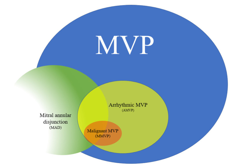
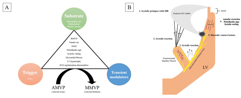
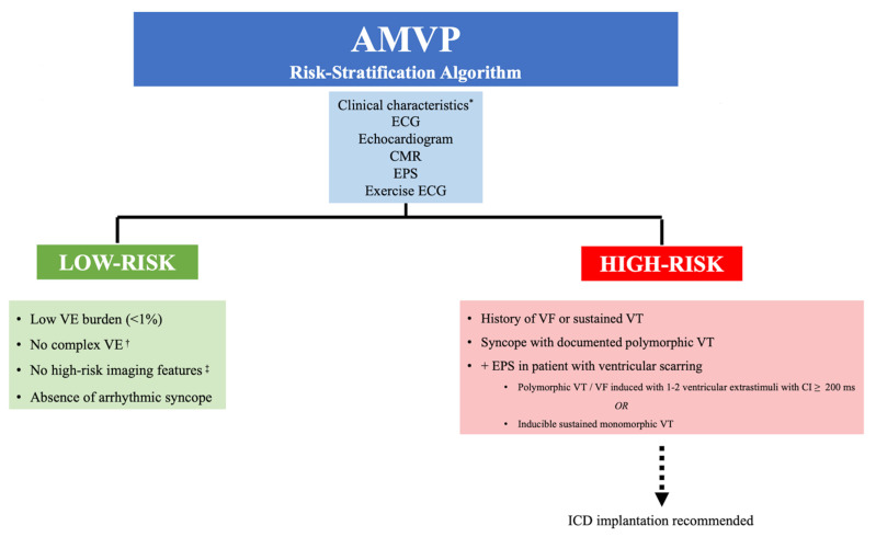
Sudden cardiac death (SCD) from ventricular fibrillation (VF) can occur in mitral valve prolapse (MVP) in the absence of other comorbidities including mitral regurgitation, heart failure or coronary disease. Although only a small proportion with MVP are at risk, it can affect young, otherwise healthy adults, most commonly premenopausal women, often as the first presentation of MVP. In this review, we discuss arrhythmic mechanisms in MVP and mechanistic approaches for sudden death risk assessment and prevention. We define arrhythmogenic or arrhythmic MVP (AMVP) as MVP associated with complex and frequent ventricular ectopy, and malignant MVP (MMVP) as MVP with high risk of SCD. Factors predisposing to AMVP are myxomatous, bileaflet MVP and mitral annular disjunction (MAD). Data from autopsy, cardiac imaging and electrophysiological studies suggest that ectopy in AMVP is due to inflammation, fibrosis and scarring within the left ventricular (LV) base, LV papillary muscles and Purkinje tissue. Postulated mechanisms include repetitive injury to these regions from systolic papillary muscle stretch and abrupt mitral annular dysmotility (excursion and curling) and diastolic endocardial interaction of redundant mitral leaflets and chordae. Whereas AMVP is seen relatively commonly (up to 30%) in those with MVP, MVP-related SCD is rare (2-4%). However, the proportion at risk (i.e., with MMVP) is unknown. The clustering of cardiac morphological and electrophysiological characteristics similar to AMVP in otherwise idiopathic SCD suggests that MMVP arises when specific arrhythmia modulators allow for VF initiation and perpetuation through action potential prolongation, repolarization heterogeneity and Purkinje triggering. Adequately powered prospective studies are needed to assess strategies for identifying MMVP and the primary prevention of SCD, including ICD implantation, sympathetic modulation and early surgical mitral valve repair. Given the low event rate, a collaborative multicenter approach is essential.

Keywords: mitral regurgitation; mitral valve prolapse; sudden cardiac death; ventricular arrhythmia.


Conflict of interest statement

The authors declare no conflict of interest.

Figures

Figure 1
Figure 1
Mitral valve prolapse (MVP) represents a common valve disorder with a subgroup at elevated risk of arrhythmias, referred to as arrhythmic MVP (or AMVP). Within AMVP, there is a subset of patients at risk for developing SCD, termed malignant MVP (or MMVP). Mitral annular disjunction (MAD) has established itself as an important risk factor for AMVP and MMVP.
Figure 2
Figure 2
Mechanisms of arrhythmogenesis. (A) An interplay of substrate, trigger and transient modulators in the setting of other high-risk features involved in MVP-related SCD. (B) Prolapse leads to traction-induced injury of the mitral valve apparatus and its associated Purkinje network.
Figure 3
Figure 3
12-lead ECG in a patient with bileaflet MVP. (A) ECG reveals TWI in multiple leads as well as bigeminal PVCs that have a posteromedial papillary muscle morphology. (B) Repeat ECG following PVC ablation demonstrates an improvement in TWI.
Figure 4
Figure 4
Patient with AMVP who has evidence of mitral annular disjunction (MAD) by CMR in a 3-chamber long-axis view in systolic frame (A) and diastolic frame (B). (C) 12-lead ECG showing PVCs with alternating axes. (D) Complex ventricular ectopy captured on ambulatory ECG monitoring.
Figure 5
Figure 5
Proposed risk-stratification algorithm for AMVP patients. AMVP: arrhythmic mitral valve prolapse. CMR: cardiac magnetic resonance imaging. CI: coupling interval. ECG: electrocardiogram. EPS: electrophysiology study. ICD: implantable cardioverter-defibrillator. VE: ventricular ectopy. VF: ventricular fibrillation. VT: ventricular tachycardia. * Clinical characteristics such as female sex and arrhythmic syncope. Complex VE is defined as having ≥1 of the following: ventricular bigeminy, PVC couplets, nonsustained ventricular tachycardia (NSVT) and sustained VT. High-risk imaging features include myxomatous bileaflet prolapse, mitral annular disjunction, abnormal annular motion (Pickelhaube sign, systolic curling, annular expansion), increased ventricular mass and CMR features of ventricular scarring.

Similar articles

Cited by

References

    1. Antoine C., Michelena H.I., Enriquez-Sarano M. Mitral valve prolapse: Where is the missing link? J. Thorac. Dis. 2016;8:2394–2396. doi: 10.21037/jtd.2016.09.17. - DOI - PMC - PubMed
    1. Obel I.W. Mitral valve billow and prolapse: A brief review at 45 years. Cardiovasc. J. Afr. 2009;20:24–26. - PMC - PubMed
    1. Freed L.A., Levy D., Levine R.A., Larson M.G., Evans J.C., Fuller D.L., Lehman B., Benjamin E.J. Prevalence and clinical outcome of mitral-valve prolapse. N. Engl. J. Med. 1999;341:1–7. doi: 10.1056/NEJM199907013410101. - DOI - PubMed
    1. Miller M.A., Dukkipati S.R., Turagam M., Liao S.L., Adams D.H., Reddy V.Y. Arrhythmic Mitral Valve Prolapse: JACC Review Topic of the Week. J. Am. Coll. Cardiol. 2018;72:2904–2914. doi: 10.1016/j.jacc.2018.09.048. - DOI - PubMed
    1. Han H.C., Ha F.J., Teh A.W., Calafiore P., Jones E.F., Johns J., Koshy A.N., O’Donnell D., Hare D.L., Farouque O., et al. Mitral Valve Prolapse and Sudden Cardiac Death: A Systematic Review. J. Am. Heart Assoc. 2018;7:e010584. doi: 10.1161/JAHA.118.010584. - DOI - PMC - PubMed

LinkOut - more resources