

Dark Adaptation and Its Role in Age-Related Macular Degeneration
- PMID: 35268448
- PMCID: PMC8911214
- DOI: 10.3390/jcm11051358
Dark Adaptation and Its Role in Age-Related Macular Degeneration
Abstract
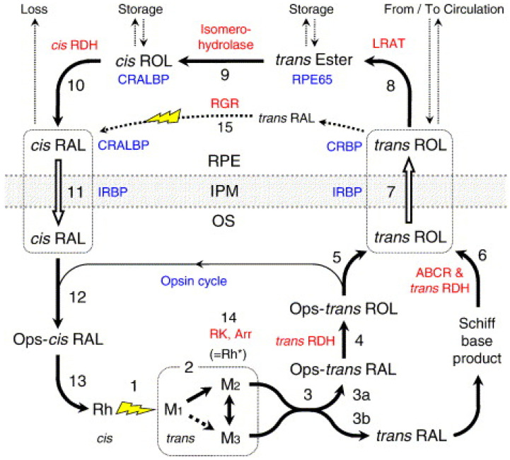
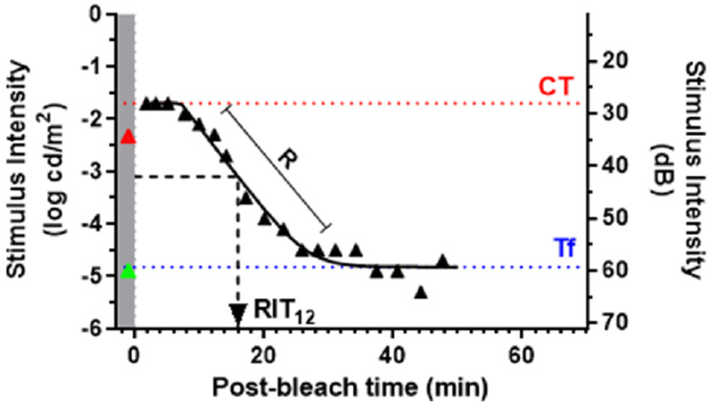
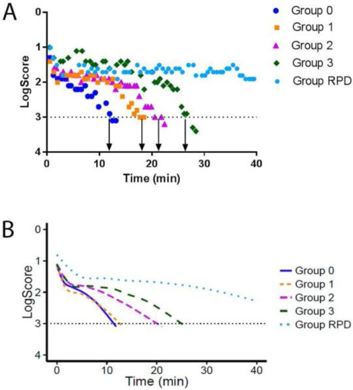
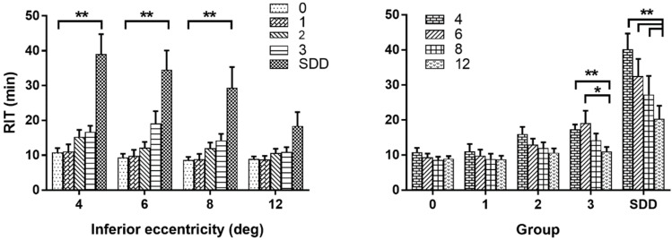
Dark adaptation (DA) refers to the slow recovery of visual sensitivity in darkness following exposure to intense or prolonged illumination, which bleaches a significant amount of the rhodopsin. This natural process also offers an opportunity to understand cellular function in the outer retina and evaluate for presence of disease. How our eyes adapt to darkness can be a key indicator of retinal health, which can be altered in the presence of certain diseases, such as age-related macular degeneration (AMD). A specific focus on clinical aspects of DA measurement and its significance to furthering our understanding of AMD has revealed essential findings underlying the pathobiology of the disease. The process of dark adaptation involves phototransduction taking place mainly between the photoreceptor outer segments and the retinal pigment epithelial (RPE) layer. DA occurs over a large range of luminance and is modulated by both cone and rod photoreceptors. In the photopic ranges, rods are saturated and cone cells adapt to the high luminance levels. However, under scotopic ranges, cones are unable to respond to the dim luminance and rods modulate the responses to lower levels of light as they can respond to even a single photon. Since the cone visual cycle is also based on the Muller cells, measuring the impairment in rod-based dark adaptation is thought to be particularly relevant to diseases such as AMD, which involves both photoreceptors and RPE. Dark adaptation parameters are metrics derived from curve-fitting dark adaptation sensitivities over time and can represent specific cellular function. Parameters such as the cone-rod break (CRB) and rod intercept time (RIT) are particularly sensitive to changes in the outer retina. There is some structural and functional continuum between normal aging and the AMD pathology. Many studies have shown an increase of the rod intercept time (RIT), i.e., delays in rod-mediated DA in AMD patients with increasing disease severity determined by increased drusen grade, pigment changes and the presence of subretinal drusenoid deposits (SDD) and association with certain morphological features in the peripheral retina. Specifications of spatial testing location, repeatability of the testing, ease and availability of the testing device in clinical settings, and test duration in elderly population are also important. We provide a detailed overview in light of all these factors.
Keywords: age-related macular degeneration (AMD); cone-rod break (CRB); dark adaptation (DA); longitudinal monitoring; phototransduction; rod-intercept time (RIT); spatial gradient; subretinal drusenoid deposits (SDD).
Conflict of interest statement
The authors declare no conflict of interest related to this article.
Figures
Similar articles
-
Biologically Guided Optimization of Test Target Location for Rod-mediated Dark Adaptation in Age-related Macular Degeneration: Alabama Study on Early Age-related Macular Degeneration 2 Baseline.Ophthalmol Sci. 2023 Jan 23;3(2):100274. doi: 10.1016/j.xops.2023.100274. eCollection 2023 Jun. Ophthalmol Sci. 2023. PMID: 36875335 Free PMC article.
-
Evaluation of Cone- and Rod-Mediated Parameters in Dark Adaptation Testing as Outcome Measures in Age-Related Macular Degeneration.Ophthalmol Retina. 2022 Dec;6(12):1173-1184. doi: 10.1016/j.oret.2022.05.018. Epub 2022 May 26. Ophthalmol Retina. 2022. PMID: 35643387
-
Relating Retinal Morphology and Function in Aging and Early to Intermediate Age-related Macular Degeneration Subjects.Am J Ophthalmol. 2016 May;165:65-77. doi: 10.1016/j.ajo.2016.02.021. Epub 2016 Mar 3. Am J Ophthalmol. 2016. PMID: 26940163 Free PMC article.
-
Tuning outer segment Ca2+ homeostasis to phototransduction in rods and cones.Adv Exp Med Biol. 2002;514:179-203. doi: 10.1007/978-1-4615-0121-3_11. Adv Exp Med Biol. 2002. PMID: 12596922 Review.
-
The role of dark adaptation in understanding early AMD.Prog Retin Eye Res. 2022 May;88:101015. doi: 10.1016/j.preteyeres.2021.101015. Epub 2021 Oct 6. Prog Retin Eye Res. 2022. PMID: 34626782 Review.
Cited by
-
Choriocapillaris Impairment, Visual Function, and Distance to Fovea in Aging and Age-Related Macular Degeneration: ALSTAR2 Baseline.Invest Ophthalmol Vis Sci. 2024 Jul 1;65(8):40. doi: 10.1167/iovs.65.8.40. Invest Ophthalmol Vis Sci. 2024. PMID: 39042400 Free PMC article.
-
Watching the human retina breath in real time and the slowing of mitochondrial respiration with age.Sci Rep. 2023 Apr 20;13(1):6445. doi: 10.1038/s41598-023-32897-7. Sci Rep. 2023. PMID: 37081065 Free PMC article.
-
The role of nutritional factors in transitioning between early, mid, and late stages of age-related macular degeneration: prospective longitudinal analysis.Am J Clin Nutr. 2024 Dec;120(6):1387-1398. doi: 10.1016/j.ajcnut.2024.08.019. Epub 2024 Aug 23. Am J Clin Nutr. 2024. PMID: 39181206 Free PMC article.
-
Intraocular Pressure After Anti-Vascular Endothelial Growth Factor Injection in Eyes With a Mineralized Bruch's Membrane Caused by Pseudoxanthoma Elasticum.Invest Ophthalmol Vis Sci. 2025 Aug 1;66(11):25. doi: 10.1167/iovs.66.11.25. Invest Ophthalmol Vis Sci. 2025. PMID: 40787936 Free PMC article.
-
[Pseudoxanthoma elasticum-Novel therapeutic approaches on the horizon?].Ophthalmologie. 2025 Jun;122(6):432-437. doi: 10.1007/s00347-025-02223-9. Epub 2025 May 28. Ophthalmologie. 2025. PMID: 40434430 Review. German.
References
Publication types
Grants and funding
LinkOut - more resources
Full Text Sources
