Review
. 2022 Feb 23;15(5):1664.
doi: 10.3390/ma15051664.

Flexible and Stretchable Bioelectronics

Affiliations
Review

Flexible and Stretchable Bioelectronics

Chandani Chitrakar et al. Materials (Basel). .

Abstract

Medical science technology has improved tremendously over the decades with the invention of robotic surgery, gene editing, immune therapy, etc. However, scientists are now recognizing the significance of 'biological circuits' i.e., bodily innate electrical systems for the healthy functioning of the body or for any disease conditions. Therefore, the current trend in the medical field is to understand the role of these biological circuits and exploit their advantages for therapeutic purposes. Bioelectronics, devised with these aims, work by resetting, stimulating, or blocking the electrical pathways. Bioelectronics are also used to monitor the biological cues to assess the homeostasis of the body. In a way, they bridge the gap between drug-based interventions and medical devices. With this in mind, scientists are now working towards developing flexible and stretchable miniaturized bioelectronics that can easily conform to the tissue topology, are non-toxic, elicit no immune reaction, and address the issues that drugs are unable to solve. Since the bioelectronic devices that come in contact with the body or body organs need to establish an unobstructed interface with the respective site, it is crucial that those bioelectronics are not only flexible but also stretchable for constant monitoring of the biological signals. Understanding the challenges of fabricating soft stretchable devices, we review several flexible and stretchable materials used as substrate, stretchable electrical conduits and encapsulation, design modifications for stretchability, fabrication techniques, methods of signal transmission and monitoring, and the power sources for these stretchable bioelectronics. Ultimately, these bioelectronic devices can be used for wide range of applications from skin bioelectronics and biosensing devices, to neural implants for diagnostic or therapeutic purposes.

Keywords: conductive polymers; fabrication of stretchable bioelectronics; flexible and stretchable bioelectronics; flexible and stretchable power sources; stretchable batteries; stretchable polymer; stretchable sensors; supercapacitors.


Conflict of interest statement

The authors declare no conflict of interest.

Figures

Figure 8
Figure 8
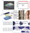
Alternative fabrication methods. (a,b) Printing method. (a). Schematics of printing of liquid metal (eGaIn) on an elastomer base layer [60]. (i) Extrusion printing of a base elastomer layer (ii) Spray printing of liquid metal (iii) Selective activation of the electrical path (iv) Extrusion printing of an encapsulation elastomer to seal the device. Reprinted with permission from Ref. [60]. Copyright 2017 Wiley-VCH Verlag GmbH & Co. (b). Fabrication steps for hydroprinted electronics. Process for application of a hydroprinted electronic tattoo over the forearm for EMG signal acquisition [113]. Reprinted with permission from Ref. [113]. Copyright 2018 American Chemical Society. (c,d) The wet spinning method of fabrication. (c). Schematic illustration of wet spinning GO fibers. Wet spinning set-up [118]. (d). Schematic illustration of the coaxial spinning process for highly stretchable fibers [122]. Reprinted with permission from Ref. [122] Copyright 2018 American Chemical Society.
Figure 1
Figure 1
(a) Young’s modulus of multiple biomaterials compared to cells [37]. (b) Young’s modulus scale comparing multiple types of bioelectronics and the moduli of multiple tissue types in the human body [38].
Figure 2
Figure 2
Stretchable Parylene-C electrodes. (a) A schematic of stretchable parylene-C electrodes with serpentine interconnects to increase the stretchability of the electrodes. A silicone rubber adhesive was used to stick the electrodes onto the elastomer substrate. Attaching the electrodes to an elastic substrate limited external deformation [43]. (b) A diagram of the strain applied to the electrodes when stretched [43]. Reprinted with permission from Ref. [43]. Copyright 2020 The Chinese Ceramic Society.
Figure 3
Figure 3
Chemical structure of polyurethanes. (left) The basic components of polyurethanes [49]. (right). Structure of the elastomer thermoplastic polyurethane which contains hard and soft segments [50]. Reprinted with permission from Ref. [50]. Copyright 2020 Springer Science Business Media.
Figure 4
Figure 4
(a) Two-dimensional graphene sheets in various shapes: green shows 1D fullerene shapes, red shows graphene rolled into nanotubes, blue shows graphene stacked into 3D graphite [67] Reprinted with permission from Ref. [67]. Copyright 2007 Nature Publishing Group.; (b) atomic arrangement of single- and double-layered graphene [68]. Reprinted with permission from Ref. [68]. Copyright 2010 Elsevier Ltd.
Figure 5
Figure 5
A chart summarizing the engineering of conductive material into flexible and stretchable format.
Figure 6
Figure 6
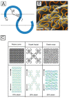
Representative geometrical configurations for hard–soft material integration. (A). Schematic representation of a serpentine unit cell. (B). Example of serpentine structured traces on an electrode array [91]. Reprinted with permission from Ref. [91]. Copyright 2015. American Chemical Society. (C). Different patterns of fractal-inspired layouts of metal wires (top), and FEM images of each structure under elastic tensile strain [90]. Reprinted with permission from Ref. [90]. Copyright 2014 Nature Publishing Group.
Figure 7
Figure 7
Fabrication methods for stretchable electronics. (ac) Chemical vapor deposition (CVD) (a). Schematics of the synthesis mechanism of CVD graphene on Cu foil (left) and the roll-to-roll transfer process of graphene (right) [95]. Reprinted with permission from Ref. [95]. Copyright 2013 American Chemical Society. (b). Modified CVD (mCVD). Schematic representation for synthesizing multilayer MoSe2 film. Schematic cross-section of a collection of MoSe2 transistors on a flexible PI/PET substrate [94]. Reprinted with permission from Ref. [94]. Copyright 2016 Wiley-VCH Verlag GmbH & Co. (c). Fabrication process of the wrinkled graphene-AgNW hybrid electrode [96]. (df) Lithography method. (d). UV Nanolithography (top) and soft lithography (bottom) [97] (e). Photolithographic methods using masked irradiation and a negative photoresist material: (i) Patterning by single exposure, (ii) patterning by layer-by-layer coating and exposure, (iii) tilted patterning by single inclined exposure, (iv) patterning by double inclined exposure, (v) tapered patterns by rotating tilted exposure [97]. Reprinted with permission from Ref. [97]. Copyright 2011 Elsevier Ltd. (f). Fabrication of highly stretchable and transparent nanomesh electrodes via grain boundary lithography [98]. A sheet of paper is laser-cut using the magnified image of Au nanomesh. The left image shows without any cutting. The middle image shows the stretching of the structure after cutting a few ligaments. The right image shows the stretching of the structure after cutting more ligaments. Reprinted with permission from Ref. [98]. Copyright 2014 Nature Publishing Group.
Figure 9
Figure 9
The proposed shoe-insole nanogenerator that can generate an open circuit voltage of ~27 V. The sensors used for movement detection responded to almost every joint movement [136]. Reprinted with permission from Ref. [136]. Copyright 2020 American Chemical Society.
Figure 10
Figure 10
Flexible CMUT step process by Pang et al. [145].
Figure 11
Figure 11
The Ag-PDMS sample with transmission line adapters seen in (a), (b) the tensile test fixture (c) graphing the different Ag volumes in comparison to the attenuation of transmission line, and (d) showing the attenuation of the transmission [148].
Figure 12
Figure 12
Stretchable batteries for powering bioelectronics. (a). Schematic showing a stretchable aqueous batteries configuration in which the HCP composite was used as a current collector. Long-term cycle performance and coulombic efficiency of the full cell at a rate of 20 C over 500 cycles [156]. Reprinted with permission from Ref. [156]. Copyright 2018 Wiley-VCH Verlag GmbH &Co. (b). Schematic showing the composition of a stretchable EGaIn battery [157] A. Photographs of the stretchable EGaIn-MnO2 battery array in series of two stretched under 0, 50, and 100% strain integrated with LEDs. B. Photograph of battery-powered strain sensor that is mounted on the wrist. Reprinted with permission from Ref. [157]. Copyright 2019 Wiley-VCH Verlag GmbH & Co. (c). Schematics of simple serpentine current collector and optical images of four serpentine-shaped batteries connected in series. Batteries continuously power an OLED while being subjected to a uniaxial strain of 100%. Schematics of a self-similar serpentine current collector and optical images of the full battery assembled around such a current collector. Geometry of the battery facilitates biaxial stretching [158].
Figure 13
Figure 13
Supercapacitors for powering bioelectronic devices. (a) (i) Schematic illustration of fabricating stretchable conducting wire by wrapping an aligned CNT sheet around a pre-stretched elastic wire. CV curves of the supercapacitors based on (ii) the bare CNT-wrapped and (iii) CNT/PEDOT-PSS-wrapped wires [160]. (b) Digital photograph of a typical wire-shaped supercapacitor with a twisted structure after being stretched from strains of 0 to 370% [160]. Reprinted with permission from Ref. [160]. Copyright 2015 Wiley-VCH Verlag GmbH & Co. (c). Stretchable Au-CNT forest electrodes: (i) SEM image of the Au-CNT forest pattern morphology generated by a uniaxial pre-strain of 300% and by applying a biaxial pre-strain of 200% × 200%. (ii) Capacitance retention of a uniaxially stretchable Au-CNT forest electrode under mechanical stretching–relaxation cyclic deformations to a strain of 200% and for 10,000 charge/discharge cycles at the relaxed state. Inset shows the CV curves measured before and after the electrochemical stability test at the scan rate of 500 mV s−1 [159]. Reprinted with permission from Ref. [159]. Copyright 2020 Elsevier Inc. (d). Digital images of stretchable rectangular-shaped supercapacitors (with geometric parameters of y = 0.7 cm, m = 0.2 cm, x = 194 µm, T = 0.5 cm) under different strain tests. The inset images (upper left) are the scheme showing the expandable honeycomb structure and the hexagonal unit cell before and after being stretched. Capacitance retention ratio of 3D stretchable supercapacitor based on PPy/BPO-CNT electrodes tested at 7.8 mA cm−2 under the cycling tensile strain of 2000% [161]. (e). Arched bridge-shaped supercapacitors acting as a 3D helmet worn on the head of an owl toy model to power a 3.0 V flexible LED strip (right) [161]. Reprinted with permission from Ref. [161]. Copyright 2018 Wiley-VCH Verlag GmbH & Co.

References

    1. Peeples L. The rise of bioelectric medicine sparks interest among researchers, patients, and industry. Proc. Natl. Acad. Sci. USA. 2019;116:24379–24382. doi: 10.1073/pnas.1919040116. - DOI - PMC - PubMed
    1. Zhang D., Liu Q. Biosensors and bioelectronics on smartphone for portable biochemical detection. Biosens. Bioelectron. 2016;75:273–284. doi: 10.1016/j.bios.2015.08.037. - DOI - PubMed
    1. Someya T., Bao Z., Malliaras G.G. The rise of plastic bioelectronics. Nature. 2016;540:379–385. doi: 10.1038/nature21004. - DOI - PubMed
    1. Jiang Y., Tian B. Inorganic semiconductor biointerfaces. Nat. Rev. Mater. 2018;3:473–490. doi: 10.1038/s41578-018-0062-3. - DOI - PMC - PubMed
    1. Yao G., Yin C., Wang Q., Zhang T., Chen S., Lu C., Zhao K., Xu W., Pan T., Gao M., et al. Flexible bioelectronics for physiological signals sensing and disease treatment. J. Mater. 2020;6:397–413. doi: 10.1016/j.jmat.2019.12.005. - DOI

LinkOut - more resources