

Novel, Inexpensive, and Scalable Amyloid Fibril Formation Method
- PMID: 35268997
- PMCID: PMC8911616
- DOI: 10.3390/ma15051766
Novel, Inexpensive, and Scalable Amyloid Fibril Formation Method
Abstract
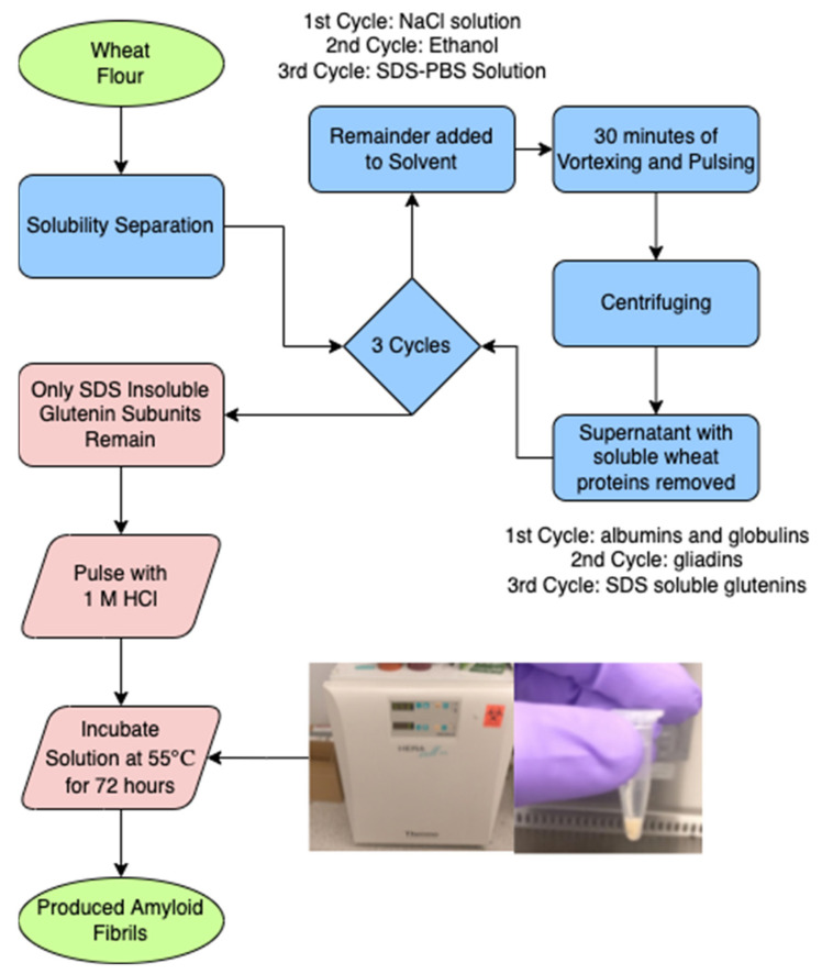
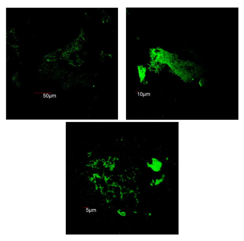
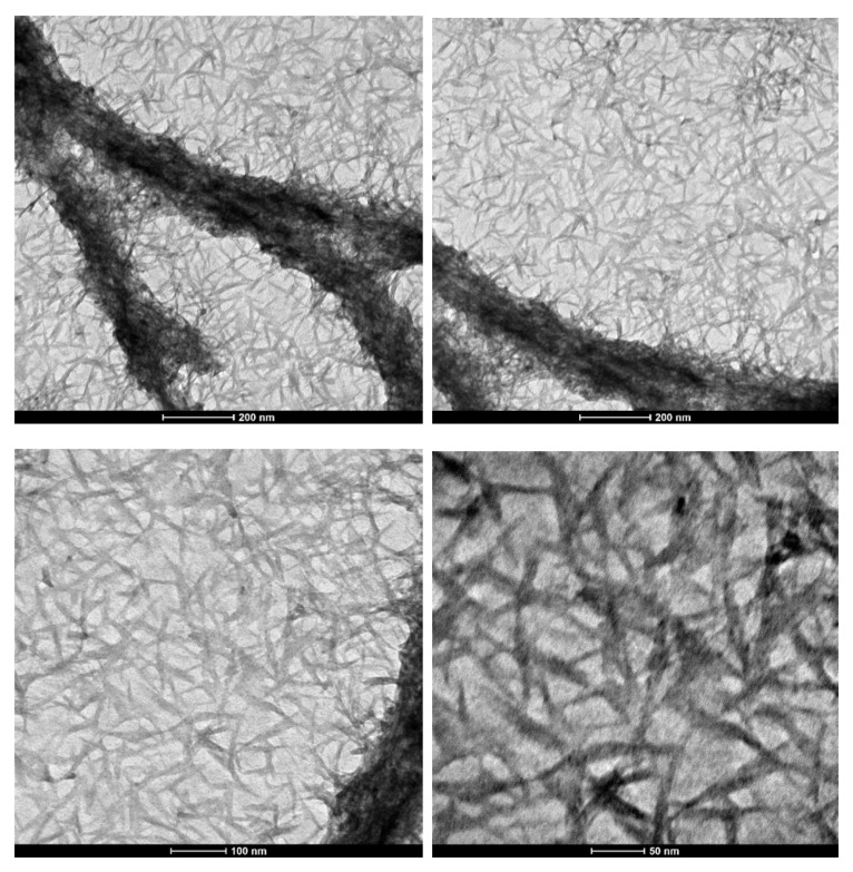
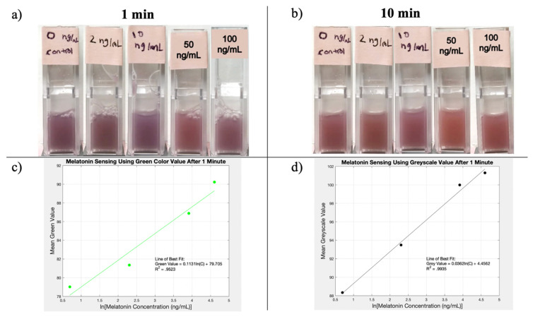
Wheat flour was used as a source of protein for the in vitro synthesis of Amyloid fibrils to develop a novel and inexpensive fabrication method. Amyloid fibrillation was confirmed by Thioflavin T Fluorescence, using confocal microscopy. A morphological study was carried out by transmission electron microscopy (TEM), which revealed the high aspect ratio of the amyloid fibrils formed via a novel process. An application of the amyloid fibers produced by the novel method is shown to be melatonin sensing. Tests showed that the amyloid samples had a measurable color variation dependent on the melatonin concentration. This newly derived process could prove to be a cost-effective tool for future nano-biomaterial applications in commercial and research settings.
Keywords: amyloid; inexpensive; melatonin biosensing; synthesis.
Conflict of interest statement
The authors declare no conflict of interest. The funders had no role in the design of the study; in the collection, analyses, or interpretation of data; in the writing of the manuscript, or in the decision to publish the results.
Figures
Similar articles
-
Hydrothermal Treatments Cause Wheat Gluten-Derived Peptides to Form Amyloid-like Fibrils.J Agric Food Chem. 2021 Feb 17;69(6):1963-1974. doi: 10.1021/acs.jafc.0c05868. Epub 2021 Feb 5. J Agric Food Chem. 2021. PMID: 33544593
-
Mechanism of thioflavin T binding to amyloid fibrils.J Struct Biol. 2005 Sep;151(3):229-38. doi: 10.1016/j.jsb.2005.06.006. J Struct Biol. 2005. PMID: 16125973
-
Amyloid fibril formation from crude protein mixtures.Biotechnol Prog. 2011 Nov-Dec;27(6):1768-76. doi: 10.1002/btpr.693. Epub 2011 Sep 9. Biotechnol Prog. 2011. PMID: 21910260
-
[Molecular mechanism of amyloid formation by Ab peptide: review of own works].Biomed Khim. 2018 Jan;64(1):94-109. doi: 10.18097/PBMC20186401094. Biomed Khim. 2018. PMID: 29460839 Review. Russian.
-
Direct observation of amyloid fibril growth, propagation, and adaptation.Acc Chem Res. 2006 Sep;39(9):663-70. doi: 10.1021/ar050074l. Acc Chem Res. 2006. PMID: 16981683 Review.
Cited by
-
Polymorphic Biological and Inorganic Functional Nanomaterials.Materials (Basel). 2022 Aug 3;15(15):5355. doi: 10.3390/ma15155355. Materials (Basel). 2022. PMID: 35955287 Free PMC article.
-
Molecularly Imprinted Polymer-Amyloid Fibril-Based Electrochemical Biosensor for Ultrasensitive Detection of Tryptophan.Biosensors (Basel). 2022 May 2;12(5):291. doi: 10.3390/bios12050291. Biosensors (Basel). 2022. PMID: 35624592 Free PMC article.
-
Wheat flour-derived amyloid fibrils for efficient removal of organic dyes from contaminated water.Bioresour Bioprocess. 2024 Feb 14;11(1):22. doi: 10.1186/s40643-024-00737-9. Bioresour Bioprocess. 2024. PMID: 38647993 Free PMC article.
References
Grants and funding
LinkOut - more resources
Full Text Sources
