. 2022 Feb 22;11(5):760.
doi: 10.3390/cells11050760.

Cerebral Organoids Maintain the Expression of Neural Stem Cell-Associated Glycoepitopes and Extracellular Matrix

Affiliations

Cerebral Organoids Maintain the Expression of Neural Stem Cell-Associated Glycoepitopes and Extracellular Matrix

Lars Roll et al. Cells. .

Abstract

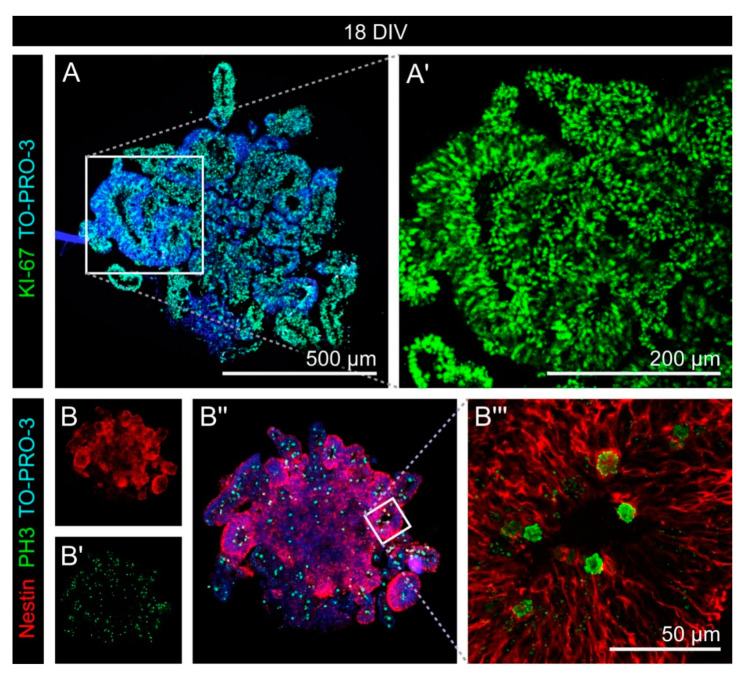
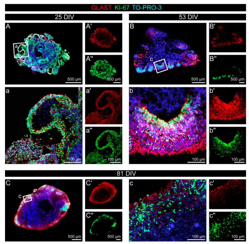
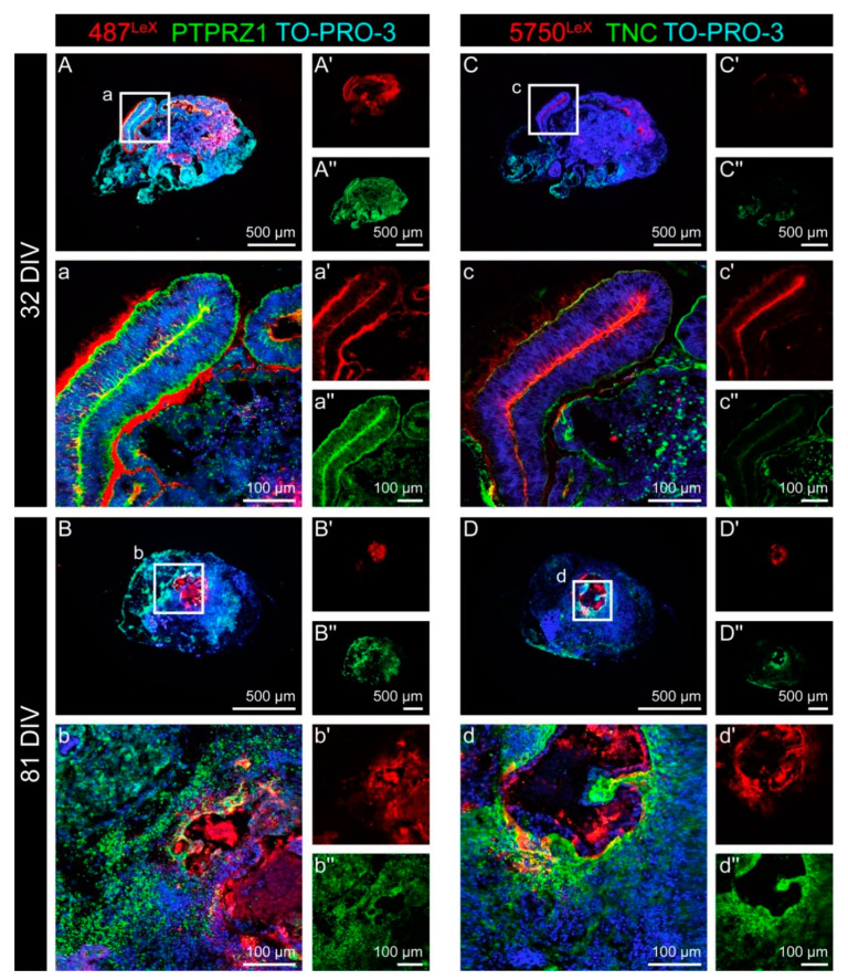
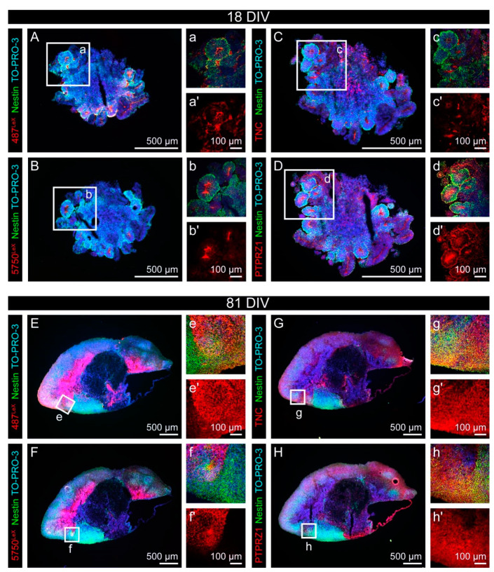
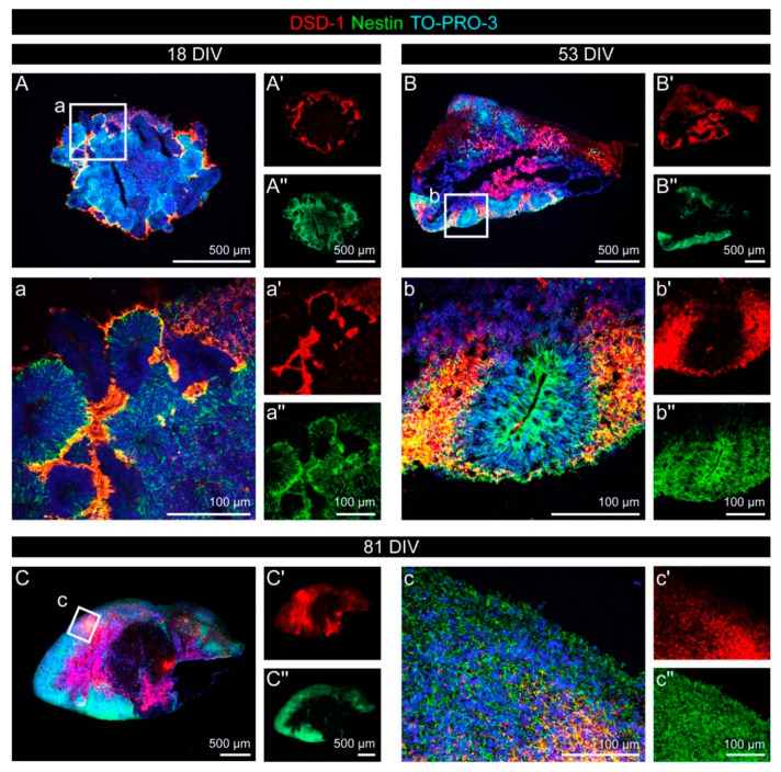
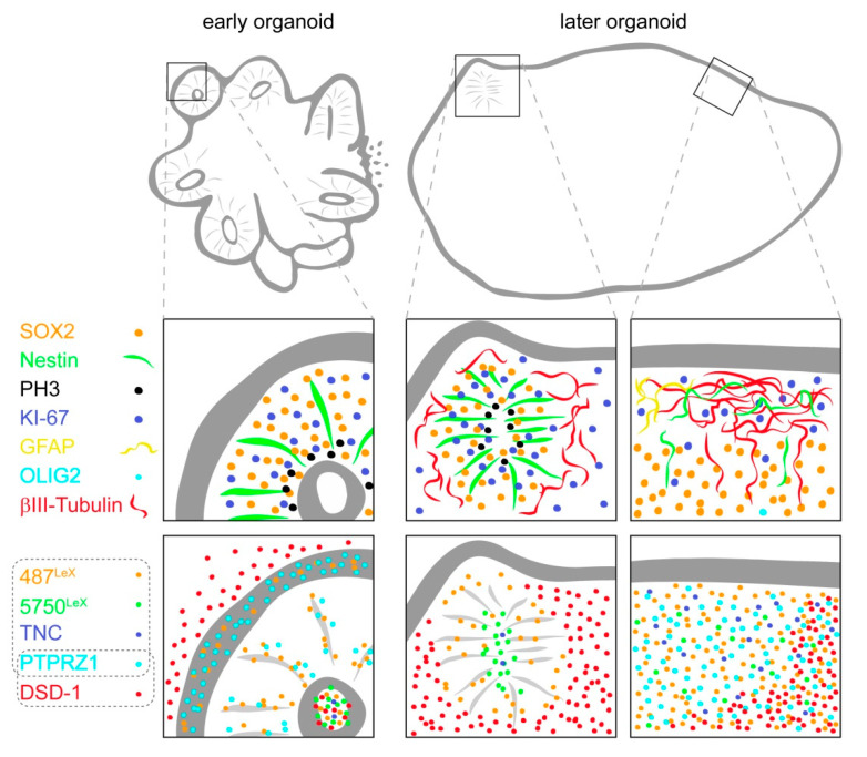
During development, the nervous system with its highly specialized cell types forms from a pool of relatively uniform stem cells. This orchestrated process requires tight regulation. The extracellular matrix (ECM) is a complex network rich in signaling molecules, and therefore, of interest in this context. Distinct carbohydrate structures, bound to ECM molecules like Tenascin C (TNC), are associated with neural stem/progenitor cells. We have analyzed the expression patterns of the LewisX (LeX) trisaccharide motif and of the sulfation-dependent DSD-1 chondroitin sulfate glycosaminoglycan epitope in human cerebral organoids, a 3D model for early central nervous system (CNS) development, immunohistochemically. In early organoids we observed distinct expression patterns of the glycoepitopes, associated with rosette-like structures that resemble the neural tube in vitro: Terminal LeX motifs, recognized by the monoclonal antibody (mAb) 487LeX, were enriched in the lumen and at the outer border of neural rosettes. In contrast, internal LeX motif repeats detected with mAb 5750LeX were concentrated near the lumen. The DSD-1 epitope, labeled with mAb 473HD, was detectable at rosette borders and in adjacent cells. The epitope expression was maintained in older organoids but appeared more diffuse. The differential glycoepitope expression suggests a specific function in the developing human CNS.

Keywords: DSD-1 epitope; LewisX epitope; PTPRZ1; Tenascin C; cerebral organoid; chondroitin sulfate; extracellular matrix; glycoepitope; neural stem cell; radial glia.


Conflict of interest statement

The funders had no role in the design of the study; in the collection, analyses, or interpretation of data; in the writing of the manuscript, or in the decision to publish the results.

Figures

Figure A1
Figure A1
Protocol to generate human cerebral organoids from hiPS cells. A detailed description of the procedure is provided in Appendix A.
Figure A2
Figure A2
Cell type-specific marker expression in early cerebral organoids. (Ab″) SOX2 (red), which labels neuroepithelial and radial glia cells, was expressed by the majority of cells, with a clear association to cells within the neural rosettes after 25 DIV. βIII-Tubulin (green) as neuronal marker was virtually undetectable after 18 DIV, but appeared in cells outside the neural rosette-like areas after 25 DIV. (Cd″) The progenitor and oligodendroglial marker OLIG2 (red) could not be detected after 18 or 25 DIV. The astrocyte marker GFAP (green) was only weakly expressed in early cerebral organoids, and restricted to very few regions within the aggregates.
Figure A3
Figure A3
Cell type-specific marker expression in later cerebral organoids. (Ab″) Expression of SOX2 (red) as neuroepithelial and radial glia marker was limited in older organoids, with a restriction to neural rosette-like regions after 53 DIV and outer regions after 81 DIV. βIII-Tubulin (green) as a neuronal marker showed an inverted image compared to SOX2. Neurons could be identified clearly outside the neural rosette structures after 53 DIV and also after 81 DIV the βIII-Tubulin-positive regions were poor in SOX2-expressing cells. (Cd″) The progenitor and oligodendroglial marker OLIG2 (red) could not be detected in terms of specific signals, even in older cerebral organoids. In contrast, the astrocyte marker GFAP (green) was found on cells at the outer regions of the organoid. The distribution was not even, instead large regions appeared free of GFAP-positive cells.
Figure A4
Figure A4
Fluorescence intensity profiles for 487LeX, 5750LeX and DSD-1 in cerebral organoids. (AB″) The signal for 487LeX was detected on the cells of neural rosette structures, including the inner and outer border. In later organoids, the distribution appeared diffuse. The yellow line indicates the pixels that were measured and depicted in the diagram (487LeX red, Nestin green, TO-PRO-3 blue). (CD″) 5750LeX signals were most prominently detected in the lumen of neural rosettes in early organoids, later the expression pattern became diffuse. The yellow line indicates the pixels that were measured and depicted in the diagram (5750LeX red, Nestin green, TO-PRO-3 blue). (EF″) The DSD-1 epitope was located at the outer border of neural rosettes and a faint signal could also be detected in the lumen. As shown for the other glycoepitopes, also the distribution of the DSD-1 epitope became diffuse with the ongoing maturation of the cerebral organoids. The yellow line indicates the pixels that were measured and depicted in the diagram (DSD-1 red, Nestin green, TO-PRO-3 blue).
Figure 1
Figure 1
Morphology of human cerebral organoids over time. (A) After neural induction, small protrusions were visible on the surface of the early cerebral organoid. (B,C) The surface became smoother, as shown after 32 DIV and 95 DIV. Here, only a few, larger protrusions could be observed.
Figure 2
Figure 2
Proliferation in early cerebral organoids. (A,A′) KI-67 (green), which labels all proliferating cells, was expressed by the vast majority of all cells labeled with nuclear TO-PRO-3 staining (blue). (BB‴) Phospho-Histone H3 (green), a marker restricted to late G2- and M-phase cells, was expressed by a minority of cells. Within the neural rosette-like structures formed by Nestin-positive cells (red), PH3 signals were primarily found at the apical side, near the lumen.
Figure 3
Figure 3
Proliferation in cerebral organoids over time. (Ac″) The ratio of proliferating cells was reduced over time, as the comparison of KI-67 (green) in an early cerebral organoid after 25 DIV (Aa″) with the situation after 53 DIV (Bb″) or even 81 DIV (Cc″) shows. GLAST (red), a marker for the radial glia type of neural stem cells, was strongly expressed in the neural rosette structures in the early organoids, whereas the signal appeared more diffuse after 81 DIV.
Figure 4
Figure 4
Double staining for the LewisX (LeX) motif and for the potential LeX carrier molecules PTPRZ1 and TNC in cerebral organoids. (Ab″) The monoclonal antibody (mAb) 487LeX binds to terminal LeX motifs (red) and was clearly enriched in the lumen and at the outer border of neural rosette structures after 32 DIV. PTPRZ1 (green) showed a similar, but not identical staining pattern. The strongest signals were found on the cells within the rosette structures. After 81 DIV, the signals of 487LeX and for PTPRZ1 appeared more diffuse and showed no clear co-localization at this stage. (Cd″) mAb 5750LeX (red) was used to detect internal LeX motif repeats. The signals were located near the lumen of neural rosettes after 32 DIV, whereas the outer borders of such areas were only weakly stained. TNC (green) was mainly labeled at the rosette borders. After 81 DIV, signals for mAb 5750LeX and for TNC could still be detected in the organoid. Here, regions with intensely labeled cells on the one hand and regions with only faint signals, on the other hand, were observed.
Figure 5
Figure 5
Expression of the LewisX (LeX) glycoepitope and of the potential LeX carriers TNC and PTPRZ1 with regard to Nestin-positive progenitor cells in cerebral organoids. (Ad′) After 18 DIV, Nestin signals were most prominent in the neural rosettes (green). Signals for 487LeX (red) were detectable at the lumen and at the outer border of neural rosettes. In contrast, 5750LeX signals (red) were most intense in the lumen of these structures. TNC (red) was also detectable on cells of the neural rosettes, although it appeared weaker compared to PTPRZ1 (red). (Eh′) After 81 DIV, the rosette-like structures disappeared and the staining patterns of the molecules changed accordingly. Nestin-positive cells were present throughout the organoid. Signals of mAb 487LeX, mAb 5750LeX, for TNC and for PTPRZ1 were detectable, without a clear expression pattern.
Figure 6
Figure 6
Expression of the neural stem cell-related DSD-1 chondroitin sulfate epitope with regard to the neural stem cell marker Nestin in cerebral organoids. (Ab″) After 18 and 53 DIV, the DSD-1 epitope (red) was highly enriched at the outer border of neural rosette structures and in the area adjacent to the rosettes. A broad overlap with Nestin-positive (green) cells could be observed. (Cc″) Later, after 81 DIV, Nestin, as well as the DSD-1 epitope, were labeled in a relatively diffuse and in part overlapping pattern.
Figure 7
Figure 7
Schematic representation of neural stem cell-associated glycoepitopes and associated extracellular matrix molecules in human cerebral organoids over time. Important cell type-specific markers are indicated as reference.

Similar articles

Cited by

References

    1. Kriegstein A., Alvarez-Buylla A. The glial nature of embryonic and adult neural stem cells. Annu. Rev. Neurosci. 2009;32:149–184. doi: 10.1146/annurev.neuro.051508.135600. - DOI - PMC - PubMed
    1. Nikolopoulou E., Galea G.L., Rolo A., Greene N.D., Copp A.J. Neural tube closure: Cellular, molecular and biomechanical mechanisms. Development. 2017;144:552–566. doi: 10.1242/dev.145904. - DOI - PMC - PubMed
    1. Long K.R., Huttner W.B. How the extracellular matrix shapes neural development. Open Biol. 2019;9:180216. doi: 10.1098/rsob.180216. - DOI - PMC - PubMed
    1. Hynes R.O., Naba A. Overview of the matrisome—An inventory of extracellular matrix constituents and functions. Cold Spring Harb. Perspect. Biol. 2012;4:a004903. doi: 10.1101/cshperspect.a004903. - DOI - PMC - PubMed
    1. Mizumoto S., Yamada S., Sugahara K. Molecular interactions between chondroitin-dermatan sulfate and growth factors/receptors/matrix proteins. Curr. Opin. Struct. Biol. 2015;34:35–42. doi: 10.1016/j.sbi.2015.06.004. - DOI - PubMed

Publication types

LinkOut - more resources