. 2022 Mar 1;11(5):841.
doi: 10.3390/cells11050841.

A Human In Vitro Model to Study Adenoviral Receptors and Virus Cell Interactions

Affiliations

A Human In Vitro Model to Study Adenoviral Receptors and Virus Cell Interactions

Raphael L Tsoukas et al. Cells. .

Abstract

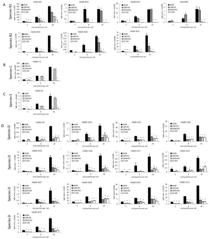
To develop adenoviral cell- or tissue-specific gene delivery, understanding of the infection mechanisms of adenoviruses is crucial. Several adenoviral attachment proteins such as CD46, CAR and sialic acid have been identified and studied. However, most receptor studies were performed on non-human cells. Combining our reporter gene-tagged adenovirus library with an in vitro human gene knockout model, we performed a systematic analysis of receptor usage comparing different adenoviruses side-by-side. The CRISPR/Cas9 system was used to knockout CD46 and CAR in the human lung epithelial carcinoma cell line A549. Knockout cells were infected with 22 luciferase-expressing adenoviruses derived from adenovirus species B, C, D and E. HAdV-B16, -B21 and -B50 from species B1 as well as HAdV-B34 and -B35 were found to be CD46-dependent. HAdV-C5 and HAdV-E4 from species E were found to be CAR-dependent. Regarding cell entry of HAdV-B3 and -B14 and all species D viruses, both CAR and CD46 play a role, and here, other receptors or attachment structures may also be important since transductions were reduced but not completely inhibited. The established human knockout cell model enables the identification of the most applicable adenovirus types for gene therapy and to further understand adenovirus infection biology.

Keywords: CAR; CD46; CD46-knockout cell line; CRISPR; adenovirus; human; knockout cell line; receptor; tight-junction-knockout cell line; virus.


Conflict of interest statement

The authors declare no conflict of interest.

Figures

Figure 1
Figure 1
Expression of surface proteins involved in adenoviral cell entry on A549 cells. For CD46, we measured 100% positive cells, and for CAR, 96.4% positive cells. The negative control was analyzed without the use of a primary antibody.
Figure 2
Figure 2
Exon structure of the CD46 and CAR genes and the CRISPR/Cas9 plasmid used for generation of the knockout cell lines. (A) Arrow-marked exons in the CD46 and CAR gene regions containing the selected target sites for the gRNAs are translated in all protein isotypes. (B) Insertion of gRNA into the customized shuttle vector pShV-CBh-Cas9.
Figure 3
Figure 3
Generation of CD46 and CAR knockout cell lines using cell sorting. (A) CD46 protein expression after the first and second round of the MACS procedure. (B) CD46 protein expression after cell singularization of the CD46 knockout cells. The chosen clone CD46-KO is displayed. (C) CAR protein expression after flow cytometric sorting and (D) after cell singularization. The chosen CAR knockout clone, CAR-KO, is displayed. The parental A549 cell line serves as a positive control, incubated with the corresponding primary and secondary antibodies. For the negative control, the parental A549 cells were incubated with the secondary antibody only. All measurements were carried out with the same monoclonal APC-linked secondary antibody.
Figure 4
Figure 4
Establishment of a CD46/CAR double knockout cell line. The selected clone CC-KO exhibits a loss of CAR and CD46 protein expression in the FACS measurement. As the positive control, the parental A549 cell line incubated with the same primary antibodies was used. For the negative control, the parental A549 cells were incubated with the secondary antibody only. All measurements were carried out with the same monoclonal APC-linked secondary antibody.
Figure 5
Figure 5
Analysis of the genome-edited CD46 and CAR loci in A549 cells. (A) To further confirm genome editing at the CD46 target locus, a T7 endonuclease I (T7EI) assay was performed. For this purpose, the genomic region of CD46 spanning the CRISPR/Cas target site was amplified by PCR and analyzed by the T7EI assay. Results are shown for the CD46 genomic region in A549, A549 KO CD46 and A549 KO CD46/CAR cells. Cleavage of the PCR product resulting in two smaller fragments indicates a CRISPR/Cas edit within the generated PCR product. The positive control was derived from the EnGen® Mutation Detection Kit (New England Biolabs, MA, USA) and the PCR products were either T7EI-digested (+) or not (−). (B) To further confirm genome editing at the CAR target locus, a T7 endonuclease I (T7EI) assay was performed. For this purpose, the genomic region of CAR spanning the CRISPR/Cas target site was PCR-amplified. Cleavage of the PCR product resulting in two smaller fragments indicates a CRISPR/Cas edit within the generated PCR product. Results are shown for the CD46 genomic region in A549, A549 KO CD46 and A549 KO CD46/CAR cells. The positive control derived from the EnGen® Mutation Detection Kit (New England Biolabs, MA, USA) and the PCR products were either T7EI-digested (+) or not (−).
Figure 6
Figure 6
Screening of receptor usage in adenovirus species B, C and E adenoviruses. Parental A549, CAR-KO, CD46-KO and CC-KO cells were transduced with respective viruses at varying viral particle numbers per cell (VP/C: 2, 8, 20). Luminescence levels were analyzed 26 h post-infection. Uninfected cells were used for the background level. Each bar represents the mean of triplicated wells (96-well plate). Error bars represent standard deviation. Each of the KO cell lines was compared to the A549 cell line. Significant luminescence reductions are marked by * (p < 0.05). Each graph represents data from one out of three independent experiments. All experiments are depicted in Supplementary Figure S4.

References

    1. Lion T. Adenovirus persistence, reactivation, and clinical management. FEBS Lett. 2019;593:3571–3582. doi: 10.1002/1873-3468.13576. - DOI - PubMed
    1. Parks R.J., Chen L., Anton M., Sankar U., Rudnicki M.A., Graham F.L. A helper-dependent adenovirus vector system: Removal of helper virus by Cre-mediated excision of the viral packaging signal. Proc. Natl. Acad. Sci. USA. 1996;93:13565–13570. doi: 10.1073/pnas.93.24.13565. - DOI - PMC - PubMed
    1. Berciaud S., Rayne F., Kassab S., Jubert C., Corte M.F.-D., Salin F., Wodrich H., Lafon M. Adenovirus infections in Bordeaux University Hospital 2008–2010: Clinical and virological features. J. Clin. Virol. 2012;54:302–307. doi: 10.1016/j.jcv.2012.04.009. - DOI - PubMed
    1. Chen S.-P., Huang Y.-C., Chiu C.-H., Wong K.-S., Huang Y.-L., Huang C.-G., Tsao K.-C., Lin T.-Y. Clinical features of radiologically confirmed pneumonia due to adenovirus in children. J. Clin. Virol. 2013;56:7–12. doi: 10.1016/j.jcv.2012.08.021. - DOI - PubMed
    1. Alharbi S., Van Caeseele P., Consunji-Araneta R., Zoubeidi T., Fanella S., Souid A.-K., Alsuwaidi A.R. Epidemiology of severe pediatric adenovirus lower respiratory tract infections in Manitoba, Canada, 1991–2005. BMC Infect. Dis. 2012;12:55. doi: 10.1186/1471-2334-12-55. - DOI - PMC - PubMed

Publication types

LinkOut - more resources