Review
. 2022 Mar 7;11(5):919.
doi: 10.3390/cells11050919.

Waste Clearance in the Brain and Neuroinflammation: A Novel Perspective on Biomarker and Drug Target Discovery in Alzheimer's Disease

Affiliations
Review

Waste Clearance in the Brain and Neuroinflammation: A Novel Perspective on Biomarker and Drug Target Discovery in Alzheimer's Disease

Kazuhiko Uchida. Cells. .

Abstract

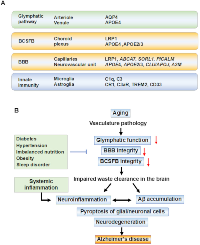
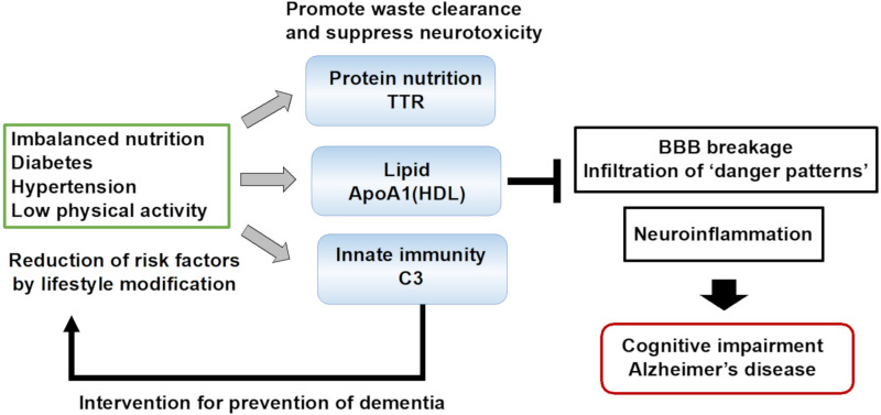
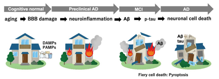
Alzheimer's disease (AD) is a multifactorial disease with a heterogeneous etiology. The pathology of Alzheimer's disease is characterized by amyloid-beta and hyperphosphorylated tau, which are necessary for disease progression. Many clinical trials on disease-modifying drugs for AD have failed to indicate their clinical benefits. Recent advances in fundamental research have indicated that neuroinflammation plays an important pathological role in AD. Damage- and pathogen-associated molecular patterns in the brain induce neuroinflammation and inflammasome activation, causing caspase-1-dependent glial and neuronal cell death. These waste products in the brain are eliminated by the glymphatic system via perivascular spaces, the blood-brain barrier, and the blood-cerebrospinal fluid barrier. Age-related vascular dysfunction is associated with an impairment of clearance and barrier functions, leading to neuroinflammation. The proteins involved in waste clearance in the brain and peripheral circulation may be potential biomarkers and drug targets in the early stages of cognitive impairment. This short review focuses on waste clearance dysfunction in AD pathobiology and discusses the improvement of waste clearance as an early intervention in prodromal AD and preclinical stages of dementia.

Keywords: Aβ clearance; blood-brain barrier; glymphatic system; innate immunity; mild cognitive impairment.


Conflict of interest statement

The author is a board member and shareholder of MCBI, Inc. (Tsukuba, Ibaraki, Japan).

Figures

Figure 1
Figure 1
Waste clearance systems and pathobiological events in Alzheimer’s disease. (A) Anatomical regions and key molecules involved in waste clearance systems. Anatomical regions and key molecules identified in the genome-wide association study (GWAS) associated with Alzheimer’s disease (AD) are shown. The molecules which have genetic variations associated with an increased risk for AD are shown as identified by GWAS. The paravascular glymphatic flow is dependent on the water channel aquaporin-4 (AQP4) at the astrocytic endfeet. The blood–cerebrospinal fluid barrier (BCSFB) at the choroid plexus and the blood-brain barrier (BBB) at the capillaries regulate the transport of molecules into and out of the brain. Microglia survey the brain to support homeostasis, and the pathogens and amyloid-beta (Aβ) aggregates are cleared by microglia through receptor-mediated phagocytosis. Astrocytes (astroglia) promote neuroinflammation. After exposure to Aβ, astroglia release NF-kB and complement components, which act through the complement receptors such as complement receptor 1 (CR1) and complement component 3a receptor (C3aR). Aβ is removed by the low-density lipoprotein (LDL) receptor-related protein-1 (LRP-1)-mediated transport. The variants of adenosine triphosphate-binding cassette transporter A7 (ABCA7), sortilin-related receptor-1 (SORL1), phosphatidylinositol-binding clathrin assembly (PICALM), clusterin (CLU/APOJ), and alpha-2 macroglobulin (A2M) affect BBB and clearance functions. Among the rare variants of genes associated with an increased risk of AD, triggering receptors expressed on myeloid cells-2 (TREM2), CD33, and CR1 are expressed on microglia and ascribed to innate immune pathways. (B) Underlined mechanisms and pathobiological events in the disease progression of AD. In sporadic AD, the aging of the vasculature may be an initial event in AD pathobiology. The waste clearance pathways contribute to brain homeostasis and its dysfunction is associated with vasculature disease. In preclinical stages, age-related vascular changes including vasomotor dysfunction, structural remodeling, and chronic inflammation, cause the breakage of tight junctions between epithelial cells in the BBB, and barrier dysfunction in the brain leads to the neuroinflammation of the parenchyma. Inflammation-related signal transduction induces beta-site amyloid precursor protein (APP) cleaving enzyme-1 (BACE1) expression, leading to Aβ production following tau pathology, resulting in amyloid plaques and neurofibrillary tangles. The accumulation of Aβ reduces capillary blood flow and impairs waste clearance. Systemic inflammation is a risk factor for AD, and recent studies have suggested that systemic inflammation can drive neuroinflammation. In these aging processes, an increase in damage-associated danger patterns, including Aβ, which has synaptotoxicity in the brain, promotes neuroinflammation in the parenchyma, and finally, caspase-1 dependent programmed cell death (pyroptosis) in the glial and neuronal cells occurs, resulting in cognitive impairment. Abbreviations: AD—Alzheimer’s disease; BBB—blood-brain barrier; BCSFB—blood–CSF barrier.
Figure 2
Figure 2
Possible roles of blood biomarker proteins in dementia prevention.
Figure 3
Figure 3
Illustration of disease progression from preclinical stages to onset of Alzheimer’s disease. DAMPs—damage-associated molecular patterns; PAMPs—pathogen-associated molecular patterns.

Similar articles

Cited by

References

    1. GBD 2016 Dementia Collaborators Global, Regional, and National Burden of Alzheimer’s Disease and Other Dementias, 1990–2016: A Systematic Analysis for the Global Burden of Disease Study 2016. Lancet Neurol. 2019;18:88–106. doi: 10.1016/S1474-4422(18)30403-4. - DOI - PMC - PubMed
    1. Gauthier S., Rosa-Neto P., Morais J.A., Webster C. World Alzheimer Report 2021: Journey through the Diagnosis of Dementia. Alzheimer’s Disease International; London, UK: 2021.
    1. Sweeney M.D., Montagne A., Sagare A.P., Nation D.A., Schneider L.S., Chui H.C., Harrington M.G., Pa J., Law M., Wang D.J.J., et al. Vascular Dysfunction-The Disregarded Partner of Alzheimer’s Disease. Alzheimers Dement. 2019;15:158–167. doi: 10.1016/j.jalz.2018.07.222. - DOI - PMC - PubMed
    1. Cummings J., Lee G., Zhong K., Fonseca J., Taghva K. Alzheimer’s Disease Drug Development Pipeline: 2021. Alzheimers Dement. 2021;7:e12179. doi: 10.1002/trc2.12179. - DOI - PMC - PubMed
    1. Alexander G.C., Knopman D.S., Emerson S.S., Ovbiagele B., Kryscio R.J., Perlmutter J.S., Kesselheim A.S. Revisiting FDA Approval of Aducanumab. N. Engl. J. Med. 2021;385:769–771. doi: 10.1056/NEJMp2110468. - DOI - PMC - PubMed