Structural Characterization and Osseointegrative Properties of Pulsed Laser-Deposited Fluorinated Hydroxyapatite Films on Nano-Zirconia for Implant Applications
- PMID: 35269557
- PMCID: PMC8910007
- DOI: 10.3390/ijms23052416
Structural Characterization and Osseointegrative Properties of Pulsed Laser-Deposited Fluorinated Hydroxyapatite Films on Nano-Zirconia for Implant Applications
Abstract
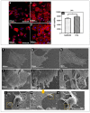
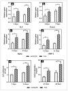
Standard zirconia implants used in restoration still present problems related to inertness and long-term stability. Various physicochemical approaches have been used to modify the implant surfaces to improve early and late bone-to-implant integration; however, no ideal surface modification has been reported. This study used pulsed laser deposition to deposit a fluorinated hydroxyapatite (FHA) film on a zirconia implant to create a biologically active surface. The film prepared was uniform, dense, and crack-free, and exhibited granular surface droplets; it also presented excellent mechanical strength and favorable biological behavior. The FHA-coated implant was implanted on the femur of Sprague-Dawley rats, and various tests and analyses were performed. Results show that the in vitro initial cell activity on the FHA-coated samples was enhanced. In addition, higher alkaline phosphatase activity and cell mineralization were detected in cells cultured on the FHA-coated groups. Further, the newly formed bone volume of the FHA-coated group was higher than that of the bare micro-adjusted composite nano-zirconia (NANOZR) group. Therefore, the FHA film facilitated osseointegration and may improve the long-term survival rates of dental implants, and could become part of a new treatment technology for implant surfaces, promoting further optimization of NANOZR implant materials.
Keywords: fluorinated hydroxyapatite; implant; nano-zirconia; osseointegration; pulsed laser deposition.
Conflict of interest statement
The authors declare that they have no known competing financial interests or personal relationship that could have appeared to influence the work reported in this paper.
Figures


Similar articles
-
Effects of magnesium-substituted nanohydroxyapatite coating on implant osseointegration.Clin Oral Implants Res. 2013 Aug;24 Suppl A100:34-41. doi: 10.1111/j.1600-0501.2011.02362.x. Epub 2011 Dec 6. Clin Oral Implants Res. 2013. PMID: 22145854
-
Biological and osseointegration capabilities of hierarchically (meso-/micro-/nano-scale) roughened zirconia.Int J Nanomedicine. 2018 Jun 8;13:3381-3395. doi: 10.2147/IJN.S159955. eCollection 2018. Int J Nanomedicine. 2018. PMID: 29922058 Free PMC article.
-
Stable sol-gel hydroxyapatite coating on zirconia dental implant for improved osseointegration.J Mater Sci Mater Med. 2021 Jun 30;32(7):81. doi: 10.1007/s10856-021-06550-6. J Mater Sci Mater Med. 2021. PMID: 34191141 Free PMC article.
-
Zirconia surface modifications for implant dentistry.Mater Sci Eng C Mater Biol Appl. 2019 May;98:1294-1305. doi: 10.1016/j.msec.2019.01.062. Epub 2019 Jan 16. Mater Sci Eng C Mater Biol Appl. 2019. PMID: 30813009 Free PMC article. Review.
-
Osseointegrative and antimicrobial properties of graphene oxide nano coated dental implants: A systematic review.F1000Res. 2024 Aug 9;13:281. doi: 10.12688/f1000research.148180.2. eCollection 2024. F1000Res. 2024. PMID: 39149510 Free PMC article.
Cited by
-
LASER as a tool for surface modification of dental biomaterials: A review.Heliyon. 2023 Jun 20;9(6):e17457. doi: 10.1016/j.heliyon.2023.e17457. eCollection 2023 Jun. Heliyon. 2023. PMID: 37408894 Free PMC article. Review.
-
Polymeric Composite Thin Films Deposited by Laser Techniques for Antimicrobial Applications-A Short Overview.Polymers (Basel). 2025 Jul 24;17(15):2020. doi: 10.3390/polym17152020. Polymers (Basel). 2025. PMID: 40808071 Free PMC article. Review.
-
Effect of Er:YAG Pulsed Laser-Deposited Hydroxyapatite Film on Titanium Implants on M2 Macrophage Polarization In Vitro and Osteogenesis In Vivo.Int J Mol Sci. 2023 Dec 26;25(1):349. doi: 10.3390/ijms25010349. Int J Mol Sci. 2023. PMID: 38203519 Free PMC article.
-
Femtosecond Laser Irradiation to Zirconia Prior to Calcium Phosphate Coating Enhances Osteointegration of Zirconia in Rabbits.J Funct Biomater. 2024 Feb 11;15(2):42. doi: 10.3390/jfb15020042. J Funct Biomater. 2024. PMID: 38391895 Free PMC article.
-
Biocompatible and Antibacterial Chemical Coatings on TiZr Dental Implants.J Funct Biomater. 2025 Mar 20;16(3):112. doi: 10.3390/jfb16030112. J Funct Biomater. 2025. PMID: 40137391 Free PMC article.
References
-
- Karoussis I.K., Salvi G.E., Heitz-Mayfield L.J., Brägger U., Hämmerle C.H., Lang N.P. Long-term implant prognosis in pa- tients with and without a history of chronic periodontitis: A 10-year prospective cohort study of the ITI Dental Implant System. Clin. Oral Implant. Res. 2003;14:329–339. doi: 10.1034/j.1600-0501.000.00934.x. - DOI - PubMed
MeSH terms
Substances
LinkOut - more resources
Full Text Sources

