Review
. 2022 Feb 22;23(5):2425.
doi: 10.3390/ijms23052425.

Adipose Tissue-Derived Mesenchymal Stem/Stromal Cells and Their Contribution to Angiogenic Processes in Tissue Regeneration

Affiliations
Review

Adipose Tissue-Derived Mesenchymal Stem/Stromal Cells and Their Contribution to Angiogenic Processes in Tissue Regeneration

Agnieszka Krawczenko et al. Int J Mol Sci. .

Abstract

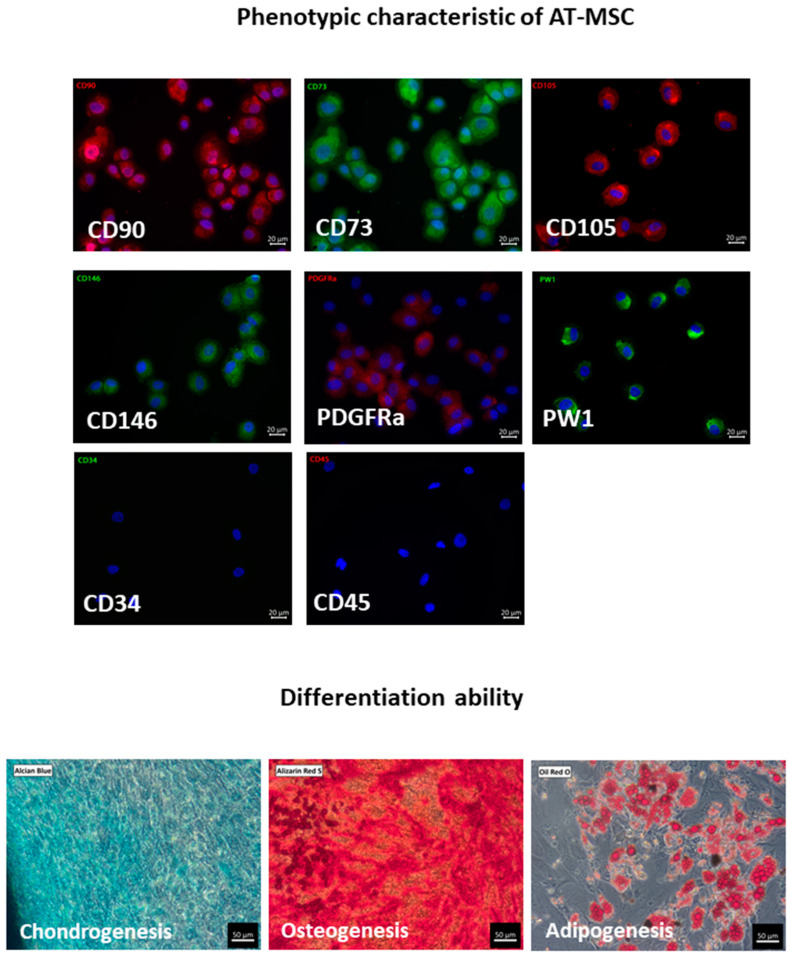
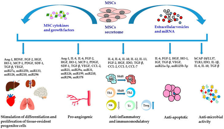
Mesenchymal stem/stromal cells (MSCs) are widely described in the context of their regenerative and immunomodulatory activity. MSCs are isolated from various tissues and organs. The most frequently described sources are bone marrow and adipose tissue. As stem cells, MSCs are able to differentiate into other cell lineages, but they are usually reported with respect to their paracrine potential. In this review, we focus on MSCs derived from adipose tissue (AT-MSCs) and their secretome in regeneration processes. Special attention is given to the contribution of AT-MSCs and their derivatives to angiogenic processes described mainly in the context of angiogenic dysfunction. Finally, we present clinical trials registered to date that concern the application of AT-MSCs and their secretome in various medical conditions.

Keywords: MSCs secretome; angiogenesis; mesenchymal stem cells; tissue regeneration.


Conflict of interest statement

The authors declare no conflict of interest.

Figures

Figure 1
Figure 1
Immunophenotyping and differentiation ability of primary AT-MSCs. Cells are positive for common MSC markers CD90, CD73, CD105 and additionally for CD146, PDGFRa, and PW1. AT-MSCs do not express CD34 and CD45 antigens (immunofluorescence staining, scale bar represents 20 μm). CD90, CD105 and PDGFRa were stained with AlexaFluor 594 (red), CD73, CD146 and PW1 were stained with AlexaFluor 488 (green). Cell nuclei were stained with DAPI. According to the minimal criteria for MSCs, cells are able to differentiate to chondrocytes, osteocytes, and adipocytes as confirmed by staining using Alcian Blue for chondrogenesis, Alizarin Red S for osteogenesis, and Oil Red O for adipogenesis (scale bar represents 50 μm). All pictures come from our own research and are available in the BINWIT open database [11].
Figure 2
Figure 2
Mesenchymal stem/stromal cells secretome and its therapeutic activity. MSCs can stimulate the differentiation and proliferation of tissue-resident progenitor cells, induce angiogenesis, modulate the inflammatory response, prevent cell apoptosis, and exert antimicrobial activity. Ang-1, angiopoietin 1; BDNF, brain-derived neurotrophic factor; CCL-2, C-C motif chemokine ligand 2; CCL-5, C-C motif chemokine ligand 5; CCL-7, C-C motif chemokine ligand 7; FGF-2, fibroblast growth factor 2; hCAP18/LL37, human cationic antimicrobial protein; HGF, hepatocyte growth factor; HO-1, heme oxygenase-1; IDO, indoleamine 2,3-dioxygenase; IGF, insulin-like growth factor; IL, interleukin; M1, M1 macrophages; M2, M2 macrophages; MCP-1, monocyte chemoattractant protein-1; miR, microRNA; MSCs, mesenchymal stem cells; NK, natural killer cells; PDGF, platelet derived growth factor; SDF-1, stromal cell-derived factor 1; Tc, cytotoxic T cells; Th1, T helper cells type 1; Th2, T helper cells type 2; TGF-β, transforming growth factor β; TLR3, toll-like receptor 3; Treg, regulatory T cells; VEGF, vascular endothelial growth factor.

Similar articles

Cited by

References

    1. Caplan A.I. Mesenchymal stem cells. J. Orthop. Res. 1991;9:641–650. doi: 10.1002/jor.1100090504. - DOI - PubMed
    1. Pittenger M.F., Discher D.E., Peault B.M., Phinney D.G., Hare J.M., Caplan A.I. Mesenchymal stem cell perspective: Cell biology to clinical progress. NPJ Regen. Med. 2019;4:22. doi: 10.1038/s41536-019-0083-6. - DOI - PMC - PubMed
    1. Dominici M., Le Blanc K., Mueller I., Slaper-Cortenbach I., Marini F., Krause D., Deans R., Keating A., Prockop D., Horwitz E. Minimal criteria for defining multipotent mesenchymal stromal cells. The International Society for Cellular Therapy position statement. Cytotherapy. 2006;8:315–317. doi: 10.1080/14653240600855905. - DOI - PubMed
    1. Brown C., McKee C., Bakshi S., Walker K., Hakman E., Halassy S., Svinarich D., Dodds R., Govind C.K., Chaudhry G.R. Mesenchymal stem cells: Cell therapy and regeneration potential. J. Tissue Eng. Regen. Med. 2019;13:1738–1755. doi: 10.1002/term.2914. - DOI - PubMed
    1. Klimczak A., Kozlowska U. Mesenchymal Stromal Cells and Tissue-Specific Progenitor Cells: Their Role in Tissue Homeostasis. Stem Cells Int. 2016;2016:4285215. doi: 10.1155/2016/4285215. - DOI - PMC - PubMed