Review
. 2022 Feb 23;23(5):2441.
doi: 10.3390/ijms23052441.

A Review of Alpha-1 Antitrypsin Binding Partners for Immune Regulation and Potential Therapeutic Application

Affiliations
Review

A Review of Alpha-1 Antitrypsin Binding Partners for Immune Regulation and Potential Therapeutic Application

Michael E O'Brien et al. Int J Mol Sci. .

Abstract

Alpha-1 antitrypsin (AAT) is the canonical serine protease inhibitor of neutrophil-derived proteases and can modulate innate immune mechanisms through its anti-inflammatory activities mediated by a broad spectrum of protein, cytokine, and cell surface interactions. AAT contains a reactive methionine residue that is critical for its protease-specific binding capacity, whereby AAT entraps the protease on cleavage of its reactive centre loop, neutralises its activity by key changes in its tertiary structure, and permits removal of the AAT-protease complex from the circulation. Recently, however, the immunomodulatory role of AAT has come increasingly to the fore with several prominent studies focused on lipid or protein-protein interactions that are predominantly mediated through electrostatic, glycan, or hydrophobic potential binding sites. The aim of this review was to investigate the spectrum of AAT molecular interactions, with newer studies supporting a potential therapeutic paradigm for AAT augmentation therapy in disorders in which a chronic immune response is strongly linked.

Keywords: alpha-1 antitrypsin; alpha-1 antitrypsin deficiency; complement C3; coronavirus disease 2019 (COVID-19); cytokines; interleukin-6; proteases.


Conflict of interest statement

The authors declare no conflict of interest. The funders had no role in the writing of the manuscript, or in the decision to publish the results.

Figures

Figure 1
Figure 1
Molecular model of glycosylated alpha-1 antitrypsin. Blue—peptide; yellow—glycans; red—reactive centre loop (peptide linkage) (residues M382-S383). Methods: Molecular modelling was performed on a Silicon Graphics Fuel workstation using InsightII and Discover software (Accelrys Inc., San Diego, USA). Figures were produced using the program Pymol [43]. Protein structures used for modelling were obtained from the pdb database and the structure of glycosylated AAT was based on the crystal structure of human alpha-1 antitrypsin as previously described [44]. The AAT molecule is post-translationally modified by N-glycosidically linked oligosaccharides at three asparagine residues at positions 70, 107 and 271.
Figure 2
Figure 2
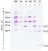
Isoelectric focusing patterns of AAT phenotypes. Healthy control MM AAT glycoforms (M2–M8) are denoted on the left. Glycoforms from an AATD patient homozygous for the Z allele (Z2, Z4 and Z6) are shown on the right.
Figure 3
Figure 3
Clinical implications of alpha-1 antitrypsin deficiency. Polymerised aggregates of Z-AAT protein are implicated in the pathogenesis of liver cirrhosis and chronic hepatitis. Accumulation of Z-AAT in hepatocytes leads to impaired secretion of the protein, with individuals homozygous for the Z mutation having 10–15% of normal circulating levels of AAT. Deficiency in AAT results in high influx of neutrophils to the airways, where increased release of serine proteases and uninhibited NE activity can cause damage to lung parenchyma, ultimately leading to emphysema and COPD. In rare cases, AATD is associated with a severe skin condition known as panniculitis and antineutrophil cytoplasmic antibody associated vasculitis (granulomatosis with polyangitis, formally Wegener’s granulomatosis). Panniculitis is characterised by intense neutrophil infiltrates in the subcutaneous tissues and resultant tissue destruction due to the low levels of antiprotease and high levels of protease.
Figure 4
Figure 4
Alpha-1 antitrypsin elastase complex. Model based on the crystal structure of alpha-1 antitrypsin complexed with porcine pancreatic elastase (pdb code 2D26) and the crystal structure of human neutrophil elastase (pdb code 3Q76).

References

    1. Schroeder W.T., Miller M.F., Woo S.L., Saunders G.F. Chromosomal localization of the human alpha 1-antitrypsin gene (PI) to 14q31–32. Am. J. Hum. Genet. 1985;37:868–872. - PMC - PubMed
    1. Hunt L.T., Dayhoff M.O. A surprising new protein superfamily containing ovalbumin, antithrombin-III, and alpha1-proteinase inhibitor. Biochem. Biophys. Res. Commun. 1980;95:864–871. doi: 10.1016/0006-291X(80)90867-0. - DOI - PubMed
    1. Van Gent D., Sharp P., Morgan K., Kalsheker N. Serpins: Structure, function and molecular evolution. Int. J. Biochem. Cell Biol. 2003;35:1536–1547. doi: 10.1016/S1357-2725(03)00134-1. - DOI - PubMed
    1. Hammond G.L., Smith C.L., Paterson N.A.M., Sibbald W.J. A role for corticosteroid-binding globulin in delivery of cortisol to activated neutrophils. J. Clin. Endocrinol. Metab. 1990;71:34–39. doi: 10.1210/jcem-71-1-34. - DOI - PubMed
    1. Huber R., Carrell R.W. Implications of the three-dimensional structure of alpha.1-antitrypsin for structure and function of serpins. Biochemistry. 1989;28:8951–8966. doi: 10.1021/bi00449a001. - DOI - PubMed