. 2022 Feb 23;23(5):2458.
doi: 10.3390/ijms23052458.

Production of an Active, Human Membrane Protein in Saccharomyces cerevisiae: Full-Length FICD

Affiliations

Production of an Active, Human Membrane Protein in Saccharomyces cerevisiae: Full-Length FICD

Minttu S Virolainen et al. Int J Mol Sci. .

Abstract

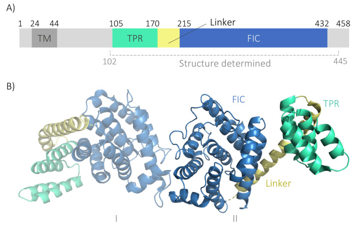
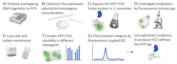
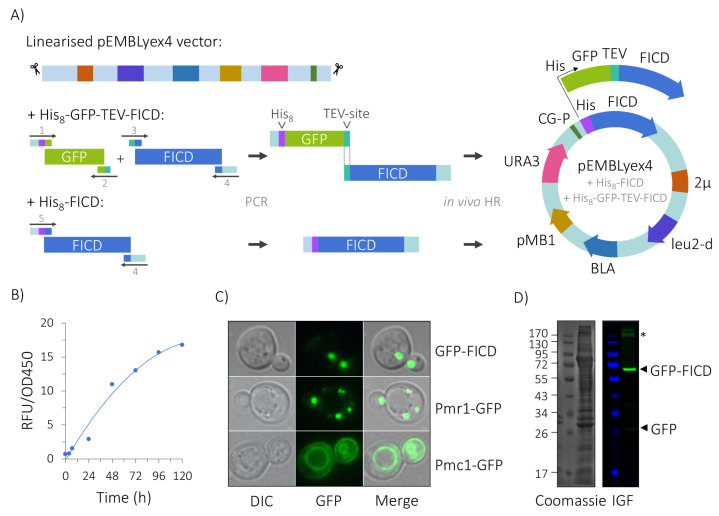
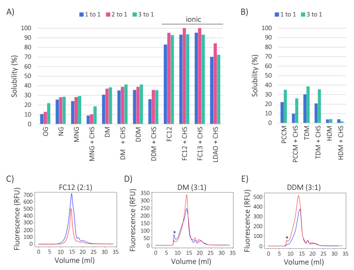
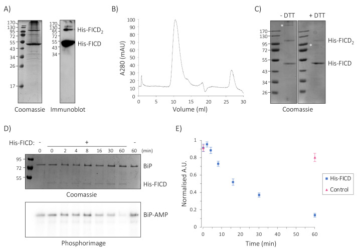
The human Fic domain-containing protein (FICD) is a type II endoplasmic reticulum (ER) membrane protein that is important for the maintenance of ER proteostasis. Structural and in vitro biochemical characterisation of FICD AMPylase and deAMPylase activity have been restricted to the soluble ER-luminal domain produced in Escherichia coli. Information about potentially important features, such as structural motifs, modulator binding sites or other regulatory elements, is therefore missing for the approximately 100 N-terminal residues including the transmembrane region of FICD. Expressing and purifying the required quantity and quality of membrane proteins is demanding because of the low yields and poor stability often observed. Here, we produce full-length FICD by combining a Saccharomyces cerevisiae-based platform with green fluorescent protein (GFP) tagging to optimise the conditions for expression, solubilisation and purification. We subsequently employ these conditions to purify milligram quantities of His-tagged FICD per litre of culture, and show that the purified, detergent-solubilised membrane protein is an active deAMPylating enzyme. Our work provides a straightforward methodology for producing not only full-length FICD, but also other membrane proteins in S. cerevisiae for structural and biochemical characterisation.

Keywords: AMPylation; FICD; Fic proteins; Saccharomyces cerevisiae; membrane protein purification; recombinant protein expression.


Conflict of interest statement

The authors declare no conflict of interest.

Figures

Figure 1
Figure 1
Schematic overview of the human FICD structure. (A) FICD is comprised of a short N-terminal cytosolic tail, a transmembrane domain (TM; dark grey), two tetratricopeptide repeats (TPR; turquoise), a linker (yellow), and the catalytic Fic domain (FIC; blue). The segment for which crystal structures have been solved (approximately residues 102 to 445 depending on the particular study) is indicated [10,11,12,22]. The numbers denote residue positions that mark the boundaries of key regions in the structure of FICD. (B) Crystal structure of the N-terminally truncated, soluble FICD103–433 dimer (PDB ID: 4U04) displays a helical conformation in which FICD forms an asymmetric, non-covalent dimer via the protomer Fic domains (I and II). Same colour scheme as Panel A. Created using PyMol [23].
Figure 2
Figure 2
Workflow for screening GFP-FICD expression and purification conditions. (A,B) The expression vector was constructed from overlapping DNA fragments using homologous recombination in S. cerevisiae. (C) PAP1500 expression system was used to express GFP-tagged FICD, (D) and membrane insertion was checked by fluorescence microscopy. (E) Cells were subsequently homogenised and the membranes isolated. (F) GFP fluorescence was used to quantify solubilisation efficiency and identify the best detergents. (G) Homogeneity of solubilised FICD was determined using fluorescence-coupled size exclusion chromatography (FSEC). LUM; ER lumen, CYT; cytosol.
Figure 3
Figure 3
Generation and expression of FICD constructs. (A) FICD was tagged with His8-GFP-TEV (where TEV denotes a tobacco etch virus protease cleavage site) or His8 alone and ligated into linearised pEMBLyex4 vector by in vivo homologous recombination (HR) in S. cerevisiae. The His8-GFP-TEV, TEV-FICD and His8-FICD DNA fragments were constructed by PCR using appropriate cDNA templates and primers containing overlapping sequences (see Table S1 for primer details). CG-P, CYC-GAL promoter; leu2-d, a poorly expressing allele of the β-isopropylmalate dehydrogenase gene; URA3, selective marker gene encoding the orotidine-5′-phosphate decarboxylase enzyme; BLA, β-lactamase gene; 2μ, 2-μm origin of replication; pMB1, bacterial origin of replication. (B) Time dependent accumulation of His8-GFP-TEV-FICD (GFP-FICD) after galactose induction at 15 °C, with whole-cell fluorescence measured in relative fluorescence units (RFU) in 1 OD450 unit of cells. (C) Cellular localisation of GFP-FICD, Pmr1-GFP (Calcium-transporting ATPase 1 (Golgi)) and Pmc1-GFP (Calcium-transporting ATPase 2 (vacuole)), imaged by fluorescence and phase contrast microscopy. (1000×). (D) SDS-PAGE analysis of GFP-FICD-containing membranes (corresponding to 200 RFU) by Coomassie staining (left) and in-gel fluorescence (IGF, right). High molecular weight species are marked with an asterisk (*).
Figure 4
Figure 4
GFP-FICD maintains a folded conformation upon solubilisation. (A) Percentage solubility of GFP-FICD in the indicated detergent conditions (see Table 1 for detergent names and properties). When noted, solubilisation was performed in the presence of CHS. Fluorescence intensity was measured in the soluble fraction after ultracentrifugation, and solubility was quantified as the proportion of fluorescence in the soluble fraction relative to total membrane fluorescence. Solubility in three different detergent to protein ratios (1:1, 2:1 and 3:1) is shown. (B) GFP-FICD solubility measured as in A, but in two detergent to protein ratios (1:1 and 3:1). (CE) FSEC profiles of GFP-FICD solubilised in the indicated detergents in the highest yielding detergent to protein ratio (FC12; 2:1 (C), DM; 3:1 (D), DDM; 3:1 (E)). An in-line fluorescence detector was used to record the elution profile of GFP-FICD. Blue line, without CHS; red line, with CHS. Fluorescent material eluting in the void volume is denoted by an asterisk (*).
Figure 5
Figure 5
Solubilised His-FICD can be purified in a correctly folded, active conformation. (A) Non-reducing SDS-PAGE gel stained with Coomassie (left) and an immunoblot developed with α-FICD antiserum (right) of the IMAC purified FICD. Monomeric His-FICD (53 kDa) and dimeric His-FICD2 (106 kDa) are annotated. (B) SEC analysis of IMAC-purified FICD. Protein absorbance (A280) is plotted against the elution volume. (C) Non-reducing (left) and reducing (right) SDS-PAGE gel analysis of SEC-purified FICD. Where indicated, proteins were reduced using 10 mM dithiothreitol (DTT). Unidentified bands are marked with an asterisk (*). (D) Time-dependent deAMPylation of radioactively labelled BiP-α-[32P]-AMP in the presence and absence of His-FICD. The reaction was sampled and quenched at indicated time-points and analysed by SDS-PAGE. Coomassie stain (top) depicts the total protein content of the reaction, and the phosphorimage (bottom) shows the radioactive BiP-AMP signal. Representative gel and phosphorimage for seven experiments. (E) Quantification of deAMPylation of BiP-AMP in the presence (blue squares) and absence (red triangles) of His-FICD. Plotted are normalised mean values ± standard error of the mean (SEM) from seven replicates, performed in four independent experiments (one single experiment and three performed in duplicate).

Similar articles

Cited by

References

    1. Garcia-Pino A., Zenkin N., Loris R. The many faces of Fic: Structural and functional aspects of Fic enzymes. Trends Biochem. Sci. 2014;39:121–129. doi: 10.1016/j.tibs.2014.01.001. - DOI - PubMed
    1. Faber P.W., Barnes G.T., Srinidhi J., Chen J., Gusella J.F., MacDonald M.E. Huntingtin interacts with a family of WW domain proteins. Hum. Mol. Genet. 1998;7:1463–1474. doi: 10.1093/hmg/7.9.1463. - DOI - PubMed
    1. Khater S., Mohanty D. In silico identification of AMPylating enzymes and study of their divergent evolution. Sci. Rep. 2015;5:10804. doi: 10.1038/srep10804. - DOI - PMC - PubMed
    1. Sanyal A., Chen A.J., Nakayasu E.S., Lazar C.S., Zbornik E.A., Worby C.A., Koller A., Mattoo S. A novel link between Fic (filamentation induced by cAMP)-mediated adenylylation/AMPylation and the unfolded protein response. J. Biol. Chem. 2015;290:8482–8499. doi: 10.1074/jbc.M114.618348. - DOI - PMC - PubMed
    1. Ham H., Woolery A.R., Tracy C., Stenesen D., Kramer H., Orth K. Unfolded protein response-regulated Drosophila Fic (dFic) protein reversibly AMPylates BiP chaperone during endoplasmic reticulum homeostasis. J. Biol. Chem. 2014;289:36059–36069. doi: 10.1074/jbc.M114.612515. - DOI - PMC - PubMed

MeSH terms

Substances

LinkOut - more resources