. 2022 Feb 23;23(5):2452.
doi: 10.3390/ijms23052452.

Secondary Structure of Influenza A Virus Genomic Segment 8 RNA Folded in a Cellular Environment

Affiliations

Secondary Structure of Influenza A Virus Genomic Segment 8 RNA Folded in a Cellular Environment

Barbara Szutkowska et al. Int J Mol Sci. .

Abstract

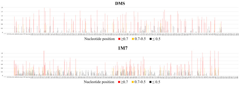
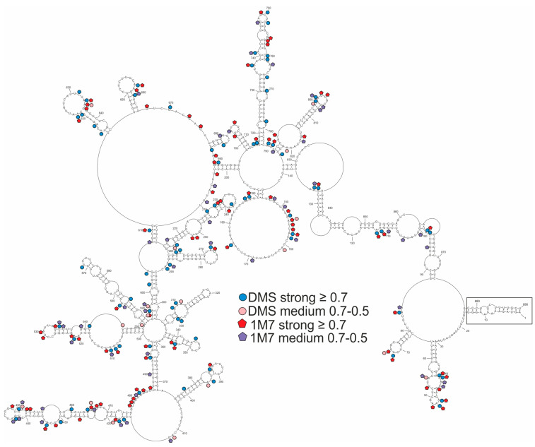
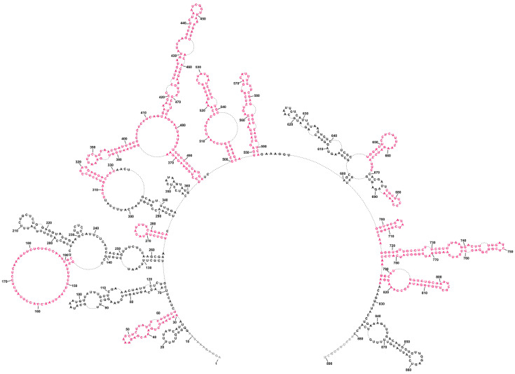
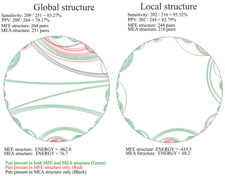
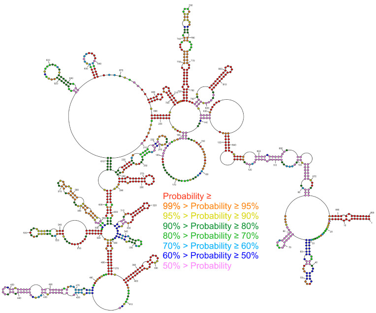
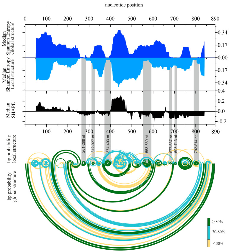
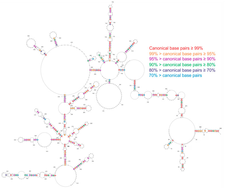
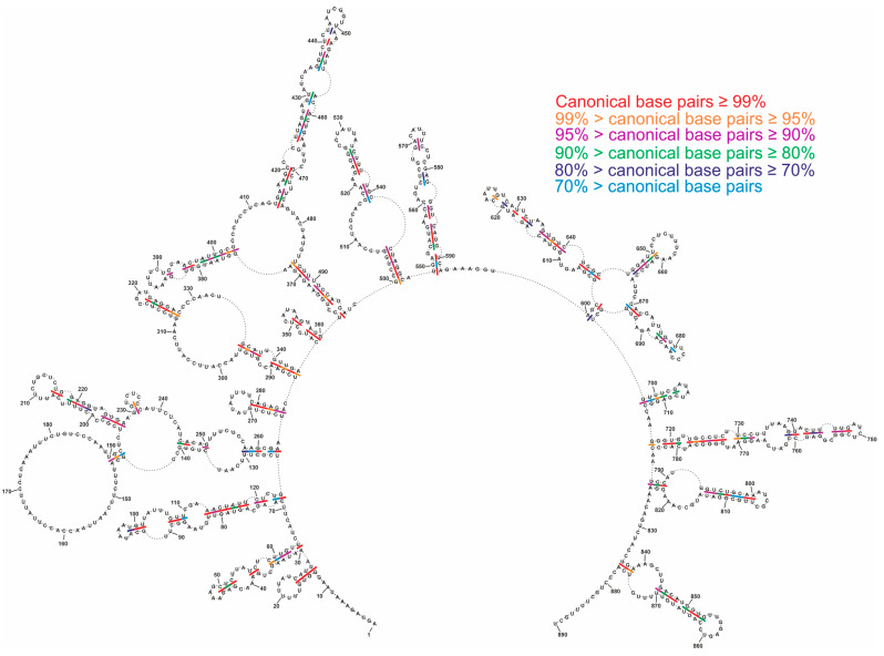
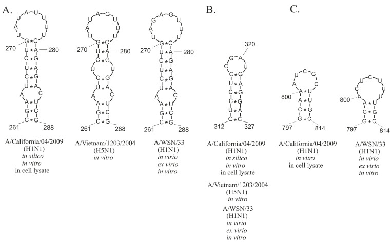
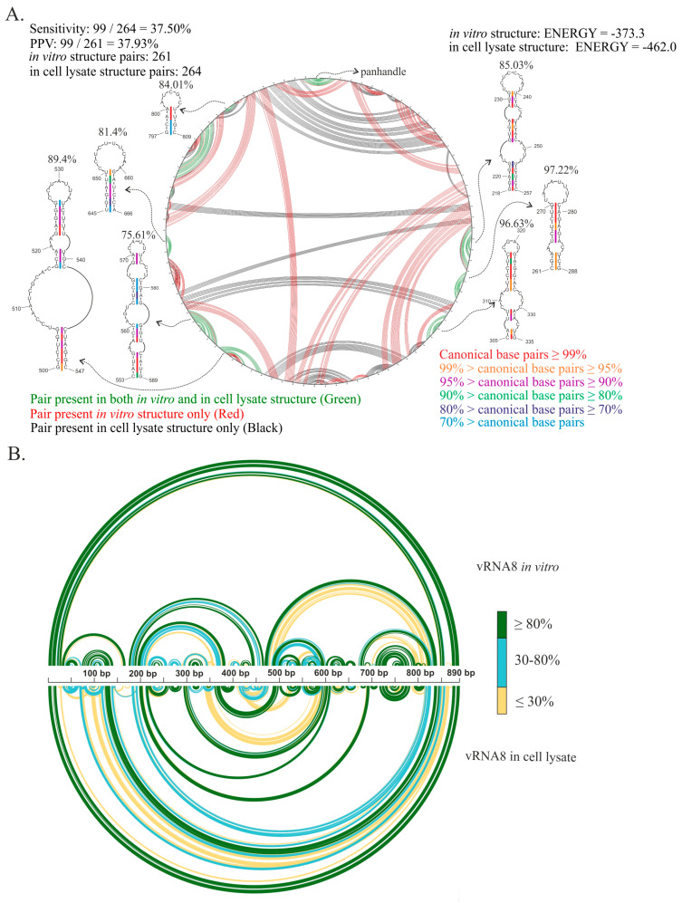
Influenza A virus (IAV) is a member of the single-stranded RNA (ssRNA) family of viruses. The most recent global pandemic caused by the SARS-CoV-2 virus has shown the major threat that RNA viruses can pose to humanity. In comparison, influenza has an even higher pandemic potential as a result of its high rate of mutations within its relatively short (<13 kbp) genome, as well as its capability to undergo genetic reassortment. In light of this threat, and the fact that RNA structure is connected to a broad range of known biological functions, deeper investigation of viral RNA (vRNA) structures is of high interest. Here, for the first time, we propose a secondary structure for segment 8 vRNA (vRNA8) of A/California/04/2009 (H1N1) formed in the presence of cellular and viral components. This structure shows similarities with prior in vitro experiments. Additionally, we determined the location of several well-defined, conserved structural motifs of vRNA8 within IAV strains with possible functionality. These RNA motifs appear to fold independently of regional nucleoprotein (NP)-binding affinity, but a low or uneven distribution of NP in each motif region is noted. This research also highlights several accessible sites for oligonucleotide tools and small molecules in vRNA8 in a cellular environment that might be a target for influenza A virus inhibition on the RNA level.

Keywords: IAV; RNA chemical mapping; RNA secondary structure; RNA virus; influenza A virus.


Conflict of interest statement

The authors declare no conflict of interest.

Figures

Figure 1
Figure 1
Average nucleotide reactivity distribution across vRNA8 A/California/04/2009. The vRNA8 chemical mapping experiments were performed in cell lysates at 37 °C with 1M7 (SHAPE) or DMS chemical reagents. The probing experiments were performed in three biological replicates of which a minimum of three technical replicates were made. High reactivities are marked with red (values ≥ 0.700), medium reactivities with yellow (values 0.500–0.700), and low reactivities (values ≤ 0.500) are marked with black. Regions of unknown reactivity due to the limitation of the read-out method were 1–38 nt and 818–890 nt (1M7); 1–38 nt and 828–890 nt (DMS).
Figure 2
Figure 2
Model of the MFE secondary structure of vRNA8 A/California/04/2009. This global folding model was predicted according to chemical probing with DMS and 1M7 (SHAPE) in cell lysates. Strong (≥0.7) and medium (0.7–0.5) reactivities are marked on the structure. The panhandle structure between the 3′ end and 5′ end is marked. Due to the limitations of reverse transcription as well as the readout method, there are no data from 1–38 nt, 818–890 nt (1M7); and 1–13 nt, 827–890 nt (DMS).
Figure 3
Figure 3
Local folding of vRNA8 A/California/04/2009 as predicted by RNAstructure using a maximum pairing distance. The structure was predicted by applying a maximum pairing distance of 150 nucleotides, with constraints from chemical probing with DMS and 1M7 (SHAPE) in cell lysates. This structure shows the preservation of 12 hairpins and a long loop region (red color) also predicted in the global structure without a distance restriction on base pairing (Figure 2).
Figure 4
Figure 4
Comparison of the predicted MFE and MEA structures of vRNA8 in cell lysates. For the comparison both global as well as local structures were compared via CircleCompare tool (RNAstructure www.rna.urmc.rochester.edu, accessed 27 January 2022).
Figure 5
Figure 5
Base-pairing probabilities of single- and double-stranded regions of the vRNA8 A/California/04/2009 structure in cell lysates. The colors indicate the percentage of pairing probability. The probabilities were calculated via the partition function in RNAstructure 6.2 using experimental data constraints.
Figure 6
Figure 6
The secondary structure of vRNA8 in cell lysate. Median Shannon entropies of global and local structures were calculated in a centered, sliding 50 nt window. Median SHAPE (1M7) reactivities were calculated in 50 nt window and plotted with respect to global median. Arc plots showing the base-pairing probabilities of predicted local (upper) and global (lower) structures were estimated using the partition function (RNAstructure). Grey shadings indicate the low Shannon entropy/SHAPE reactivity regions of the most probable, well-defined structural motifs predicted in both global and local vRNA8 secondary structures. Calculations for regions of 50 nt of vRNA8 from both ends (Median Shannon, SHAPE) were excluded from visualization. No reactivity data were obtained in regions: 1–38 nt and 818-890 nt (1M7).
Figure 7
Figure 7
Conservation of vRNA8 global structure across type A influenza. Base pair conservations were marked based on an analysis of 34,248 IAV strains sequences obtained from the NCBI Influenza Virus Database.
Figure 8
Figure 8
Conservation of vRNA8 local structure across type A influenza. Base pair conservations were marked based on the analysis of 34,248 IAV strains obtained from the NCBI Influenza Virus Database.
Figure 9
Figure 9
Conserved structural motifs of vRNA8 within different IAV strains. The motifs were predicted based on bioinformatics analysis and chemical probing experiments in different conditions. (A) Motif 261–288 nt, (B) Motif 312–327 nt, (C) Motif 797–814 nt.
Figure 10
Figure 10
Comparison of vRNA8 A/California/04/2009 structures predicted based on experimental results in vitro and in cell lysates. (A) CircleCompare comparison of structures shows differences (red color—pair predicted in vitro, black color—pair predicted in cell lysate) and similarities (green color) in base-pairing of vRNA8 between in vitro and in cell lysate. Low sensitivity and PPV values indicated a low resemblance between the vRNA8 structure probed in different environments. Structural motifs predicted in both environments are shown and the base-pairing conservation according to a color scale and average percentage conservation of each motif is indicated above. (B) Comparison of vRNA8 structures predicted in vitro and in cell lysates with the base-pairing probabilities calculated with partition function (RNAstructure). The colors indicate the probability of each base pair in a three-point color scale.

References

    1. Su S., Fu X., Li G., Kerlin F., Veit M. Novel Influenza D virus: Epidemiology, pathology, evolution and biological characteristics. Virulence. 2017;8:1580–1591. doi: 10.1080/21505594.2017.1365216. - DOI - PMC - PubMed
    1. Mostafa A., Abdelwhab E.M., Mettenleiter T.C., Pleschka S. Zoonotic potential of influenza A viruses: A comprehensive overview. Viruses. 2018;10:497. doi: 10.3390/v10090497. - DOI - PMC - PubMed
    1. World Health Organisation . Global Influenza Strategy Summary 2019–2030 Influenza. World Health Organisation; Geneva, Switzerland: 2019. pp. 1–2.
    1. Francis M.E., King M.L., Kelvin A.A. Back to the future for influenza preimmunity—Looking back at influenza virus history to infer the outcome of future infections. Viruses. 2019;11:122. doi: 10.3390/v11020122. - DOI - PMC - PubMed
    1. Duwe S. Influenza viruses-antiviral therapy and resistance. GMS Infect. Dis. 2017;5:Doc04. doi: 10.3205/id000030. - DOI - PMC - PubMed

LinkOut - more resources