Review
. 2022 Feb 25;23(5):2536.
doi: 10.3390/ijms23052536.

Mechanisms Underlying Dichotomous Procoagulant COAT Platelet Generation-A Conceptual Review Summarizing Current Knowledge

Affiliations
Review

Mechanisms Underlying Dichotomous Procoagulant COAT Platelet Generation-A Conceptual Review Summarizing Current Knowledge

Lucas Veuthey et al. Int J Mol Sci. .

Abstract

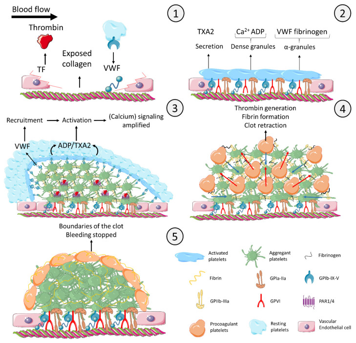
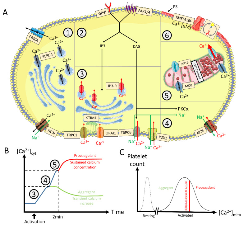
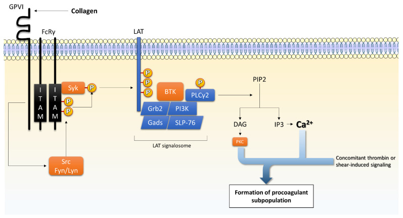
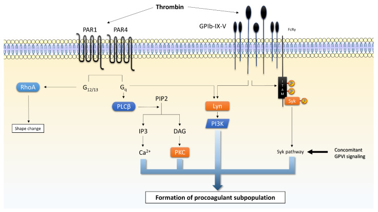
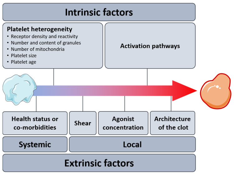
Procoagulant platelets are a subtype of activated platelets that sustains thrombin generation in order to consolidate the clot and stop bleeding. This aspect of platelet activation is gaining more and more recognition and interest. In fact, next to aggregating platelets, procoagulant platelets are key regulators of thrombus formation. Imbalance of both subpopulations can lead to undesired thrombotic or bleeding events. COAT platelets derive from a common pro-aggregatory phenotype in cells capable of accumulating enough cytosolic calcium to trigger specific pathways that mediate the loss of their aggregating properties and the development of new adhesive and procoagulant characteristics. Complex cascades of signaling events are involved and this may explain why an inter-individual variability exists in procoagulant potential. Nowadays, we know the key agonists and mediators underlying the generation of a procoagulant platelet response. However, we still lack insight into the actual mechanisms controlling this dichotomous pattern (i.e., procoagulant versus aggregating phenotype). In this review, we describe the phenotypic characteristics of procoagulant COAT platelets, we detail the current knowledge on the mechanisms of the procoagulant response, and discuss possible drivers of this dichotomous diversification, in particular addressing the impact of the platelet environment during in vivo thrombus formation.

Keywords: COAT; hemostasis; heterogeneity; procoagulant platelets; regulation; signaling.


Conflict of interest statement

The authors declare no conflict of interest.

Figures

Figure 1
Figure 1
Model of time-and agonist-dependent formation of the thrombus and diversification of platelets at the site of injury. Panel 1. Initiation: The injured endothelial cells express TF and secrete VWF. The subendothelial collagen becomes exposed. Initial thrombin is produced due to TF expression. Platelet GPIb-IX-V complex interacts with the VWF deposited on collagen. Panel 2. Secretion and shape change: Platelets bind to collagen via the receptor GPIa-IIa. Even though GPIb-IX-V and GPIa-IIa trigger intracellular signaling, platelet activation is mainly mediated by the collagen receptor GPVI. Activated platelets synthesize and secrete TXA2. Dense granules containing Ca2+ and ADP and α-granules containing VWF and fibrinogen are secreted. Panel 3. Aggregation and endoluminal thrombus growth: Platelets aggregate by means of fibrinogen bridging activated GPIIb-IIIa receptors (integrin αIIbβ3). Secreted TXA2 and ADP activate platelets that are recruited into the growing thrombus by VWF secreted from α-granules. Initial thrombin generation in combination with collagen further activates platelets in the thrombus core. Panel 4. Procoagulant platelets: As the thrombus is growing and the platelets become more and more activated, the increasing cytosolic level of calcium eventually creates procoagulant platelets. Procoagulant platelets downregulate their GPIIb-IIIa receptor, are coated with fibrinogen and other adhesive proteins to be retained in the thrombus, and express negatively charged phospholipids binding coagulation complexes, which localize and enhance thrombin generation. Thrombin converts fibrinogen into fibrin, which consolidates the platelet clot. In parallel, the outside-in signaling of aggregating platelets inducing clot retraction (blue arrows) squeezes procoagulant platelets out of core of the thrombus (red arrows). Panel 5. Termination: The final configuration completely stops the bleeding and defines the boundaries of the clot to avoid any undesired expansion. The figure uses modified images from Servier Medical Art under a Creative Commons Attribution 3.0 Unported License (http://smart.servier.com, accessed on 31 January 2021). This figure is inspired from [14,15,16,17,18]. Legend: ADP: adenosine diphosphate; Ca2+: calcium; GP: glycoprotein; PAR: protease activated receptor; TF: tissue factor; TXA2: thromboxane A2; VWF: von Willebrand factor.
Figure 2
Figure 2
Calcium mobilization mechanisms and intracellular profile leading to procoagulant COAT platelets. Panel (A). Cytosolic calcium regulation. 1. Downregulation: The intracellular level of calcium (Ca2+) is negatively regulated (Ca2+ in black) by PMCA and the forward mode of NCX (Ca2+ efflux towards the extracellular space) respectively compartmentalized by SERCA in intracellular stores (granules, DTS) and MCU in mitochondria. 2. Initial activation: Engagement of GPVI (by collagen) and PAR1/4 (by thrombin) mediate IP3 and DAG production. 3. Internal Ca2+ storage: IP3 triggers the release of Ca2+ from the internal stores through its receptor while the stores are refilled by ORAI1 mediated by STIM1 (Ca2+ sensor). TRPC1 is either agonist or storage depletion dependent and increases cytosolic Ca2+ and sodium (Na+) when activated. TRPC6 activation is mediated by DAG. ATP released from granules activates the P2X1 receptor increasing cytosolic Ca2+ and Na+. 4. NCX reversing: Increased cytosolic Na+ and PKCα contribute to the reverse mode of NCX. 5. Mitochondrial permeation: Cytosolic Ca2+ is transferred into mitochondria through MCU. At a given threshold, and in some platelets, mitochondrial Ca2+ triggers mPTP opening, releasing an important amount of Ca2+ in the cytosol. 6. PS exposure: Sustained micromolar levels of cytosolic Ca2+ are necessary to activate low Ca2+-sensitive actors such as calpain and TMEM16F. The latter scrambles membrane phospholipids inducing the expression of PS in the outer part of the cytosplamic membrane. Panel (B). Model of stepwise Ca2+ mobilization. After initial [Ca2+]cyt increase, some platelets do not trigger NCX reversing, and will remain aggregant decreasing their [Ca2+]cyt (green). Other platelets undergo NCX reversing, thus further increasing [Ca2+]cyt. Platelets become procoagulant (red) approx. 2 min after activation, when Ca2+ concentrations in cytoplasm and mitochondria reach a threshold level to induce mitochondrial depolarization, supramaximal [Ca2+]cyt, and subsequent PS exposure. Panel (C). Mitochondrial Ca2+ profile at the population level. The level of accumulated mitochondrial Ca2+ depends on previous activation of the Ca2+ mobilization sources following platelet activation. Mitochondria of a platelet subpopulation accumulate enough Ca2+ to open their PTP and induce a procoagulant response in those platelets. The figure uses modified images from Servier Medical Art under a Creative Commons Attribution 3.0 Unported License (http://smart.servier.com, accessed on 31 January 2021). This figure is inspired from [22,26,27]. Legend: [Ca2+]cyt/mito: cytosolic/mitochondrial calcium concentration; DAG: diacylglycerol; DTS: dense tubular system; IP3: inositol trisphosphate; GP: glycoprotein; PAR: protease activated receptors; MCU: mitochondrial calcium uniporter; NCXf/r: sodium calcium exchanger forward or reverse mode, respectively; PMCA: plasma membrane calcium ATPase; PS: phosphatidylserine; mPTP: mitochondrial permeability transition pore; SERCA: sarco-endoplasmic reticulum calcium ATPase; STIM: stromal interaction molecule; TMEM: transmembrane; TRPC: transient receptor potential C.
Figure 3
Figure 3
Collagen-mediated signaling via glycoprotein (GP) VI. In addition to adhesion mediated by GPIa-IIa (integrin α2β1), collagen induces platelet activation via GPVI. It triggers the formation of IP3 releasing Ca2+ from internal stores. PKC isoforms also participate in further platelet activation. This figure is inspired from [138,139,140]. Legend: BTK: Bruton’s kinases; Ca2+: calcium; DAG: diacylglycerol; FcRγ: Fc receptor γ-chain; IP3: inositol trisphosphate; ITAM: immunoreceptor tyrosine activation motif; LAT: linker for activation of T cells; MAPK: mitogen-activated protein kinases; PI3K: phosphatidylinositide-3-kinase; PIP2: phosphatidylinositol-4,5-bisphosphate; PLCγ2: phospholipase C γ2; Rap1: Ras-related protein 1; Syk: spleen tyrosine kinase; TXA2: thromboxane A2.
Figure 4
Figure 4
Thrombin-mediated signaling. Thrombin activates platelets through two types of receptors acting in a synergistic way: the G protein-coupled receptors PAR 1/4 and the GPIbα from the GPIb-IX-V complex. This figure is inspired, simplified, and adapted from [24,140,150]. Legend: Ca2+: calcium, DAG: diacylglycerol; G: guanine nucleotide-binding proteins; IP3: inositol trisphosphate; ITAM: immuno-receptor tyrosine activation motif; PAR: protease-activated receptors; PKC: protein kinase C; PI3K: phosphatidylinositide-3-kinase; PIP2: phosphatidylinositol-4,5-bisphosphate; PLC: phospholipase C; Rap1: Ras-related protein 1; RhoA: Ras homolog family member A; Syk: spleen tyrosine kinase.
Figure 5
Figure 5
Intrinsic and extrinsic factors potentially driving the procoagulant response of platelets.

References

    1. Alberio L., Safa O., Clemetson K.J., Esmon C.T., Dale G.L. Surface expression and functional characterization of α-granule factor V in human platelets: Effects of ionophore A23187, thrombin, collagen, and convulxin. Blood. 2000;95:1694–1702. doi: 10.1182/blood.V95.5.1694.005k24_1694_1702. - DOI - PubMed
    1. Aliotta A., Calderara D.B., Zermatten M.G., Marchetti M., Alberio L. Thrombocytopathies: Not Just Aggregation Defects-The Clinical Relevance of Procoagulant Platelets. J. Clin. Med. 2021;10:894. doi: 10.3390/jcm10050894. - DOI - PMC - PubMed
    1. Agbani E.O., Poole A.W. Procoagulant platelets: Generation, function, and therapeutic targeting in thrombosis. Blood. 2017;130:2171–2179. doi: 10.1182/blood-2017-05-787259. - DOI - PubMed
    1. Chu Y., Guo H., Zhang Y., Qiao R. Procoagulant platelets: Generation, characteristics, and therapeutic target. J. Clin. Lab. Anal. 2021;35:e23750. doi: 10.1002/jcla.23750. - DOI - PMC - PubMed
    1. Podoplelova N.A., Nechipurenko D.Y., Ignatova A.A., Sveshnikova A.N., Panteleev M.A. Procoagulant Platelets: Mechanisms of Generation and Action. Hämostaseologie. 2021;41:146–153. doi: 10.1055/a-1401-2706. - DOI - PubMed