. 2022 Feb 25;23(5):2546.
doi: 10.3390/ijms23052546.

Methyl Donors Reduce Cell Proliferation by Diminishing Erk-Signaling and NFkB Levels, While Increasing E-Cadherin Expression in Panc-1 Cell Line

Affiliations

Methyl Donors Reduce Cell Proliferation by Diminishing Erk-Signaling and NFkB Levels, While Increasing E-Cadherin Expression in Panc-1 Cell Line

Eva Kiss et al. Int J Mol Sci. .

Abstract

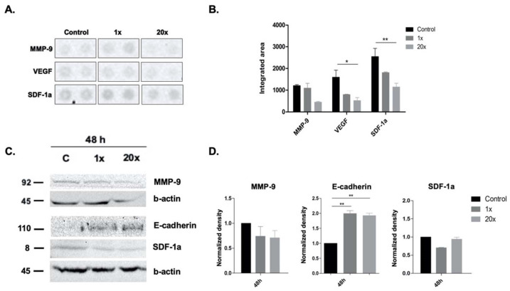
Pancreatic cancer is an aggressive malignancy with high metastatic potential. There are several lifestyle-related determinants in its etiology, including diet. Methyl donors are dietary micronutrients which play an important role in fueling vital metabolic pathways, and as bioactive food components provide methyl groups as substrates and cofactors. The imbalanced nutritional status of methyl donors has recently been linked to pathological conditions. Therefore, we hypothesized that dietary methyl donors may improve the physiology of cancer patients, including those with pancreatic cancer, and could be used for intervention therapy. In this study, methyl-donor treatment (L-methionine, choline chloride, folic acid and vitamin B12) of an aggressive pancreatic adenocarcinoma cell line (Panc-1) resulted in significantly increased p21WAF1/Cip1 cyclin-dependent kinase inhibitor levels, along with apoptotic SubG1 fractions. At the same time, phospho-Erk1/2 levels and proliferation rate were significantly reduced. Though methyl-donor treatments also increased the pro-apoptotic protein Bak, Puma and Caspase-9, it failed to elevate cleaved Caspase-3 levels. In addition, the treatment significantly reduced the production of the pro-inflammatory cytokine IL-17a and the transcription factor NFkB. Similarly, a significant decrease in VEGF and SDF-1a levels were detected, which may indicate reduced metastatic potential. As expected, E-cadherin expression was inversely associated with these changes, showing elevated expression after methyl-donor treatment. In summary, we found that methyl donors may have the potential to reduce aggressive and proliferative phenotype of Panc-1 cells. This suggests a promising role of dietary methyl donors for complementing relevant cancer therapies, even in treatment-resistant pancreatic adenocarcinomas.

Keywords: E-cadherin; apoptosis; cell cycle; methyl donors.


Conflict of interest statement

The authors declare no conflict of interest.

Figures

Figure 1
Figure 1
Proliferation and cell cycle analysis of Panc-1 cells after methyl-donor treatment. Proliferation of Panc-1 cells decreased significantly both at 48 h and 72 h after 1× and 20× methyl-donor treatments (A). SubG1 fraction of Panc-1 cells were significantly increased after 20× methyl-donor treatments at 48 h compared to untreated control (B,C). p21 (p21WAF1/Cip1) growth inhibitory protein significantly increased, while p-Erk 1/2 protein level significantly decreased after both 1× and 20× methyl-donor treatments at 48 h compared to untreated control (D,E). Each bar represents the average number of positive cells or normalized density in cell cycle analysis or Western blot, respectively; from at least 3 repeats ± SD. Statistical significance: *: p < 0.05; **: 0.01< p < 0.05; ***: p < 0.001. 1× and 20×: concentrations of methyl donors.
Figure 2
Figure 2
Detection of apoptosis in Panc-1 cell line. Early apoptotic cells (red-squared highlighted areas) did not change significantly; however, the number of live cells (lower left square) decreased significantly (A,B). Each bar represents the average percentage of positive cells in early apoptotic, late apoptotic, necrotic, and live cells area from at least 3 repeats ± SD. Statistical significance was plotted as **: p < 0.01. Early: early apoptotic cells; Late: late apoptotic cells; Necrotic: necrotic cells; Live: live cells. 1× and 20×: concentrations of methyl donors.
Figure 3
Figure 3
Western blot analysis of apoptotic proteins in methyl-donor-treated Panc-1 cells. Pro-apoptotic Bak, Puma and Caspase-9 were significantly increased in Panc-1 cells after 72 h of 20× methyl-donor treatment (AD). Each bar represents the average normalized density from at least 3 repeats ± SD. Statistical significances plotted as **: p < 0.01. 1× and 20×: concentration of methyl donors.
Figure 4
Figure 4
Cytokine array and Western blot analysis of metastatic potential-related markers in Panc-1 cell line. Significantly decreased VEGF and SDF-1a level by cytokine array and SDF-1a level by Western blot were detected in Panc-1 cells after 48 h of 20× methyl-donor treatment (AD). Expectedly, E-cadherin level changed inversely, and its significant increase was detected after 48 h of 20× methyl-donor treatment (C,D). Unexpectedly, we could not measure significantly decreased MMP-9 level after the methyl-donor treatments. Each bar represents the average normalized density from at least 3 repeats ± SD. Statistical significance are plotted as *: p < 0.05; **: p < 0.01. 1× and 20×: concentrations of methyl donors.
Figure 5
Figure 5
Cytokine array and Western blot analysis of inflammation-related markers in Panc-1 cell line. Significantly decreased level of IL-17a measured by cytokine array and NFkB by Western blot were detected after 48 h of 20× methyl-donor treatment (AD). Decreased protein level of CD30 did not reach significance after 48 h methyl-donor treatment, either by cytokine array (A,B) or by Western blot, similar to STAT3 protein level (C,D). Each bar represents the average normalized density from at least 3 repeats ± SD. Statistical significance are plotted as *: p < 0.05; **: p < 0.01. 1× and 20×: concentration of methyl donors.
Figure 6
Figure 6
Measurement of plasma IL-8 and IL-6 cytokine levels of pancreatic cancer patients. Significantly increased levels of IL-8 and IL-6 were detected by ELISA from 35 pancreatic cancer patients compared to non-cancerous controls (A,B). Each dot represents a concentration of either IL8 or IL-6 cytokine level in a pancreatic cancer patient where boxes show the median value with the interquartile range. Statistical significances are plotted as **: p < 0.01; ****: p < 0.0001.

Similar articles

Cited by

References

    1. Ferlay J., Partensky C., Bray F. More deaths from pancreatic cancer than breast cancer in the EU by 2017. Acta Oncol. 2016;55:1158–1160. doi: 10.1080/0284186X.2016.1197419. - DOI - PubMed
    1. Rawla P., Sunkara T., Gaduputi V. Epidemiology of Pancreatic Cancer: Global Trends, Etiology and Risk Factors. World J. Oncol. 2019;10:10–27. doi: 10.14740/wjon1166. - DOI - PMC - PubMed
    1. Kleeff J., Korc M., Apte M., La Vecchia C., Johnson C.D., Biankin A.V., Neale R.E., Tempero M., Tuveson D.A., Hruban R.P., et al. Pancreatic cancer. Nat. Rev. Dis. Primers. 2016;2:16022. doi: 10.1038/nrdp.2016.22. - DOI - PubMed
    1. Arnold M., Abnet C.C., Neale R.E., Vignat J., Giovannucci E.L., McGlynn K.A., Bray F. Global Burden of 5 Major Types of Gastrointestinal Cancer. Gastroenterology. 2020;159:335–349.e15. doi: 10.1053/j.gastro.2020.02.068. - DOI - PMC - PubMed
    1. Clare C.E., Brassington A.H., Kwong W.Y., Sinclair K.D. One-Carbon Metabolism: Linking Nutritional Biochemistry to Epigenetic Programming of Long-Term Development. Annu. Rev. Anim. Biosci. 2019;7:263–287. doi: 10.1146/annurev-animal-020518-115206. - DOI - PubMed