. 2022 Feb 26;23(5):2584.
doi: 10.3390/ijms23052584.

Mycobacterium tuberculosis Acetyltransferase Suppresses Oxidative Stress by Inducing Peroxisome Formation in Macrophages

Affiliations

Mycobacterium tuberculosis Acetyltransferase Suppresses Oxidative Stress by Inducing Peroxisome Formation in Macrophages

Ananyaashree Behera et al. Int J Mol Sci. .

Abstract

Mycobacterium tuberculosis (Mtb) inhibits host oxidative stress responses facilitating its survival in macrophages; however, the underlying molecular mechanisms are poorly understood. Here, we identified a Mtb acetyltransferase (Rv3034c) as a novel counter actor of macrophage oxidative stress responses by inducing peroxisome formation. An inducible Rv3034c deletion mutant of Mtb failed to induce peroxisome biogenesis, expression of the peroxisomal β-oxidation pathway intermediates (ACOX1, ACAA1, MFP2) in macrophages, resulting in reduced intracellular survival compared to the parental strain. This reduced virulence phenotype was rescued by repletion of Rv3034c. Peroxisome induction depended on the interaction between Rv3034c and the macrophage mannose receptor (MR). Interaction between Rv3034c and MR induced expression of the peroxisomal biogenesis proteins PEX5p, PEX13p, PEX14p, PEX11β, PEX19p, the peroxisomal membrane lipid transporter ABCD3, and catalase. Expression of PEX14p and ABCD3 was also enhanced in lungs from Mtb aerosol-infected mice. This is the first report that peroxisome-mediated control of ROS balance is essential for innate immune responses to Mtb but can be counteracted by the mycobacterial acetyltransferase Rv3034c. Thus, peroxisomes represent interesting targets for host-directed therapeutics to tuberculosis.

Keywords: Mycobacterium tuberculosis; Rv3034c; acetyltransferase; macrophages; oxidative stress; peroxisome.


Conflict of interest statement

The authors have no conflict of interest.

Figures
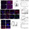
Figure 1
Figure 1
Determination of peroxisome formation in Mtb-infected mice and BMDM and determination of survival, expression, and acetyltransferase activity of Rv3034c. (A) Immunofluorescence analysis to determine the expression of peroxisome markers (ABCD3 and PEX14p) in control and Mtb-infected C57BL/6 mice (n = 5, male). Fluorescence intensity quantification has been represented in bar graph form. (B) Determination of different peroxisome-related markers in control and Mtb-infected BMDM using confocal microscopy. (C) Determination of intracellular survival of Mtb H37Rv and MtbRv3034c conditional mutants in mouse peritoneal macrophages. Mycobacterial strains grown in the presence and absence of pristinamycin are designated as “+pptr” and “−pptr”, respectively. Mouse peritoneal macrophages were infected with Mtb H37Rv (−pptr), Mtb H37Rv (+pptr), Mtb::Rv3034c (−pptr), and Mtb::Rv3034c (+pptr). The cells were lysed 0, 24, 48, and 72 h post-infection and the bacterial survival was determined by CFU assay. (D) qRT-PCR analysis to determine the intracellular expression of Rv3034c. Total RNA was isolated from Mtb H37Rv-(−pptr)-, Mtb-H37Rv-(+pptr)-, Mtb::Rv3034c-(−pptr)-, and Mtb::Rv3034c-(+pptr)-infected THP-1 cells. (E and F) Acetyltransferase activity was determined in protein lysates prepared from (E) Mtb H37Rv (±pptr) and Mtb::Rv3034c conditional mutants grown in absence (−pptr) and presence (+pptr) of pristinamycin in 7H9 medium for 24 h, and (F) purified rRv3034c protein. BSA was used as a negative control. 60 µg of protein lysates were added to the substrate and the absorbance was measured at 412 nm. The absorbance values plotted were obtained after deduction from the absorbance value of the control sample. (G) Immunofluorescence studies were performed to determine the expression of PEX5p and ABCD3 in Msm-pSMT3- and Msm-Rv3034c-infected macrophages after 24 h. The puncta per cell were represented in bar graph form. Experiments were performed in duplicates. Statistical significance was performed with one-way ANOVA. For acetyltransferase activity, statistical significance was performed with two-way ANOVA Bonferroni post-tests. Data represent mean ± SD; * for p ≤ 0.05, ** for p ≤ 0.01, and *** for p ≤ 0.001.
Figure 2
Figure 2
Determination of ROS production and expression of peroxisomal catalase in Mtb-infected BMDM and THP-1 cells. (A) ROS production was determined in Msm-pSMT3- and Msm-Rv3034c-infected and Msm-Rv3034c-plus-zymosan- (200 μg/mL) treated macrophages by DCFH-DA staining using flow cytometry. (B) Determination of peroxisomal catalase in control and Mtb-infected BMDM using confocal microscopy. (C) qRT-PCR of catalase was performed from total RNA isolated from THP-1 cells infected with Mtb H37Rv (−pptr), Mtb H37Rv (+pptr), Mtb::Rv3034c mutant (−pptr), and Mtb::Rv3034c (+pptr) after 24 h. The expression values were normalized with the GAPDH gene. Experiments were performed in duplicates. Statistical significance was performed with one-way ANOVA. Data represent mean ± SD; *** for p ≤ 0.001.
Figure 3
Figure 3
Analysis of expression of peroxisomal β-fatty acid oxidation and bacterial glyoxylate cycle intermediates in Mtb-infected BMDM and THP-1 cells and determination of Rv3034c survival, ROS production, and expression of peroxisomal proteins after treatment with peroxisomal β-oxidation inhibitor. (A) Determination of different peroxisomal β-fatty acid oxidation markers in control and Mtb-infected BMDM using confocal microscopy. (B,C) qRT-PCR analysis to determine the expression of peroxisomal β-fatty acid oxidation (B) and glyoxylate genes (C) from the total RNA isolated from THP-1 cells infected with Mtb H37Rv (−pptr), Mtb H37Rv (+pptr), Mtb::Rv3034c (−pptr), and Mtb::Rv3034c (+pptr). The expression values were normalized with GAPDH and sigA genes. (D) Intracellular survival of MsmWT, Msm pSMT3, and Msm Rv3034c strains in untreated and TZ-treated macrophages after 24 h. (E) Determination of ROS production by DCFH-DA staining in untreated (upper panel) and TZ-treated (lower panel) macrophages. Macrophages were infected with Msm pSMT3 and Msm Rv3034c for 24 h in presence of TZ. (F) Western blot analysis was performed to check the expression of peroxisome markers (PEX5p and ABCD3) in RAW264.7 macrophages infected with Msm pSMT3 and Msm Rv3034c in absence and presence of peroxisome inhibitor (TZ) after 24-h infection. Experiments were performed in duplicates. Statistical significance was performed with two-way ANOVA. Data represent mean ± SD; ** for p≤ 0.01 and *** for p ≤ 0.001.
Figure 4
Figure 4
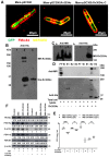
Cellular localization of Rv3034c and analysis of Mtb Rv3034c and mannose receptor interaction and expression of peroxisomal proteins in infected THP-1 cells. (A) Localization of Rv3034c in Msm-pSC300, Msm-pSC300:Rv3034c, and Msm-pSC300:Rv3034c-C strains using FM4-64 membrane staining dye by fluorescence microscopy. (B) Western blot analysis was performed to check the interaction of Mtb Rv3034c with mannose receptor (MR) using anti-His Tag antibody. Lane 1- whole cell protein (WCP) isolated from mouse macrophages (RAW264.7) expressing mannose receptor (MR), Lane 2- purified Rv3034c protein (containing His Tag) mixed with WCP isolated from RAW264.7 cells, and Lane 3- purified Rv3034c protein. (C) Western blot analysis to determine the interaction between Rv3034c and MR using MR antibody. Lane 1-whole cell protein isolated from RAW264.7 cells and Lane 2- purified Rv3034c protein (containing His Tag) mixed with WCP lysate of RAW264.7. The membrane was probed with anti-MR antibody (a) and anti-His tag antibody (b). (D) Ni-NTA affinity pull-down assay was performed to check the interaction of Mtb Rv3034c with MR using anti-His Tag antibody. First five lanes correspond to only WCP lysates isolated from RAW264.7 cells (expressing MR) and the last five lanes correspond to WCP lysates and purified Rv3034c-His protein (MR-Rv3034c-His protein) incubated with Ni-NTA beads. The eluted fractions were separated on 12% SDS-PAGE and Western blot was done using anti-MR and His-Tag antibodies. (E) Intracellular survival of Mtb H37Rv and Mtb Rv3034c conditional mutants in presence and absence of MR blocker in mouse peritoneal macrophages. Macrophages were infected with Mtb strains in presence and absence of pristinamycin and anti-MR antibody for 24 h. Intracellular bacterial survival was determined by CFU assay. (F) Determination of expression of peroxisomal proteins in presence and absence of MR blocker and different Mtb-strain-infected cells. THP-1 cells were infected with Mtb H37Rv, Mtb::Rv3034c (−pptr), and Mtb::Rv3034c (+pptr) in presence and absence of mannose receptor blocker. Protein lysates were prepared after 24 h of infection, and the expression of PEX11β, PEX19p, PEX5p, and ABCD3 was determined by Western blot analysis. Experiments were performed in duplicates. For Mtb survival, statistical analysis was performed with one-way ANOVA. Data represent mean ± SD; *** for p ≤ 0.001 and ns for non-significant.
Figure 5
Figure 5
Diagrammatic representation of the role of Rv3034c in modulating host immune responses by inhibiting ROS production through induction of peroxisomal proteins and increased bacterial persistence by induction of glyoxylate pathway. Here, we studied that Mtb Rv3034c interacts with the mannose receptor present on the host macrophages. This leads to transcriptional activation of peroxisomal proteins (PEX5p, PEX11β, PEX19p) and related enzymes (such as ACOX1 and ACAA1), as well as expression of antioxidative enzyme catalase. It is considered that ROS generated during peroxisomal β-oxidation is counterbalanced by the overexpression of catalase during MtbRv3034c infection. The growth and proliferation of pre-peroxisomal vesicles are mediated via PEX11β and PEX19p expression, which further recruits ABCD3. The final maturation occurs via the expression of PEX5p and other peroxins. Peroxisomal β-fatty acid oxidation pathway helps in the metabolism of long-chain fatty acids into short-chain fatty acids and acetylCoA. Induction of peroxisomes post-infection also activates the glyoxylate pathway. Thus, Mtb-Rv3034c-mediated events help in promoting persistence of Mtb in macrophages. The diagram was created with BioRender.com.

References

    1. World Health Organization . Global Tuberculosis Report 2020. World Health Organization; Geneva, Switzerland: 2020.
    1. Zammarchi L., Bartalesi F., Bartoloni A. Tuberculosis in tropical areas and immigrants. Mediterr. J. Hematol. Infect. Dis. 2014;6:e2014043. doi: 10.4084/mjhid.2014.043. - DOI - PMC - PubMed
    1. Smith T., Wolff K.A., Nguyen L. Molecular Biology of Drug Resistance in Mycobacterium tuberculosis. In: Pieters J., McKinney J.D., editors. Pathogenesis of Mycobacterium tuberculosis and Its Interaction with the Host Organism. Springer; Berlin/Heidelberg, Germany: 2013. pp. 53–80. - PMC - PubMed
    1. Ehrt S., Schnappinger D. Mycobacterial survival strategies in the phagosome: Defence against host stresses. Cell. Microbiol. 2009;11:1170–1178. doi: 10.1111/j.1462-5822.2009.01335.x. - DOI - PMC - PubMed
    1. Guirado E., Schlesinger L.S., Kaplan G. Macrophages in tuberculosis: Friend or foe. Semin. Immunopathol. 2013;35:563–583. doi: 10.1007/s00281-013-0388-2. - DOI - PMC - PubMed

Substances

LinkOut - more resources