Review
. 2022 Feb 26;23(5):2594.
doi: 10.3390/ijms23052594.

Identification of Neoantigens in Cancer Cells as Targets for Immunotherapy

Affiliations
Review

Identification of Neoantigens in Cancer Cells as Targets for Immunotherapy

Masahiro Okada et al. Int J Mol Sci. .

Abstract

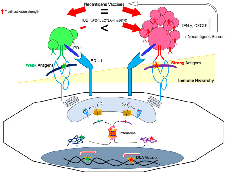
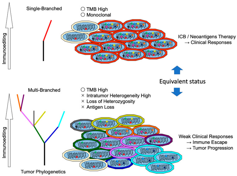
The clinical benefits of immune checkpoint blockage (ICB) therapy have been widely reported. In patients with cancer, researchers have demonstrated the clinical potential of antitumor cytotoxic T cells that can be reinvigorated or enhanced by ICB. Compared to self-antigens, neoantigens derived from tumor somatic mutations are believed to be ideal immune targets in tumors. Candidate tumor neoantigens can be identified through immunogenomic or immunopeptidomic approaches. Identification of neoantigens has revealed several points of the clinical relevance. For instance, tumor mutation burden (TMB) may be an indicator of immunotherapy. In various cancers, mutation rates accompanying neoantigen loads may be indicative of immunotherapy. Furthermore, mismatch repair-deficient tumors can be eradicated by T cells in ICB treatment. Hence, immunotherapies using vaccines or adoptive T-cell transfer targeting neoantigens are potential innovative strategies. However, significant efforts are required to identify the optimal epitopes. In this review, we summarize the recent progress in the identification of neoantigens and discussed preclinical and clinical studies based on neoantigens. We also discuss the issues remaining to be addressed before clinical applications of these new therapeutic strategies can be materialized.

Keywords: ICB; neoantigens; vaccines.


Conflict of interest statement

The authors declare no conflict of interest.

Figures

Figure 1
Figure 1
Neoantigen presentation and T cell responses. Cellular proteins are degraded by the Ub-proteasome. Some peptide products are transported and further processed in the ER, then loaded onto MHC-I, and presented on the cell surface. (a) Autologous T cells cannot recognize self-antigens. In contrast, (b) mutant proteins resulting from tumor somatic mutations yield mutant peptides, which facilitate MHC-I interaction or TCR recognition depending on the mutant position. By responding to the neoantigen, T cells proliferate and show activated phenotypes with tumoricidal capacity.
Figure 2
Figure 2
Neoantigen identification by immunogenomic or immunopeptidomic method. Tumor biopsy samples are analyzed by immunogenomic or immunopeptidomic method. (a) In immunogenomic method, tumor and matched germinal tissues are subjected to exome and tumor RNA-seq to detect somatic mutations in expressed genes. Overlapping missense or indel mutation peptide sequences are analyzed to predict affinity to each MHC-I allele. (b) In the immunopeptidomic method, tumor tissues are lysed, and peptide/MHC-I complexes are purified by immunoprecipitation using anti-MHC-I antibodies. Binding peptides are eluted and separated by size. Then, mass spectrometry is performed to determine molecular weight and identifying corresponding mutated peptides. Using candidate neoantigen peptides, T cell responses are investigated by evaluating cytokine production, activation marker expression, and tetramer staining. Validated neoantigen peptide data are subjected to machine learning analysis.
Figure 3
Figure 3
Neoantigen hierarchy. Most tumors present dominant and subdominant antigens on the surface. (i) PD-L1 on tumors suppresses neoantigen-reactive T cells. (ii) Immune checkpoint blockage preferentially reinvigorates dominant antigens. The subdominant neoantigens are also discovered by a highly sensitive neoantigen screen. Subdominant neoantigen-specific T cells can be activated by vaccines and dominant ones and can attack tumors.
Figure 4
Figure 4
Relation between neoantigen clonality and intratumor heterogeneity. Immunoediting shapes tumor evolution. Immune cells eliminate neoantigens expressing tumor cells at early stages. (a) High mutation burden with monoclonal tumors is responsive to immunotherapy. Whereas (b) in progressed tumors, gain or loss mutations in tumor cells evade immune pressure. Beyond the equilibrium point, tumors aggressively proliferate, and heterogenous cancerous cells compose the tumor tissue. This ostensible high mutation burden with multiclonal tumors is resistant to immunotherapy.

References

    1. Schumacher T.N., Schreiber R.D. Neoantigens in Cancer Immunotherapy. Science. 2015;348:69–74. doi: 10.1126/science.aaa4971. - DOI - PubMed
    1. Bobisse S., Foukas P.G., Coukos G., Harari A. Neoantigen-Based Cancer Immunotherapy. Ann. Trans. Med. 2016;4:262. doi: 10.21037/atm.2016.06.17. - DOI - PMC - PubMed
    1. Gupta R.G., Li F., Roszik J., Lizée G. Exploiting Tumor Neoantigens to Target Cancer Evolution: Current Challenges and Promising Therapeutic Approaches. Cancer Discov. 2021;11:1024–1039. doi: 10.1158/2159-8290.CD-20-1575. - DOI - PMC - PubMed
    1. Hodi F.S., O’Day S.J., McDermott D.F., Weber R.W., Sosman J.A., Haanen J.B., Gonzalez R., Robert C., Schadendorf D., Hassel J.C., et al. Improved Survival with Ipilimumab in Patients with Metastatic Melanoma. N. Engl. J. Med. 2010;363:711–723. doi: 10.1056/NEJMoa1003466. - DOI - PMC - PubMed
    1. Wolchok J.D., Chiarion-Sileni V., Gonzalez R., Rutkowski P., Grob J.-J., Cowey C.L., Lao C.D., Wagstaff J., Schadendorf D., Ferrucci P.F., et al. Overall Survival with Combined Nivolumab and Ipilimumab in Advanced Melanoma. N. Engl. J. Med. 2017;377:1345–1356. doi: 10.1056/NEJMoa1709684. - DOI - PMC - PubMed